

 PDF
PDF ePub
ePub Citation
 Print
Print


INTRODUCTION
A thin and damaged endometrium (Asherman's syndrome) has been the most challenging problem in the management of infertile women who present with various clinical symptoms, such as amenorrhea, hypomenorrhea, recurrent pregnancy loss, or pregnancy complications.12 Asherman's syndrome is usually characterized by poor growth of the glandular epithelium with little stroma, intrauterine adhesions or fibrosis, poor vascular development, and altered expression of adhesion-related cytokines.3
Several treatments have been tried to restore endometrial function, which include exogenous estrogen, aspirin, vaginal sildenafil citrate, vitamin E, and pentoxifylline.4567 Nonetheless, clinical outcomes are still poor.2 Recent studies have reported that bone marrow-derived stem cells (BMDSCs) can improve regeneration of damaged endometrium in murine models.891011 However, some issues are still unresolved with respect to the safety and usability of BMDSCs, including immunogenic reactions and effect on endometrial proliferative disorders, such as endometriosis, infections, and invasive process of harvesting.1213
Platelet-rich plasma (PRP) is composed of serum containing a platelet concentration of more than 1,000,000 platelets per cubic microliter, which is a rich source of growth factors having angiogenic and mitogenic properties, including platelet-derived growth factor (PDGF), transforming growth factor-beta (TGF-β), epidermal growth factor (EGF), fibroblast growth factor (FGF), and vascular endothelial growth factor (VEGF).1415 These growth factors have been demonstrated to play a role in modulation of endometrial cell proliferation and differentiation. 161718 Therefore, PRP is expected to have beneficial effects on damaged endometrium. Moreover, the use of PRP is considered safe based on long-term clinical experience, and it can be much more easily obtained, compared to BMDSCs.19
Several papers describing the specific use of ethanol in animal models have been published.102021 In the present study, we investigated whether PRP treatment can have a beneficial effect in a murine model of thin and damaged endometrium.
Go to : 
MATERIALS AND METHODS
Experimental animals
Female, adult Sprague-Dawley rats (n=60), weighing approximately 201.7 g (range, 183.0−215.6 g) each, were used. In our laboratory, two rats were housed together in a standard animal cage (420×270×180 mm). Rats had free access to water and pellets, were housed in a light-controlled room with 13 hours of light (7 AM−8 PM) and 11 hours of darkness, and were maintained at a temperature of 22 to 24℃. All procedures were supervised by a veterinarian. The study was approved by the Korea University School of Medicine Institutional Animal Care and Use Committee (KUIACUC-2014-194).
Groups and treatments
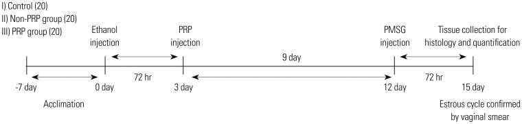
The rats were randomly assigned into three groups: sham-operated (control) group I, injection of physiological saline in the uterine horns; ethanol group II, injection of 95% ethanol into the uterine horns to induce damage, without PRP treatment; or PRP-treated group III, administration of PRP at 72 hours after damage as described in Fig. 1. After 7 days of acclimation, the animals were anesthetized for the surgical procedures with intramuscular injection of a mixture of 0.07 mL of Tiletamine plus Zolazepam (Zoletil 50, Virbac, Carros, France), 0.05 mL of xylazine (Rompun, Bayer Korea, Seoul, Korea), and 0.1 mL of normal saline. The uterine horn was exteriorized, and small curved hemostatic forceps were used to clamp each uterine horn near the cervix. While rats in the experimental groups (the ethanol and PRP-treated groups) were administered 0.5 mL of 95% ethanol into each uterine cavity with a 1 mL-syringe with a 30-gauge needle, each rat in the control group was administered normal saline. After 3 days of endometrial damage, each rat in the PRP-treated group was administered 0.25 mL of PRP into both uterine cavities as the same manner initially, while rats in the control and ethanol groups were administered normal saline. After 9 days of post-modeling, all the animals were treated intramuscularly with pregnant mare serum gonadotropin, which has been shown to induce ovulation at any stage of the estrous cycle in adult rats, and the estrous cycle was verified by daily vaginal smears within 3 days.22 Uterine horns were immediately excised after the animals were sacrificed and placed into 4% paraformaldehyde for further research. The samples were taken from the mid-uterine horn; the marginal portions of the upper and lower parts of the uterine horn were disregarded.
Blood collection and preparation of PRP
A total of 1.5 mL of whole blood was withdrawn into a sterile tube containing 0.35 mL of 10% sodium citrate via the retro-orbital plexus puncture in the anesthetized rat using a glass capillary tube. The blood underwent the first centrifugation at 160 g for 20 minutes at a temperature of 22℃ and then separated into three layers: the plasma, the buffy coat (platelets and leukocytes), and the erythrocytes. The layer just above the line dividing two fractions of the buffy coat and the red blood cells was then extracted and transferred to a new sterile tube. This layer underwent another centrifugation at 400 g for 15 minutes, and the platelets were allowed to sediment at the bottom of the tube. The lower half of the PRP layer was carefully collected using a pipette, and the upper half of the platelet-poor plasma layer was discarded. To activate the PRP, 0.15 mL of 3% calcium chloride was added into the liquid PRP, which then changed into a semi-solid, jelly-like structure. The autologous PRP was then carefully collected and administered into both uterine cavities of rats in the PRP-treated group. Our preliminary study with 20 collected blood samples demonstrated that, by using this method, we could obtain relatively constant platelet concentrations of 2,018,000±33,941.1/µL and a total white blood cell count of 1,480±565.6/µL, with 4.8±6.7% neutrophils, using an XE-5000 cell counter (Sysmex Co., Kobe, Japan). The PRP produced and used in this study was classified as P4-x-Bβ according to the PAW (platelet, activation, and white cells) classification system.23
Analysis of H&E staining
The biopsy specimens were fixed for 24 hours in 10% formalin, embedded in paraffin, cut into 4-µm thick sections and stained with hematoxylin-eosin (H&E). Endometrial morphology was analyzed by H&E staining, and images were captured using the Olympus BX43 microscope (Olympus Co., Tokyo, Japan) equipped with a DP27 camera (Olympus Co.). Images were captured at magnifications of ×40 and ×400. To determine the area of the endometrium (µm2), each slide was analyzed in a double-blinded manner by two experts using image analysis software (ImageJ 1.49p, National Institutes of Health, Bethesda, MD, USA) by performing the following steps: 1) The basal endometrial zone was outlined using area selection tools. 2) ‘Clear Outside’ was pressed to make the area outside the outlined endometrium part of the white background. 3) ‘Color Threshold’ was opened with the following parameters: Thresholding method, Default; Threshold color, Black & White; Color space, Lab; Dark background, checked. 4) To measure the total endometrial area, the brightness bar was adjusted to a point at which the total endometrial area was selected. 5) ‘Select’ was pressed to select the appropriate area.
The selected area was measured. The global scale of the image analysis was set at 6.21 pixels per µm, at a pixel ratio of 1.
MT staining
Collagen accumulation (mostly found in fibrous tissues) is known to be a major pathologic feature of fibrosis.24 Collagen fibers stain blue with Masson's trichrome (MT), highlighting degrees of fibrosis, which can be used to determine a variety of pathologic fibrotic processes.925 To investigate the histological assessment of the effect of PRP 12 days after the injection, we used MT to stain the collagen-rich fibrotic regions of paraffin-embedded tissue sections to assess and visualize extents of fibrosis. To objectively quantify the severity of fibrosis in sections, five random sections of endometrial tissue slides were electronically scanned into a tagged image file format (TIFF) file at a magnification of ×400 and subsequently analyzed in a double-blind manner by two experts using a computerized image analysis system as previously described.9 The amount of fibrosis (density and area) was then estimated from the TIFF image files.
IHC analysis
The growth of epithelial, stromal, and vascular cells was evaluated by immuno-histochemical (IHC) for cytokeratin (CK), homeobox A10 (HOXA10), VEGF, and Ki-67 using a diaminobenzidine-based staining system (Golden Bridge International Inc., Bothell, WA, USA). After deparaffinization and rehydration, antigen retrieval was performed with 0.01 M sodium citrate buffer (pH 6.0) using a water bath. The tissue sections were incubated in peroxide blocking buffer (ScyTek, Logan, UT, USA) for 10 min. Sections were then incubated with mouse monoclonal anti-pan CK antibody (ab7753; Abcam, Cambridge, UK) at a dilution of 1:250, rabbit monoclonal anti-Ki67 antibody (ab16667; Abcam) at a dilution of 1:100, rabbit polyclonal anti-HOXA10 antibody (orb13476; Biorbyt, Cambridge, UK) at a dilution of 1:250, and mouse monoclonal anti-VEGF antibody (ab1316; Abcam) at a dilution of 1:400. This was followed by a Polink-2 Plus horseradish-peroxidase (HRP) Anti-Mouse or Rabbit 3,3′-diaminobenzidene tetrahydrochloride (DAB) Detection kit (Golden Bridge International, Inc., Bothell) according to the manufacturer's instructions. For quantitative assessment of cytoplasmic or nuclear protein expressions of CK, HOXA10, and VEGF IHC staining, five randomly selected fields per section of endometrial tissue slides from at least 15 rats per group were electronically scanned into a TIFF image file at a magnification of ×400. This assessment was subsequently analyzed in a double-blind manner by two experts using an open-source plug-in (IHC Profiler) compatible with t ImageJ software (1.49p, National Institutes of Health) as previously described.26 The total percentage intensity (sum of high positive intensity, medium positive intensity, and low positive intensity) was used for the assessment of DAB images. For the quantitative analysis of nuclear markers for Ki-67 IHC staining, publicly available ImmunoRatio software with an adjusted Advanced Mode setting was used along with threshold values for hematoxylin (+30) and DAB (-30).27
RNA extraction and quantitative real-time PCR
Uterine tissue (30−50 mg) was homogenized with the Precellys® 24 tissue homogenizer (Bertin Technologies, Saint-Quentin-en-Yvelines, France) twice for 20 seconds at a speed of 6000 rpm with a 20-second pause between the homogenization steps. Total RNA was extracted using RNAiso Plus (9109; Takara Biotechnology Inc., Shiga, Japan) and treated with DNase I (18068-015; Invitrogen, Carlsbad, CA, USA). RNA yield was determined by a NanoDrop™ ND-1000 spectrophotometer at 260/280 nm (NanoDrop Technologies Inc., Wilmington, DE, USA). First-strand cDNA was synthesized from 1 µg of RNA with a PrimeScript™ RT reagent kit (RR037A; Takara Biotechnology Inc.) according to the manufacturer's instructions. Real-time polymerase chain reaction (PCR) measurement was performed on a CFX96 Touch qPCR system (Bio-Rad Laboratories, Foster City, CA, USA). Each PCR reaction contained 10 ng of cDNA, iQ SYBR green Supermix (170-888; Bio-Rad Laboratories), and 0.5 µM of each primer in a 20-µL reaction volume. The temperature profile was 95℃ for 3 minutes followed by 40 cycles of amplification (95℃ for 30 seconds, annealing temperature for 45 seconds, and 72℃ for 30 seconds). Values were normalized to the housekeeping gene (β-actin). Gene expression was calculated manually by the 2-ΔΔCT method. As a result, we investigated the fold change in expression of the target genes including HOXA10, VEGF-A, c-Kit (CD117), octamer-binding transcription factor 4 (Oct-4), interleukin (IL)-1β, IL-10, and nuclear factor-κB (NF-κB) relative to the internal control gene (β-actin). Primer sequences and annealing temperature are listed in Table 1.
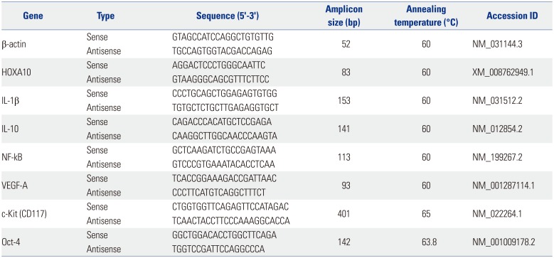
Table 1
Primers Used for Real-Time PCR
![]()
Statistical analyses
All data are expressed as the mean±standard error of the mean for continuous variables and as numbers or percentages for categorical variables. The Kruskal-Wallis test was used to compare differences in mean values for the three groups, and the Mann-Whitney test was used to compare two groups when the variable was continuous. Chi-square (χ2) tests were used to test independence of the categorical variables. p-values <0.05 were considered statistically significant, and all statistical analyses were performed using IBM SPSS Statistics software (version 20, IBM Corp., Armonk, NY, USA).
Go to :
RESULTS
H&E staining
To evaluate endometrial damage and the effect of PRP in our murine model of damaged endometrium, H&E staining and MT staining were performed. Section from the uterus of rats in the ethanol group showed narrowed endometrial lumen lined by atrophic columnar epithelium with degenerative changes and loss of endometrial glands. However, in the PRP-treated group, we found that mitotic activities in the functional layer and endometrial basal layer were distinctively increased with prominent nucleoli, proliferated endometrial glands, and endometrial stromal cells, compared to the ethanol group. Meanwhile, the surface epithelium was atrophic, compared to the control group (Fig. 2A).
MT staining
MT staining was used to assess the extent of fibrosis. This staining indicated that the PRP-treated group showed inhibition of excessive collagen deposition up to the level of the control group. However, MT staining confirmed significantly increased collagen deposition (light blue color staining) in the ethanol group, compared to the PRP-treated group (p<0.001) (Fig. 2B and D).
 | Fig. 2Comparison of the endometrial morphology, area, and extent of fibrosis. (A) On H&E staining, distinctive morphological changes in the endometrial characteristics were found in the glands, epithelial lining, and stroma between the ethanol group and the PRP-treated group. (B) On MT staining, the collagen-deposited fibrotic regions were shown in blue, and the extent of these regions was increased in the ethanol group compared to the other groups (left, control group; middle, ethanol group; right, PRP-treated group). (C) Comparison of the area of the endometrium. (D) Comparison of the extent of endometrial fibrosis (blue area on MT staining). Statistical comparisons were performed among the groups using the Kruskal-Wallis test and Mann-Whitney test. All images were captured at magnifications of ×40. H&E, hematoxylin-eosin; PRP, platelet-rich plasma; MT, Masson Trichrome.
|
Comparison of the endometrial area
The ethanol group and the PRP-treated group showed a significant decrease in the area of the endometrium, compared to the control group (Fig. 2C). The endometrial areas in the control, ethanol, and PRP-treated groups were as follows: 305.82±15.77 µm2, 212.83±15.84 µm2, and 262.34±12.33 µm2, respectively (Fig. 2C). The endometrial area in the PRP-treated group showed an increasing trend, compared to the endometrial area in the ethanol group; however, the difference was not statistically significant (p=0.065).
IHC staining for CK, HOXA10, VEGF, and Ki-67
In the control group, IHC results showed that the expression of CK was mainly localized in the cytoplasm of the endometrial epithelial cells and endometrial glands; whereas, the expressions of VEGF and HOXA10 were observed in the endometrial stromal cells, endometrial glands, and endometrial epithelial cells. IHC nuclear staining for Ki-67 was mainly observed in the endometrial stromal cells and endometrial glands (Fig. 3A, B, C, D). Compared to the control group, quantitative comparison of IHC staining in the ethanol group showed significantly decreased expressions of CK, HOXA10, VEGF, and Ki-67. The expression of these factors was significantly higher in the PRP-treated group, compared to the ethanol group. In addition, there was no significant difference in the expression of CK, HOXA10, and Ki-67 between the PRP-treated group and the control group (Fig. 3E and F).
 | Fig. 3Expression of (A) CK, (B) HOXA10, (C) VEGF, and (D) Ki-67 with IHC staining (left, control group; middle, ethanol group; right, PRP-treated group). (E) Comparisons of the expression of CK, HOXA10, and VEGF. Quantitative assessment of five random sections of endometrial tissue slides are shown as four zones with total/high/medium/low positive intensity. (F) Quantitative analysis of nuclear markers for Ki-67 IHC staining. Statistical comparisons of groups are based on the chi-square (χ2) tests, with statistical significance defined as p<0.05. *Significant when compared to the control, †Significant when compared between the ethanol group and the PRP-treated group. All images were captured at magnifications of ×400. CK, cytokeration; HOXA10, homeobox A10; VEGF, vascular endothelial growth factor; IHC, immuno-histochemical; PRP, platelet-rich plasma; DAB, diaminobenzidene tetrahydrochloride.
|
Real-time PCR assays for the expression of HOXA10, VEGF-A, c-Kit, Oct-4, IL-1β, IL-10, and NF-κB mRNA
The real-time PCR results for HOXA10 and VEGF-A showed significantly increased expression of HOXA10 (p=0.007) in the PRP-treated group, compared to the ethanol group and control group; however, no significant change in the expression of VEGF-A was noted (Fig. 4A). In addition, we found a significant up-regulation of c-Kit mRNA in the PRP-treated group, compared to the ethanol group (p=0.001). Expression of Oct-4 mRNA was significantly decreased in both the ethanol and PRP-treated groups, compared to the control group (p<0.05); however, there was no difference between the PRP-treated group and the ethanol group (Fig. 4B). The expression of pro-inflammatory cytokine, IL-1β mRNA, was found to be significantly up-regulated in the ethanol group, compared to the PRP-treated group (p=0.002) (Fig. 4C). IL-10 mRNA was prominently up-regulated in both the ethanol and PRP-treated groups, compared to the control group; no difference was observed upon comparing the PRP-treated group to the ethanol group. No significant change was found in the expression of NF-κB among the three groups.
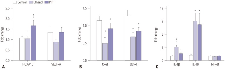 | Fig. 4The quantitative fold changes in the expression of the target gene (HOXA10, VEGF-A, c-Kit, Oct-4, IL-1β, IL-10, and NF-κB) relative to the internal control gene (β-actin) were analyzed using real-time PCR. (A) Significantly increased expression of HOXA10 in the PRP-treated group compared to the ethanol group, but no significant change in the expression of VEGF-A. (B) Significantly increased expression of the c-Kit gene in the PRP-treated group compared to the ethanol group, but no significant change in the expression of the Oct-4 gene. (C) Significantly increased expression of IL-1β in the ethanol group when compared to the other groups, but no significant change in the expressions of IL-10 and NF-κB on comparison of the ethanol group and the PRP-treated group. Statistical comparisons of groups are based on the Mann-Whitney test, with statistical significance defined as p<0.05. *Significant when compared to the control, †Significant when compared between the ethanol group and the PRP-treated group. HOXA10, homeobox A10; VEGF-A, vascular endothelial growth factor-A; Oct-4, octamer-binding transcription factor 4; IL, interleukin; NF-κB, nuclear factor-κB; PCR, polymerase chain reaction; PRP, platelet-rich plasma.
|
Go to :
DISCUSSION
The results of this study suggest that intrauterine administration of autologous PRP exerts proliferative and anti-fibrotic effects on damaged endometrium. PRP is known to contain a number of growth factors and cytokines that may aid in accelerating cell proliferation, angiogenesis, and cell migration, resulting in rapid healing and tissue regeneration.1415161718 Although there have been no specific studies assessing the effects of PRP on the endometrium, its major growth factor, PDGF, has been demonstrated to play several important roles in cellular proliferation within the endometrium.16172829 Other growth factors, including EGF, FGF, TGF-α, and TGF-β, insulin-like growth factor, and colony-stimulating factor, and their receptors have been identified and are considered to be involved in an autocrine and paracrine role in modulation of endometrial cell gr-owth, differentiation, and implantation.161718
In addition, to the best of our knowledge, this is the first study to show, using a model of damaged endometrium, that autologous PRP can enhance proliferation of the endometrium, and also decrease the degree of fibrosis. There has been a study demonstrating the anti-fibrotic effect of PRP on skeletal muscle healing after injury.25 The results of the present study, however, indicated that PRP inhibited excessive collagen deposition, suggesting that treatment with PRP may decrease the pro-gression of fibrosis in damaged endometrium.
Our analysis on the expression of Ki-67, CK, VEGF, and HOXA10 showed significant semi-quantitative differences between the PRP-treated and ethanol groups. In the present study, an increased level of Ki-67 expression was noted in the PRP-treated group, compared to the ethanol group, indicating cellular proliferation in the endometrium. The staining intensity of CK was also significantly stronger in the PRP-treated group, compared to the ethanol group, and was predominantly located in the epithelial cells lining the glands and the layer of columnar epithelium. Since CK is used as a cellular marker for the epithelium, our results indicated that PRP may accelerate induction of endometrial epithelial differentiation. Our analysis also included checking the status of the stromal endometrium for the possible use of vimentin (data not shown), a stromal cell marker. However, we found that vimentin was localized in the stromal cells and in endometrial epithelial cells, making it a relatively non-specific marker for immunostaining of this animal model.
On the other hand, HOXA10 is found to be an important transcription factor for many target genes involved in regulating endometrial function and development during the menstrual cycle, along with endometrial receptivity for establishing the necessary conditions for implantation in humans and mice.30 Additionally, VEGF is known to play a central role in angiogenesis and in the regulation of vasculogenesis; it is also found to be expressed and to optimize blastocyst implantation by mediating vascular permeability.31 In the present study, the expressions of HOXA10 and VEGF in the PRP-treated group were significantly increased on IHC staining, compared to those in the ethanol group. Similar results were also observed for mRNA expression of HOXA10 in real-time PCR. These results provided correlative evidence for the possible use of PRP in achieving implantation with improvement in uterine vascularization and endometrial receptivity.
PRP has been demonstrated to have an anti-inflammatory potential.32 In the present study, using real-time PCR, we detected the gene expression of IL-1β mRNA, IL-10 mRNA, and NF-kB mRNA. Since excessive damage to the endometrium caused by 95% ethanol induced a high inflammatory response, the results of these markers showed generally up-regulated gene expressions, especially IL-1β and IL-10 mRNA, in the treated groups, compared to the control group. Although no statistically significant differences were found in IL-10 and NF-kB mRNA between the ethanol-treated and PRP-treated groups, the expression level of pro-inflammatory cytokine, IL-1β mRNA, was found to be significantly decreased in the PRP-treated group, compared to the ethanol group, suggesting an anti-inflammatory potency of PRP. Further investigation is needed for proper elucidation of immunomodulatory effects of PRP since ethanol may affect the nuclear translocation of NF-κB and inflammatory cytokine production.3334
The stem/progenitor cells detected in the basal layer of endometrium are thought to be associated with regeneration of damaged endometrium.35 More recently, several studies reported that BMDSCs can be recruited to and have a therapeutic effect on damaged endometrium.891011 However, the exact mechanisms are not known, and it is uncertain whether these cells engraft the endometrium or whether they cooperate with other factors that stimulate endometrial stem cells and aid in endometrial repair.9 In the present study, the PRP-treated group showed a statistically significant increase in the mRNA level of c-Kit, compared to the ethanol group; in another study, c-Kit was found to be a typical stem cell marker in the human endometrium.36 These results are in line with previous findings and led us to consider, based on our own study, that endometrial stem cells may be involved in the proliferative and anti-fibrotic effects of PRP on damaged endometrium.37 PRP may exert these potential beneficial effects by stimulating the endometrial stem cells and promoting regeneration; hence, the combined use of PRP and stem cells may improve the outcome of treatment for damaged endometrium.
A few studies have already been published showing the beneficial effect of PRP in the endometrial growth and pregnancy outcome.383940 We are currently doing a preliminary clinical trial of PRP in infertile patients with a thin, homogenous, poorly developed endometrial lining. To date, the trial shows improvement of sonographic appearances such as a triple-line endometrial pattern and endometrial thickness (data not shown). In conclusion, this study showed positive effects of PRP as a potential novel treatment for women with a poor endometrium or a thin uterine lining unresponsive to standard treatments. Obviously, the clinical application of PRP and its effects on poor endometrium are still at a preliminary stage, and a lack of standardized preparation of PRP makes it difficult to establish an appropriate application. Further investigations and clinical trials are warranted for the optimization of PRP preparation along with a larger randomized study to determine the effect of PRP in poorly responding and repeated implantation failure patients due to thin and damaged endometrium.
Go to :
 XML Download