Journal List > Yonsei Med J > v.58(6) > 1032071

Moon, Choi, Chung, Sung, Cho, Chung, Kim, Yoo, Lee, and Park: Normative Values of Physical Examinations Commonly Used for Cerebral Palsy

Abstract

Purpose

The aim of this study was to establish normative values and to identify age-related change in physical examinations that are commonly used while evaluating patients with cerebral palsy (CP).

Materials and Methods

One hundred four healthy volunteers (mean age 36 years, standard deviation 15 years) were enrolled and divided into four age groups: 13−20, 21−35, 36−50, and 51 years and older. The eighteen physical examination tests for CP were selected by five orthopedic surgeons in consensus-building session. The measurements were taken by three orthopedic surgeons.

Results

There was no significant difference in the measures of physical examination among all the age groups, except for the Staheli test (p=0.002). The post hoc test revealed that the mean hip extension was 2.7° higher in the 13−20-year-old group than in the other age groups. The bilateral popliteal angle had a tendency to increase in those over 36-years-old. There were 31 participants (30%) with a unilateral popliteal angle greater than 40°.

Conclusion

We documented normative values that can be widely used for evaluating CP in patients 13 years and older.

INTRODUCTION

Cerebral palsy (CP) is primarily a permanent disorder that involves movement and posture, causing limitations in activity due to non-progressive disturbances that occur in the developing fetal or immature infant brain. Medical treatment is focused on younger patients with CP; however, this disease is a lifelong condition. The overall survival rate among all children with CP until 20 years of age is 90%.1 Cathels and Reddihough2 reported that young adults with CP had considerable, continuing impairment and disability. Thus, they require ongoing and periodic treatment.
To precisely assess the patient and prepare management plans, a medical history should be obtained, and a physical examination, functional assessment, medical imaging studies, and gait analysis should be performed. A physical examination is the most useful and crucial factor for determining the presence or absence of an underlying pathology. Therefore, clinicians need to know the normal ranges of physical examination variables to evaluate a patient for CP.
The physical examination of a child with CP has been studied extensively.345678910 In addition, numerous authors have reported the normal range of motion (ROM) in healthy adults.11121314151617 However, these reports do not include essential components needed for evaluating CP during a physical examination, such as the popliteal angle and Staheli test or Silfverskiöld test. For example, the clinical significance of a popliteal angle of 45° in a 5-year-old patient is different from that in a 35-year-old patient. It has been reported that the popliteal angle tends to increase with age in younger populations.18 However, there are few such studies including both adolescents and adults.
The purpose of this study was to establish normative values and evaluates age-related changes of the physical examination variables, which are commonly used for patients with CP.

MATERIALS AND METHODS

This prospective study was approved by the Institutional Review Board of the Seoul National University Bundang Hospital (IRB No. B-1511/321-003). Informed consent was obtained from all participants or their legal guardians.

Participants

This study was conducted with healthy adolescents and adults. One hundred four healthy volunteers were enrolled and divided into four age groups: 13−20, 21−35, 36−50, and 51 years and older. There were 26 participants in each group with an equal number of men and women (Table 1). A medical history and lower extremity teleroentgenogram were obtained for each patient. Participants with musculoskeletal diseases, a previous orthopedic surgery, or a medical or neurologic disease capable of affecting normal gait were excluded.

Physical examination

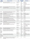
A consensus-building session was held by five orthopedic surgeons, each of whom had 5−15 years of orthopedic experience, to select the appropriate components of a physical examination for patient with CP. A literature review focusing on the physical examination used to diagnose CP was performed. From the pooled articles, the physical examination components that were considered reliable by several authors were chosen.371920212223242526 Table 2 displays the studies that evaluated the physical examination tests for CP and their reliability. All decisions regarding inclusion of a physical examination and associated measurement protocols required unanimous agreement.
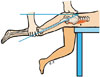
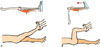
Selected items were: Thomas test, Staheli test, hip flexion, internal rotation of the hip, external rotation of the hip, adduction of the hip, abduction with hip extension, abduction with the hip at 90° flexion, trochanteric prominence angle, flexion contracture of the knee, knee flexion, unilateral popliteal angle, bilateral popliteal angle, ankle dorsiflexion with knee extension, ankle dorsiflexion with the knee at 90° flexion, and ankle plantar flexion. Hamstring shifting was defined as the difference between the bilateral popliteal angle and unilateral popliteal angle.
The measurements were taken by three orthopedic surgeons (SJM, BCC, and MKC) and each surgeon evaluated part of the patients. A rater or orthopedic surgeon positioned the patient, and a research assistant maintained each patient's position. Measurements were performed by the rater using a standard universal goniometer with an arm length of 18 cm and 1° increments. The method used to obtain physical measurements is described in detail in Table 2.

Statistical analysis

All data were analyzed with SPSS, version 22 (IBM, Corp., Armonk, NY, USA). We used the average value of the bilateral measurement for statistical independence when performing statistical analysis.27 One-way analysis of variance (ANOVA) with Bonferroni post hoc analysis was used to compare the age groups. p values less than 0.05 were considered statistically significant.

RESULTS

Participants' mean age was 36.1 years [standard deviation (SD) 15.2 years; range 13−69 years]. There were 52 male participants and 52 female participants. Participants' average body mass index was 23.5 kg/m2 (SD 3.0 kg/m2; range 15.9−30.7 kg/m2).
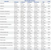
The Thomas test yielded a mean angle of 0.2° (SD 0.9°), whereas the Staheli test yielded a mean angle of -18.4° (SD 3.9°). The mean unilateral popliteal angle was 35.2° (SD 9.1°), and the mean bilateral popliteal angle was 25.5° (SD 8.5°). The mean hamstring shift was 9.7° (SD 4.6°). The mean angle of ankle dorsiflexion with knee extension was 11.3° (SD 4.8°), and the mean angle of ankle dorsiflexion with the knee at 90° flexion was 19.8° (SD 5.2°). The difference between the ankle dorsiflexion with knee extension and the ankle dorsiflexion with the knee at 90° flexion was 8.5°. The mean hip internal rotation was 39.1° (SD 9.0°), and the mean hip external rotation was 41.3° (SD 8.3°). The difference between the internal and external rotations was 2.2°; both rotations were almost symmetrical. The mean trochanteric prominence angle was 18.4° (SD 4.9°). The mean thigh-foot angle was 13.7° (SD 5.5°) (Table 3).
The Staheli test was statistically significantly different among the different age groups (p=0.002), according to one-way ANOVA. The post hoc test showed that the mean hip extension was 2.7° higher in the 13−20-year-old group than in the 21−35-year-old group (p=0.046), 3.8° higher in the 13−20-year-old group than in the 36−50-year-old group (p=0.002), and 2.8° higher in the 13−20-year-old group than in the over 51 years old group (p=0.037).

DISCUSSION

Our study presents normative data that can be widely used as a reference for evaluating patients aged 13 years and older with CP. There was no significant difference in the measures of physical examination among all the age groups, except for the Staheli test.
There was a statistically significant difference in the Staheli test. The increase in the 13−20-year-old group was 2.7°, which was low compared to the other age groups. This result may be explained by physiological change in the musculoskeletal system that occurs as part of normal ageing. Such changes include the loss in the resilience of cartilage, decreased strength of the skeletal muscle, reduced elasticity of the ligaments, and fat redistribution.29 However, this small difference seems to be clinically insignificant.
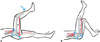
The present study showed that the bilateral popliteal angle had a tendency to increase in those over 36-years-old. Previous studies have demonstrated that the popliteal angle is increased in children and adolescents.518 The age of the cohorts in these previous studies ranged from 1−5 years. The unilateral popliteal angle with lordosis and an anteriorly tilted pelvis is a measure of functional hamstring contracture, and the bilateral popliteal angle with a loss of lordosis and neutral pelvis is a measure of the true hamstring contracture (Fig. 3). A hamstring shift greater than 20° is usually indicative of excessive anterior tilt from tight hip flexor musculature, weak abdominals, or a weak hip extensor.30 Popliteal angles are commonly used during physical examination to determine the necessity for distal hamstring lengthening in patients with CP. Some authors suggest that a popliteal angle >40−50° is an indication for distal hamstring lengthening.3132 However, Katz, et al.5 investigated the normal ranges of popliteal angle in 482 healthy children and showed that the ranges were up to 50° at ≥5 years of age. The current study also showed that 31 participants (30%) had a unilateral popliteal angle greater than 40°. Therefore, it is important to know that a popliteal angle of 40° or more is not an absolute indication of distal hamstring release. In addition, the popliteal angle should be interpreted with caution when determining whether surgery is necessary and age-related changes should be considered because weakening the hamstrings increases anterior pelvic tilt, which may actually worsen knee flexion.3334
The Silfverskiöld test is used to measure the range of ankle dorsiflexion with the knee extended and with the knee flexed 90°. If ankle dorsiflexion is decreased with the knee extended and the knee flexed 90°, a contracture of the gastrocnemius and soleus muscles is confirmed. If ankle dorsiflexion increases within the normal range with the knee flexed, an isolated contracture of the gastrocnemius muscle is confirmed. DiGiovanni, et al.35 defined the gastrocnemius contracture as less than 5° of dorsiflexion with the knee in extension, and the gastrocnemius-soleus contracture was defined as less than 10° of dorsiflexion with the knee in 90° flexion. In our study, the mean value of ankle dorsiflexion with knee extension was 11.3° [95% confidence interval (CI): 10.4−12.3°], and that of ankle dorsiflexion with the knee 90° flexion was 19.8° (95% CI: 18.8−20.8°). The mean difference between these values was 8.5°, and there was no significant difference among all the age groups. A similar result has been reported in children,18 suggesting that normal gastrocnemius muscle length is not affected by age in healthy people.
Our study has several strengths. We used a well-designed study model. Each age group consisted of an equal number and ratio of men and women. Several studies have investigated the normative value of a few items that do not apply to all kinds of physical examinations for evaluating patients with CP. However, we comprehensively suggest normative data of physical examinations that are primarily used to evaluate patients with CP.
There is a limitation to this study. We used a goniometer to measure the ROM during the physical examinations, and we held a consensus-building session to reduce variability in the value. Several authors have introduced different tools to improve the accuracy and reliability of ROM measurements such as ultrasonography or an inertial sensor, which is an optoelectronic system used to measure three-dimensional orientation.36 We need to consider using these tools to obtain a more accurate value when we conduct further research. Nevertheless, goniometric measurements are frequently used in the clinical setting, therefore, our results may be useful to clinicians.
We documented normative values that can be widely used for evaluating CP in patients 13 years and older. Further research is needed to determine correlations between physical examination findings and gait kinematic variables in healthy adolescent and adult populations.

Figures and Tables

Fig. 1

The Staheli test is performed with the participant in the prone position on the edge of the examination table. The examiner places one hand on the pelvis and gradually extends the thigh with the other. The point at which the pelvis begins to rise indicates the end of hip motion. At this point, the horizontal-thigh angle is measured. The psoas muscle (arrow) is primarily responsible for hip flexion contracture.

ymj-58-1170-g001
Fig. 2

The Silfverskiöld test. (A) The participant is placed in the supine position with the knee extended. (B) The hip and knee flexed at 90°. The angle is measured between parallel to long axis of fibula and parallel to long axis of 5th metatarsal. Arrows indicate gastrocnemius muscle.

ymj-58-1170-g002
Fig. 3

(A) Unilateral popliteal angle: the participant is in the supine position with the contralateral hip and knee in extension. The pelvis is tilted anteriorly accentuating lumbar lordosis. The tested limb is flexed to 90° at the hip and the knee is extended passively. The angle between the longitudinal axis of the leg and vertical line passing through the thigh is defined as the unilateral popliteal angle. (B) Bilateral popliteal angle: the same test is performed with the contralateral and knee flexed to neutroalize the anterior pelvic tilt, which decreases lumbar lordosis. Black arrows indicate hamstring muscle.

ymj-58-1170-g003
Table 1

Summary of Demographic Data in Study's Cohort

ymj-58-1170-i001
Age groups
13–20 years 21–35 years 36–50 years Over 51 years
No. of participants 26 26 26 26
Sex (M/F) 13/13 13/13 13/13 13/13
Age 17.2 (2.2) 28.4 (4.0) 43.1 (4.8) 56.0 (3.9)
Height (cm) 165.5 (8.2) 166.4 (9.2) 166.3 (8.0) 161.3 (9.4)
Weight (kg) 60.9 (12.5) 65.8 (12.7) 67.5 (9.2) 62.1 (9.6)
BMI (kg/m2) 22.1 (3.5) 23.7 (3.2) 24.4 (2.7) 23.8 (2.4)

BMI, body mass index.

Values are presented as mean (standard deviation).

Table 2

Methods of Measurement and Previous Studied on the Reliability of the Measurement of the Physical Examinations

ymj-58-1170-i002
Physical examination Method of measurement Study Characteristics of subjects Reliability (ICC)
Thomas test Supine, contralateral limb was flexed.
Angle between the longitudinal axis of the thigh and a horizontal line
Glanzman, et al.22 25 CP 0.98 (intra)
Kilgour, et al.23 25 CP 0.17–0.66 (intra)
25 control 0.09–0.91 (intra)
Mutlu, et al.24 38 CP 0.73–0.99 (intra), 0.92–0.95 (inter)
Lee, et al.7 36 CP 0.51 (inter)
37 control 0.21 (inter)
Staheli test (Fig. 1) Prone, one hand on the pelvis. Gradually extended the thigh with the other until pelvis begin to rise.
Angle between the longitudinal axis of the thigh and the horizontal line
Glanzman, et al.22 25 CP 0.98 (intra)
Kilgour, et al.23 25 CP 0.78–0.91 (intra), 0.55–0.80 (inter)
25 control 0.80–0.92 (intra), 0.04–0.20 (inter)
Lee, et al.7 36 CP 0.20 (inter)
37 control 0.11 (inter)
Clapper, et al.21 20 control 0.83 (intra)
Hip flexion Supine, contralateral hip & knee extended. And hip is flexed passively.
Angle between axis of the thigh and horizontal line
Sankar, et al.28 252 control >0.81
Trochanteric prominence angle test Prone, the greatest prominence of the greater trochanter can be palpated laterally.
Angle between a vertical line and the long axis of the leg
Chung, et al.3 36 CP 0.81 (inter)
Souza, et al.20 18 control 0.88–0.90 (intra), 0.83 (inter)
Shultz, et al.19 16 control 0.77–0.97 (intra), 0.58 (inter)
Hip internal rotation Prone, the leg rotated inward maximally.
Angle between a vertical line and the long axis of the leg
Chung, et al.3 36 CP 0.89 (inter)
Kouyoumdjian, et al.14 120 control 0.83 (inter)*
Hip external rotation Prone, examiner grasped the both ankles and push them apart so the leg maximally rotated outward.
Angle between a vertical line and the long axis of the leg
Chung, et al.3 36 CP 0.53 (inter)
Kouyoumdjian, et al.14 120 control 0.66 (inter)*
Knee flexion Supine, contralateral limb extended and knee flexed passively.
Angle between an extension line of thigh axis and axis of lower leg
Clapper, et al.21 20 control 0.95 (intra)
Knee flexion contracture Supine, contralateral limb extended and knee extended passively until no longer extension.
Angle between an extension line of thigh axis and axis of lower leg
Kilgour, et al.23 25 CP 0.97–0.99 (intra), 0.89–0.92 (inter)
25 control 0.79–0.87 (intra), 0.34–0.67 (inter)
Unilateral popliteal angle Supine, contralateral hip extended. Tested limb is flexed to 90° at the hip and knee is extended passively.
Angle between the longitudinal axis of the leg and vertical line
Glanzman, et al.22 25 CP 0.97 (intra)
Kilgour, et al.23 25 CP 0.96–0.99 (intra), 0.58–0.74 (inter)
25 control 0.87–0.97 (intra), 0.57–0.76 (inter)
Ten Berge, et al.25 15 CP 0.77 (intra), 0.68 (inter)
15 control 0.72 (intra), 0.82 (inter)
Lee, et al.18 47 control 0.66 (inter)
Bilateral popliteal angle Supine, Contralateral hip flexed to 90°. Tested limb is flexed to 90° at the hip and knee extended passively.
Angle between the longitudinal axis of the leg and vertical line
Kilgour, et al.23 25 CP 0.87–0.97 (intra), 0.57–0.76 (inter)
25 control 0.96–0.97 (intra), 0.58–0.74 (inter)
Lee, et al.18 47 control 0.63 (inter)
Ankle dorsiflexion with knee extension (Fig. 2A) Supine, ankle was dorsiflexed with the knee extended.
Angle between the long axis of the foot and the lower leg
Kilgour, et al.23 25 CP 0.96–0.99 (intra), 0.63–0.69 (inter)
25 control 0.95–0.98 (intra), 0.51–0.66 (inter)
Lee, et al.18 47 control 0.43 (inter)
Clapper, et al.21 20 control 0.92 (intra)
Glanzman, et al.22 25 CP 0.97–0.98 (intra)
Ankle dorsiflexion with knee 90° flexion (Fig. 2B) Supine, ankle was dorsiflexed with the knee flexed 90°.
Angle between the long axis of the foot and the lower leg
Kilgour, et al.23 25 CP 0.98–0.99 (intra), 0.75–0.90 (inter)
25 control 0.97–0.98 (intra), 0.70–0.75 (inter)
Lee, et al.18 47 control 0.36 (inter)
Thigh-foot angle Prone, knee flexed 90°, ankle in neutral position, sole parallel to the floor
Angle between the longitudinal axis of the thigh and longitudinal axis of the foot
Lee, et al.26 18 CP 0.74 (inter)

ICC, interclass correlation coefficient; CP, cerebral palsy; intra, intra-rater reliability; inter, inter-rater reliability.

*Lin's concordance correlation coefficient.

Table 3

One-Way Analysis of Variance, Mean, SD, 95% CIs and p Values for the Difference of Age Groups

ymj-58-1170-i003
Examination Mean (SD) [95% CI] (degrees) p value
13–20 years 21–35 years 36–50 years Over 51 years Total
Thomas test 0.0 (0.2)
[0.0 to 0.1]
0.3 (0.9)
[−0.1 to 0.6]
0.3 (1.2)
[−0.1 to 0.8]
0.3 (1.0)
[−0.1 to 0.7]
0.2 (0.9)
[0.1 to 0.4]
0.591
Staheli test −20.9 (4.6)
[−18.7 to −22.8]
−18.0 (3.2)
[−16.7 to −19.3]
−17.0 (2.9)
[−15.8 to −18.2]
−18.0 (3.6)
[−16.5 to −19.4]
−18.4 (3.9)
[−17.7 to −19.2]
0.002*
Hip flexion 126.8 (7.6)
[123.7 to 129.8]
126.6 (6.4)
[124.0 to 129.2]
125.8 (4.6)
[123.9 to 127.6]
125.5 (6.1)
[123.1 to 128.0]
126.2 (6.2)
[125.0 to 127.4]
0.850
Hip abduction with extension 47.6 (6.2)
[45.1 to 50.1]
46.3 (6.1)
[43.8 to 48.7]
49.3 (4.9)
[47.3 to 51.2]
47.3 (5.6)
[45.0 to 49.6]
47.6 (5.7)
[46.5 to 48.7]
0.298
Hip abduction with 90° flexion 55.6 (7.5)
[52.6 to 58.7]
53.3 (6.7)
[50.6 to 56.0]
56.1 (5.3)
[53.9 to 58.2]
54.0 (8.9)
[50.4 to 57.6]
54.8 (7.2)
[53.3 to 56.2]
0.472
Adduction of hip 28.6 (8.9)
[25.0 to 32.2]
30.9 (7.6)
[27.8 to 33.9]
33.3 (6.5)
[30.7 to 36.0]
32.4 (7.7)
[29.3 to 35.5]
31.3 (7.8)
[29.8 to 32.8]
0.146
Hip external rotation 40.1 (8.5)
[36.7 to 43.6]
41.0 (8.8)
[37.4 to 44.6]
43.5 (7.3)
[40.6 to 46.5]
40.5 (8.3)
[37.2 to 43.9]
41.3 (8.3)
[39.7 to 42.9]
0.445
Hip internal rotation 40.1 (11.1)
[35.6 to 44.5]
39.2 (8.3)
[35.8 to 42.5]
39.4 (8.7)
[35.9 to 42.9]
37.6 (7.8)
[34.4 to 40.7]
39.1 (9.0)
[37.3 to 40.8]
0.788
Trochanteric prominence angle test 17.6 (4.5)
[15.7 to 19.4]
18.9 (4.3)
[17.2 to 20.6]
19.1 (4.6)
[17.2 to 20.9]
17.9 (6.1)
[15.4 to 20.3]
18.4 (4.9)
[17.4 to 19.3]
0.611
Knee flexion contracture 1.0 (1.8)
[0.3 to 1.8]
0.4 (1.2)
[-0.1 to 0.9]
1.7 (2.0)
[0.9 to 2.5]
0.8 (1.9)
[0.0 to 1.6]
1.0 (1.8)
[0.6 to 1.3]
0.088
Knee flexion 136.5 (5.5)
[134.3 to 138.8]
137.6 (5.8)
[135.3 to 140.0]
137.0 (5.4)
[134.8 to 139.2]
137.1 (5.2)
[135.0 to 139.2]
137.1 (5.4)
[136.0 to 138.1]
0.917
Unilateral popliteal angle 33.8 (10.3)
[29.7 to 37.9]
33.1 (8.9)
[29.5 to 36.7]
35.9 (8.8)
[32.3 to 39.5]
38.0 (7.9)
[34.8 to 41.2]
35.2 (9.1)
[33.4 to 37.0]
0.203
Bilateral popliteal angle 24.3 (9.1)
[20.6 to 28.0]
22.5 (9.6)
[18.7 to 26.4]
27.0 (8.2)
[23.7 to 30.3]
28.1 (6.3)
[25.6 to 30.6]
25.5 (8.5)
[23.8 to 27.1]
0.074
Hamstring shift 9.5 (4.1)
[7.9 to 11.2]
10.6 (5.2)
[8.5 to 12.7]
8.9 (4.6)
[7.1 to 10.8]
9.9 (4.6)
[8.0 to 11.7]
9.7 (4.6)
[8.8 to 10.6]
0.649
Thigh-foot angle 12.4 (5.5)
[10.2 to 14.6]
12.8 (6.6)
[10.1 to 15.4]
15.5 (4.4)
[13.7 to 17.3]
14.0 (5.0)
[12.0 to 16.0]
13.7 (5.5)
[12.6 to 14.7]
0.169
Ankle dorsiflexion with knee extension 11.3 (4.7)
[9.5 to 13.2]
12.2 (4.5)
[10.4 to 14.1]
11.0 (5.8)
[8.7 to 13.3]
10.8 (4.2)
[9.1 to 12.5]
11.3 (4.8)
[10.4 to 12.3]
0.714
Ankle dorsiflexion with knee 90° flexion 19.6 (4.5)
[17.8 to 21.4]
21.1 (5.0)
[19.1 to 23.1]
18.3 (5.7)
[16.0 to 20.5]
20.3 (5.6)
[18.1 to 22.6]
19.8 (5.2)
[18.8 to 20.8]
0.244
Ankle plantar flexion 49.4 (9.2)
[45.7 to 53.1]
47.2 (6.5)
[44.6 to 49.9]
46.7 (8.7)
[43.2 to 50.2]
45.2 (8.3)
[41.8 to 48.5]
47.1 (8.3)
[45.5 to 48.7]
0.320

SD, standard deviation; CI, confidence interval.

*Statistically significant p<0.05.

ACKNOWLEDGEMENTS

This research was supported by Projects for Research and Development of Police Science and Technology under Center for Research and Development of Police Science and Technology and Korean National Police Agency funded by the Ministry of Science and ICT (Grant No. PA-C000001-2015-202).

Notes

The authors have no financial conflicts of interest.

References

1. Evans PM, Evans SJ, Alberman E. Cerebral palsy: why we must plan for survival. Arch Dis Child. 1990; 65:1329–1333.
crossref
2. Cathels BA, Reddihough DS. The health care of young adults with cerebral palsy. Med J Aust. 1993; 159:444–446.
crossref
3. Chung CY, Lee KM, Park MS, Lee SH, Choi IH, Cho TJ. Validity and reliability of measuring femoral anteversion and neck-shaft angle in patients with cerebral palsy. J Bone Joint Surg Am. 2010; 92:1195–1205.
crossref
4. Davids JR, Benfanti P, Blackhurst DW, Allen BL. Assessment of femoral anteversion in children with cerebral palsy: accuracy of the trochanteric prominence angle test. J Pediatr Orthop. 2002; 22:173–178.
crossref
5. Katz K, Rosenthal A, Yosipovitch Z. Normal ranges of popliteal angle in children. J Pediatr Orthop. 1992; 12:229–231.
crossref
6. Price CT. Lower-extremity rotational problems in children. Normal values to guide management. J Bone Joint Surg Am. 1985; 67:823–824.
crossref
7. Lee KM, Chung CY, Kwon DG, Han HS, Choi IH, Park MS. Reliability of physical examination in the measurement of hip flexion contracture and correlation with gait parameters in cerebral palsy. J Bone Joint Surg Am. 2011; 93:150–158.
crossref
8. Park N, Lee J, Sung KH, Park MS, Koo S. Design and validation of automated femoral bone morphology measurements in cerebral palsy. J Digit Imaging. 2014; 27:262–269.
crossref
9. Novacheck TF, Trost JP, Sohrweide S. Examination of the child with cerebral palsy. Orthop Clin North Am. 2010; 41:469–488.
crossref
10. Johari R, Maheshwari S, Thomason P, Khot A. Musculoskeletal evaluation of children with cerebral palsy. Indian J Pediatr. 2016; 83:1280–1288.
crossref
11. Ahlberg A, Moussa M, Al-Nahdi M. On geographical variations in the normal range of joint motion. Clin Orthop Relat Res. 1988; 234:229–231.
crossref
12. Boone DC, Azen SP. Normal range of motion of joints in male subjects. J Bone Joint Surg Am. 1979; 61:756–759.
crossref
13. James B, Parker AW. Active and passive mobility of lower limb joints in elderly men and women. Am J Phys Med Rehabil. 1989; 68:162–167.
crossref
14. Kouyoumdjian P, Coulomb R, Sanchez T, Asencio G. Clinical evaluation of hip joint rotation range of motion in adults. Orthop Traumatol Surg Res. 2012; 98:17–23.
crossref
15. Hu H, Li Z, Yan J, Wang X, Xiao H, Duan J, et al. Measurements of voluntary joint range of motion of the Chinese elderly living in Beijing area by a photographic method. Int J Ind Ergon. 2006; 36:861–867.
crossref
16. Roach KE, Miles TP. Normal hip and knee active range of motion: the relationship to age. Phys Ther. 1991; 71:656–665.
crossref
17. Hallaçeli H, Uruç V, Uysal HH, Ozden R, Hallaçeli C, Soyuer F, et al. Normal hip, knee and ankle range of motion in the Turkish population. Acta Orthop Traumatol Turc. 2014; 48:37–42.
crossref
18. Lee SY, Lee SH, Chung CY, Park MS, Lee KM, Akhmedov B, et al. Age-related changes in physical examination and gait parameters in normally developing children and adolescents. J Pediatr Orthop B. 2013; 22:153–157.
crossref
19. Shultz SJ, Nguyen AD, Windley TC, Kulas AS, Botic TL, Beynnon BD. Intratester and intertester reliability of clinical measures of lower extremity anatomic characteristics: implications for multicenter studies. Clin J Sport Med. 2006; 16:155–161.
crossref
20. Souza RB, Powers CM. Concurrent criterion-related validity and reliability of a clinical test to measure femoral anteversion. J Orthop Sports Phys Ther. 2009; 39:586–592.
crossref
21. Clapper MP, Wolf SL. Comparison of the reliability of the Orthoranger and the standard goniometer for assessing active lower extremity range of motion. Phys Ther. 1988; 68:214–218.
crossref
22. Glanzman AM, Swenson AE, Kim H. Intrarater range of motion reliability in cerebral palsy: a comparison of assessment methods. Pediatr Phys Ther. 2008; 20:369–372.
crossref
23. Kilgour G, McNair P, Stott NS. Intrarater reliability of lower limb sagittal range-of-motion measures in children with spastic diplegia. Dev Med Child Neurol. 2003; 45:391–399.
crossref
24. Mutlu A, Livanelioglu A, Gunel MK. Reliability of goniometric measurements in children with spastic cerebral palsy. Med Sci Monit. 2007; 13:CR323–CR329.
25. Ten Berge SR, Halbertsma JP, Maathuis PG, Verheij NP, Dijkstra PU, Maathuis KG. Reliability of popliteal angle measurement: a study in cerebral palsy patients and healthy controls. J Pediatr Orthop. 2007; 27:648–652.
26. Lee SH, Chung CY, Park MS, Choi IH, Cho TJ. Tibial torsion in cerebral palsy: validity and reliability of measurement. Clin Orthop Relat Res. 2009; 467:2098–2104.
crossref
27. Park MS, Kim SJ, Chung CY, Choi IH, Lee SH, Lee KM. Statistical consideration for bilateral cases in orthopaedic research. J Bone Joint Surg Am. 2010; 92:1732–1737.
crossref
28. Sankar WN, Laird CT, Baldwin KD. Hip range of motion in children: what is the norm? J Pediatr Orthop. 2012; 32:399–405.
29. Freemont AJ, Hoyland JA. Morphology, mechanisms and pathology of musculoskeletal ageing. J Pathol. 2007; 211:252–259.
crossref
30. Delp SL, Arnold AS, Speers RA, Moore CA. Hamstrings and psoas lengths during normal and crouch gait: implications for muscletendon surgery. J Orthop Res. 1996; 14:144–151.
crossref
31. Reimers J. Contracture of the hamstrings in spastic cerebral palsy. A study of three methods of operative correction. J Bone Joint Surg Br. 1974; 56:102–109.
32. Okawa A, Kajiura I, Hiroshima K. Physical therapeutic and surgical management in spastic diplegia. A Japanese experience. Clin Orthop Relat Res. 1990; 253:38–44.
33. Saraph V, Zwick EB, Zwick G, Steinwender C, Steinwender G, Linhart W. Multilevel surgery in spastic diplegia: evaluation by physical examination and gait analysis in 25 children. J Pediatr Orthop. 2002; 22:150–157.
crossref
34. Chang WN, Tsirikos AI, Miller F, Lennon N, Schuyler J, Kerstetter L, et al. Distal hamstring lengthening in ambulatory children with cerebral palsy: primary versus revision procedures. Gait Posture. 2004; 19:298–304.
crossref
35. DiGiovanni CW, Kuo R, Tejwani N, Price R, Hansen ST Jr, Cziernecki J, et al. Isolated gastrocnemius tightness. J Bone Joint Surg Am. 2002; 84-A:962–970.
crossref
36. van den Noort JC, Scholtes VA, Harlaar J. Evaluation of clinical spasticity assessment in cerebral palsy using inertial sensors. Gait Posture. 2009; 30:138–143.
crossref
TOOLS
Similar articles