

 PDF
PDF ePub
ePub Citation
 Print
Print


INTRODUCTION
Most microorganisms in natural ecosystems exist in the form of biofilms. The biofilms of microorganisms are formed over many stages, the first two of which are adherence to a biotic or abiotic surface and production of a structure to increase adherence. In the maturing biofilm, extracellular polymers are formed by microorganisms in the structure and are known to protect microorganisms from changes in the surrounding environment, to participate in supplying nutrients and discharging metabolic waste, and to gather cells in closer proximity in order to facilitate cell-to-cell interactions. Biofilms can consist of multi-species, including coexisting bacteria and fungi.123
Candida albicans (C. albicans) is a resident species of healthy human mucous membranes that is also an opportunistic pa-thogen that induces superficial and systemic infection via the mucous epithelium when a patient suffers from severe disease or when the immune state is deficient. C. albicans has virulence factors that allow it to invade host tissue and to evade the host defense mechanism.45 C. albicans grows in three different forms: budding yeast (or blastoconidia), pseudohyphae, and hy-phae. The expression of C. albicans genes differ according to the C. albicans form. Candida hyphae are known to be essential to pathogenicity and disease dissemination,678 and several hyphae-specific genes are known, including hwp1, als3, als8, ece1, and sap4-6.91011 The yeast-specific genes include rbe1, ywp1, and nrg1, and filament formation inhibiting genes include tup1 and nrg1.12131415
Proteus vulgaris (P. vulgaris) and Proteus mirabilis (P. mirabilis) exist in both human and animal small intestines and in the natural environment. P. vulgaris and P. mirabilis also can form biofilms on surfaces of various objects, including insertion apparatuses in humans. Proteus infections, especially urinary tract infections, are common in immunosuppressed patients.161718 In recent years, C. albicans has been highlighted as one of the most common etiologic agents of acquired hospital infections, and Candida biofilms play an important role in initiating infections. Biofilms that exist in the human body are comprised of hundreds of different bacterial species: fungi and bacteria can also co-exist in these biofilms.19 Both the microorganism relationships with the host and the interactions between various microorganisms are important, the latter of which is not being actively researched.2021
We previously reported that bacteria had a negative effect on the formation of C. albicans biofilms, and a more distinct decrease in C. albicans biofilm formation was shown when cultivated with P. vulgaris.22 In the present study, the influence of co-culture of C. albicans, a chief agent of hospital-acquired infection, with P. vulgaris and P. mirabilis on the formation of C. albicans biofilm and its underlying mechanisms were examined.
MATERIALS AND METHODS
Organisms
Clinical isolates of C. albicans were obtained: one commensal strain was isolated from the blood of a patient, and P. vulgaris and P. mirabilis were isolated from the urine of another patient. The identity of each microorganism was confirmed with the commercially-available identification systems (BioMeriéux, Marcy I'Etoile, France): API 32C for C. albicans and API 20E for P. vulgaris and P. mirabilis.
Culture conditions and experimental conditions
Prior to each experiment, C. albicans isolates were cultured at 30℃ for 48 hours on Sabouraud's dextrose agar (SDA, Difco™, Becton Dickinson, Spark, MD, USA), and one colony of yeast was inoculated into yeast nitrogen base (Difco™, Becton Dickinson) medium supplemented with 50 mM glucose. P. vulgaris and P. mirabilis were first subcultured at 37℃ for 18 hours on tryptic soy agar. One colony each of P. vulgaris and P. mirabilis was then inoculated into tryptic soy broth (Difco™, Becton Dickinson) and incubated at 37℃ for 18 hours. The experimental conditions were as follows: 1) the microorganism was cultured alone; 2) C. albicans was co-cultured with live P. vulgaris or P. mirabilis; 3) C. albicans was co-cultured with P. vulgaris or P. mirabilis killed at 100℃ for 30 minutes; or 4) C. albicans was treated with bacteria-cultured supernatants of P. vulgaris or P. mirabilis diluted four times, in which the bacteria were removed.
XTT reduction assays
Biofilm formation was quantified using the method developed by Ramage, et al.23 Biofilms were formed on commercially available pre-sterilized, polystyrene, flat-bottomed, 96-well microtiter plates (Costar, Cambridge, MA, USA). Microorganisms were prepared for each condition and transferred to selected wells of a microtiter plate. The plate was incubated for 90 minutes at 37℃ in an orbital shaker at 75 rpm. After the initial adhesion phase, the cell suspensions were aspirated, and each well was washed twice with phosphate-buffered saline (PBS) to remove loose adherent cells. A volume of 200 µL of medium was added to each well, and the plate was then incubated for another 72 hours. After biofilm formation, the medium was aspirated, and non-adherent cells were removed by thoroughly washing the biofilm three times with PBS. A quantitative measure of biofilm formation was calculated using the XTT [2,3-bis(2-methyoxy-4-nitro-5-sulfo-phenyl)-2H-tetrazolium-5-carboxanilide] reduction assay. A 200-µL aliquot of XTT (1 mg/mL, Sigma, St. Louis, MO, USA) and menadione (0.4 mM, Sigma) solution was added to each well containing the prewashed biofilm and the control well. The plates were then incubated in the dark for up to 3 hours at 37℃. A colorimetric change resulting from XTT reduction was measured using a microtiter plate reader (EMax, Molecular Devices, Sunnyvale, CA, USA) at 490 nm.
C. albicans cell counts
After biofilm formation, the medium was aspirated, and non-adherent cells were removed by thoroughly washing the biofilm three times with PBS. Then, 1 mL of PBS was transferred to each well, and biomass was meticulously scraped off. The resultant solution containing the detached biofilm cells was gently vortexed for 1 minute to disrupt the aggregates and inoculated on an SDA plate. The colony forming units (CFUs) of C. albicans were quantified after 48 hours of incubation at 30℃.
Scanning electron microscopy
We developed biofilms from single species, in addition to Candida biofilms that were co-cultured with bacteria on polystyrene coverslips as described. The coverslips were washed twice with PBS and placed in PBS with a fixative of 2.5% glutaraldehyde (Sigma) for 20 hours. Next, they were washed for 5 minutes in PBS and then placed in 1% osmium tetroxide for 30 minutes. After a series of alcohol washes, a final drying step was performed using the critical point drying method. Biofilms were then mounted and gold coated. Samples were imaged with a scanning electron microscope (TM-1000, Hitachi, Tokyo, Japan) in a high-vacuum mode at 15 kV.
Relative quantitation by real-time reverse transcriptase polymerase chain reaction
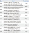
RNA was isolated from C. albicans cells using the MasterPure Yeast RNA Extraction kit (Epicentre Biotechnologies, Madison, WI, USA). RNA was treated with amplification grade DNase I (Epicentre Biotechnologies) and used for cDNA synthesis with random hexamer primer (Invitrogen Life Technologies, Carlsbad, CA, USA) using Superscript II reverse transcriptase reagents (Invitrogen Life Technologies). Each reaction contained 1 µg of total RNA, 1 µL of 50 µM hexamer, and 1 µL of 10 mM dNTP in a final volume of 10 µL. Reactions were incubated at 65℃ for 5 minutes and cooled on ice. To each reaction tube, 10 µL of the following mixture was added: 4 µL of 5x First-Strand Buffer, 2 µL of 10 mM MgCl2, 2 µL of 0.1 M DTT, 1.4 µL of RNase inhibitor, and 1 µL of Superscript II. Reactions were incubated at 42℃ for 50 minutes and then at 70℃ for 15 minutes. Real-time polymerase chain reaction (PCR) contained 10 µL of Power SYBR Green Master Mix (Applied Biosystems, Foster City, CA, USA), as well as forward and reverse primers (1 µL of each) (Table 1)22 and sterile water, at a final volume of 20 µL. The PCR was run on MicroAmp® Optical 384-well reaction plates in an ABI 7900 Real-Time PCR system (Applied Biosystems). Real-time PCR reactions were performed at 95℃ for 5 minutes, followed by 40 cycles of 15 seconds at 95℃ and 1 minute at 60℃. Dissociation curves were analyzed for all reactions to verify single peaks/products. Expression levels were analyzed using ABI 7900 System SDS software (Applied Biosystems). Real-time PCR data were normalized with the geometric mean of two reference genes. The ACT1 and PMA1 genes were used for this purpose.
Statistical analysis
All experiments were performed in triplicate on three different occasions. All data are expressed as mean values with corresponding standard deviations (SDs). Student's t-tests and Mann-Whitney U-tests were used to compare the differences between Candida only and Candida co-cultured with P. vulgaris or P. mirabilis. All p-values <0.05 were considered statistically significant.
RESULTS
The effect of P. vulgaris or P. mirabilis on C. albicans biofilm formation
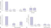
The biofilm value generated when each type of microorganism was incubated separately was 0.07±0.007 for P. vulgaris, 0.120±0.004 for P. mirabilis, and 1.403±0.103 for C. albicans. When culturing C. albicans and P. vulgaris or P. mirabilis together, biofilm formation was reduced by more than 80%, compared to the C. albicans culture alone. To assess the mechanism by which the bacteria impeded Candida biofilm formation, bacteria were initially treated for 30 minutes at 100℃ to eliminate biological activity, followed by co-culture with C. albicans. Biofilm formation of C. albicans cultured together with heat-killed P. vulgaris or P. mirabilis elicited a 70% reduction, compared to the control group. These data indicate that dead bacteria interfere with the structural formation of C. albicans biofilms (Fig. 1A).
When C. albicans and P. vulgaris or P. mirabilis were co-cultured, biofilm formation of C. albicans was significantly reduced. In order to determine if this reduction was due to the effect of the bacteria or of secretory products when cultured, P. vulgaris and P. mirabilis were cultured for 72 hours, and remaining bacteria were removed by filtration. As a result of treatment with P. vulgaris and P. mirabilis culture supernatants, C. albicans biofilm formation was reduced by 60–70%, compared to C. albicans cultured alone (Fig. 1B). To determine whether this effect was due to a depletion of nutrients in the medium or to the secretory products of the bacteria, we diluted the cultured supernatants and tested the concentration effect on C. albicans biofilm formation. C. albicans biofilm formation decreased in proportion to the concentration of the bacterial-cultured supernatants (data not shown). To determine whether P. vulgaris and P. mirabilis inhibited the growth of C. albicans, C. albicans and P. vulgaris or P. mirabilis or bacterial-cultured supernatants were cultured together for 72 hours, and C. albicans CFUs were calculated. When C. albicans was cultured alone, the count was 2.85×10⁸ CFU/mL; when cultured together with P. vulgaris, the count of C. albicans was reduced to 9×10⁶ CFU/mL; and when C. albicans was cultured together with P. mirabilis, the count of C. albicans dropped to 2.4×10⁷ CFU/mL (Fig.1C). It seems that P. vulgaris and P. mirabilis inhibit C. albicans biofilm formation and also interfere with its growth. As a result, we investigated the C. albicans CFUs after treating C. albicans with bacterial-cultured supernatants and culturing for 72 hours. In the case of culturing C. albicans alone, the count was 2.85×10⁸ CFU/mL. Meanwhile, the count from the treated culture supernatants of P. vulgaris was 3.6×10⁷ CFU/mL, and the count from the treated culture supernatants of P. mirabilis was reduced to 2.0×10⁷ CFU/mL (Fig. 1C).
The effect of P. vulgaris or P. mirabilis on C. albicans morphology-related gene expression
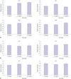
C. albicans morphology changes to the hyphae form from the yeast form as biofilm formation progresses, and the expression pattern of genes related to morphology at these stages was identified. Previous studies have shown that expression patterns of Candida species-related genes are significantly increased during hyphae-specific gene expression over the duration of biofilm formation. On the contrary, the expression of genes suppressing the formation of yeast-specific genes and filament are decreased.22 These results suggest that Candida present in yeast form adhere to a surface to increase the expression of hyphae-related genes and promote the formation of biofilms by decreasing the expression of genes that inhibit the yeast form and filament formation. To clarify the effect of co-cultures on biofilm formation, we analyzed changes in C. albicans gene expression levels in biofilms co-cultured with P. vulgaris or P. mirabilis. In contrast to C. albicans cultured alone, als3 and hwp1 showed a reduction in expression by 80% and ece1 and sap5 by 90% (Fig. 2A). When C. albicans was cultured together with P. vulgaris or P. mirabilis, the expression of tup1 and nrg1, which are genes known to suppress filament formation, increased by about two-fold, compared to when C. albicans was cultured alone (Fig. 2B). Regarding the expression of yeast-specific rhd1 and rbe1, the expression levels of rhd1 increased by 2.5-fold and rbe1 by about more than 3-fold (Fig. 2C).
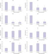
When C. albicans was co-cultured with heat-killed P. vulgaris and P. mirabilis, biofilm formation was decreased. We confirmed that these results were due to changes in the expression of biofilm-related genes in C. albicans. First, ece1, hwp1, and sap5 gene levels, which are associated with the formation of hyphae, did not increase. While als3 level was slightly increased, the change was not significant (Fig. 3A). There were also no significant changes in tup1 and nrg1, which are filament formation inhibiting genes (Fig. 3B). Yeast-specific genes also showed no difference in expression levels (Fig. 3C). The reduction in C. albicans biofilm formation in co-cultures with killed bacteria is considered to be caused not by changes in gene expression, but by the interference of killed bacteria acting as small particles that fit between C. albicans cells and, thus, suppress the structural formation of biofilm.
The effect of cultured P. vulgaris or P. mirabilis supernatants on expression of C. albicans morphology-related genes
As described above, treatment of P. vulgaris and P. mirabilis cultured supernatants inhibited C. albicans biofilm formation. We examined how this treatment affected the expression of various genes involved in biofilm formation. The hyphae-specific genes als3, ece1, hwp1, and sap5 all showed a significant reduction in expression in contrast to the cultured supernatants in which C. albicans was cultured alone (Fig. 4A). In contrast, the filament formation inhibiting genes tup1 and nrg1 slightly increased (Fig. 4B). The yeast-specific gene rhd1 increased by 2-fold, and rbe1 slightly increased (Fig. 4C). This suggests that secretory products that are formed and released with growth of P. vulgaris and P. mirabilis inhibit the growth of C. albicans and regulate the expression of biofilm-related genes, thereby inhibiting biofilm formation in C. albicans.
Scanning electron microscopy of biofilms
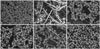
It is known that biofilms are not formed with a simple structure and that Candida exist in the yeast form in the basal layer and in the hyphae and pseudohyphae form in the layer above. These layers form a network with a three-dimensional structure. The structural difference between the biofilm when C. albicans is cultivated separately and when co-cultured with P. vulgaris or P. mirabilis was investigated via scanning electron microscopy (Fig. 5). The biofilm of C. albicans when cultured alone was high in density and was a multi-layer solid (Fig. 5A and B). The biofilm formed when C. albicans and P. vulgaris or P. mirabilis were cultured together showed that the P. vulgaris or P. mirabilis were attached to the hyphae of C. albicans and inserted between the C. albicans cells. The thickness of the biofilm in these cases also appeared to be thin with a low density (Fig. 5C and E). The structure of biofilms formed after culturing heat-killed bacteria and C. albicans together was thinner and showed lower density than biofilms formed after separate culture of C. albicans (Fig. 5D and F). However, the heat-killed bacteria biofilm was thicker than the biofilm formed after C. albicans had been cultured together with live bacteria, and more of the C. albicans was in the yeast form. The above results agree with the outcome of XTT reduction assays, which quantitatively confirmed biofilm formation (Fig. 1A).
DISCUSSION
Microorganisms that are fixed inside a biofilm show resistance to the immune system and have a strong tolerance to antibiotics relative to planktonic microorganisms.24 Many human infections are the result of microorganisms in biofilms. Most studies on biofilm formation and the interrelation between microorganisms in them have focused on bacteria.25 However, biofilms affected by the relationship between bacteria and fungi are clinically crucial, because these biofilms increase the morbidity and mortality of infections.1926
The research presented here verified the influence of the coexistence of C. albicans and P. vulgaris and P. mirabilis on C. albicans biofilm formation and whether the correlation between C. albicans and P. vulgaris and P. mirabilis was competitive or symbiotic. The architecture and functioning of complex biofilms are very intricate and were not clearly elucidated. In addition, the correlation between microorganisms inside the complex cultivated biofilm was not identified.
When C. albicans was cultivated with P. vulgaris and P. mirabilis, both biofilm formation and number of C. albicans cells decreased, compared to when C. albicans was cultured alone (Fig. 1). Even the diluted supernatants of P. vulgaris and P. mirabilis cultivation were confirmed to hinder biofilm formation, which implies that it was not the depletion of nutrients due to mixed culture of P. vulgaris and P. mirabilis plus C. albicans that decreased biofilm formation, but that the bacteria directly hindered formation due to secretory products (Fig. 1). The Proteus-specific products inhibiting the growth of C. albicans or biofilm formation are not yet clarified, and further research is needed.
Interestingly, even when C. albicans was cultivated with heat-treated P. vulgaris and P. mirabilis, biofilm formation decreased (Fig. 1A). It is considered that both the secretory products from P. vulgaris and P. mirabilis and the bacterial architecture itself induced structural changes and hindered the ability of C. albicans to form biofilms. Further, we verified biofilm structures of C. albicans alone or when cultivated with P. vulgaris and P. mirabilis via scanning electron microscopy. The biofilm formed normally only when C. albicans was cultured alone, showing high density and numerous layers (Fig. 5A). In contrast, biofilms that formed when P. vulgaris and P. mirabilis were cultured together showed low-density, thin biofilms with bacteria among the C. albicans cells, and a noticeably decreased number of mycelia (Fig. 5). It is clear that both living and dead bacteria particles influenced structural maturation of the biofilm. Thus, P. vulgaris and P. mirabilis suppress the growth of C. albicans and can function as structural obstructive factors to the maturation of Candida biofilms.
The formation of hyphae is essential to C. albicans biofilm formation, so it is also important to understand the genetic basis of the morphological changes in C. albicans.1011 The mature biofilm enables the Candida yeast to fix the biofilm on to the extracellular surface, and the hyphae form a cross-sectional structure with structural frames.27 Inhibition of the hyphae transgenes of C. albicans led to biofilm formation with the basal layer only, whereas inhibition of the yeast transgene of C. albicans led to biofilm formation with only the outer layer among the existing biofilm structures.1128 In this study, the expression of hyphae-related genes C. albicans was significantly inhibited in the presence of live Proteus or by Proteus-cultured supernatant. The expression of both yeast-related and filament formation inhibiting genes in C. albicans was up-regulated by treatment with live Proteus or Proteus-cultured supernatant (Figs. 2 and 4); however, the expression of morphology-related genes was not affected by heat-killed P. vulgaris and P. mirabilis (Fig. 3).
These results suggest that secretory products of P. vulgaris and P. mirabilis regulate the expression of genes that are related to morphologic changes, which could be the crucial factor in C. albicans biofilm formation, inhibiting hyphal transition from the yeast form to the hyphal form. Due to an increase in only the yeast form and the lack of hyphal form, the C. albicans biofilm would not form a solid 3D structure, but only a thick, basal-layered structure.

 XML Download