

 PDF
PDF ePub
ePub Citation
 Print
Print


INTRODUCTION
Congenital orbital fibrosis is a distinct non-familial, non-progressive unilateral clinical entity characterized by the presence of diffuse infiltrative orbital lesions resulting in variable symptoms because of the cicatricial process. Although several possible causes of congenital orbital fibrosis have been postulated, the etiology of the disease remains unknown. To our best knowledge, this study presents the first report on the findings of mutational analysis of a patient with congenital orbital fibrosis.
CASE REPORT
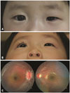
A 3-year-old girl presented with persistent asymmetric palpebral fissure height since birth. The patient had no significant medical or family history, and her birth had been uneventful and at full term. Ophthalmic examination revealed right enophthalmos with ipsilateral upper and lower lid retraction and entropion (Fig. 1A and B). Extraocular movement was unremarkable. Retinoscopy revealed refractive errors of +4.50 OD (right eye) and -1.25-1.00×180° OS (left eye) and best-corrected visual acuities of 20/400 OD and 20/30 OS. Slit-lamp biomicroscopy findings were unremarkable, and fundus examination revealed elevated right optic disc, with blurred margins (Fig. 1C).
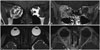
Magnetic resonance (MR) imaging findings revealed infiltrative enhancement of the right orbital connective/adipose tissue. The right recti muscles exhibited increased volume with enhancement. The posterior aspect of the right eyeball was flattened in the forward direction (Fig. 2). Based on the presence of non-progressive clinical signs since birth and enhancing infiltrative fibrotic lesions on MR imaging, the patient was diagnosed with congenital orbital fibrosis.
Because of the congenital presentation of heterogenic clinical manifestations, clinical whole exome and mitochondrial genome sequencing were performed to identify possible disease-causing mutations for molecular diagnosis. Whole-exome sequencing was performed using the TruSight® One panel of 4813 genes (Illumina, San Diego, CA, USA) for screening for pathologic mutations in 291 genes which are known to be related to eye disorders. Three novel heterozygous mutations were identified in MFRP, MTHFR, and RBP4, which are known to cause isolated microphthalmia 5 (OMIM#611040), homocystinuria (OMIM#236250), and microphthalmia with coloboma 10 (OMIM#616428) or retinal dystrophy with iris coloboma(OMIM#615147), respectively (Table 1). However, orbital fibrosis is not the main phenotype in any of these diseases. No pathologic mutations were observed in known causative genes for congenital fibrosis of extraocular muscles (CFEOM), including KIF21A, PHOX2A, TUBB3, TUBB2B, FEOM4, and TUKLS.
The findings of whole mitochondrial genome sequencing revealed no known mitochondrial diseases.
DISCUSSION
Unilateral fibrosis, blepharoptosis, and enophthalmos syndrome were first described by Leone and Weinstein1 in 1972. Since then, however, a very few cases have been reported in literature. Mavrikakis, et al.2 who described this syndrome as a distinct clinical entity termed it “congenital orbital fibrosis”. Previous studies have reported varying clinical manifestations of the condition–eyelid position has been reported to be normal, retracted, or ptotic; eyeball position has been reported to be symmetric, proptotic, or enophthalmic.234 In the present case, non-progressive unilateral enophthalmos with eyelid retraction and MR imaging findings of diffuse infiltrating lesion with enlarged extraocular muscles were appropriate for diagnosis of congenital orbital fibrosis. Unlike other reported cases, our patient exhibited an elevated right optic disc with blurred margins, accompanied with flattening of the posterior aspect of the right eyeball in the forward direction. Optic disc elevation is considered to be a secondary change due to fibrosis and contraction of orbital tissue.
Congenital orbital fibrosis has traditionally been classified as a subtype of CFEOM.5 However, CFEOM is now known as a genetically transmitted dysinnervation syndrome, in which myopathic changes are secondary to aberrant innervation of extraocular muscles.6
On the other hand, the etiology of congenital orbital fibrosis remains unknown. Although prenatal penetrating injury to the orbit, such as maternal amniocentesis, had been suggested as a possible cause of the disease, eyelid scarring possibly associated with prenatal trauma was reported only in a small number of cases.3478 With regard to the genetic aspect, earlier reports on congenital orbital fibrosis as a non-hereditary disease are not based on thorough mutational analysis of the disease.
Herein, in an isolated case of congenital orbital fibrosis, we performed molecular genetic analysis, by whole exome and mitochondrial genome sequencing. Given the facts that there are no reported cases of inheritance of this disease and that no pathologic mutation was identified in the present case, genetic background and environmental factors influencing the development of congenital orbital fibrosis need to be investigated.

 XML Download