

 PDF
PDF ePub
ePub Citation
 Print
Print


INTRODUCTION
Percutaneous coronary intervention (PCI) for chronic total occlusion (CTO) has a lower initial success rate than non-CTO lesions, however, it has many clinical benefits. Current meta-analysis reported that a successful PCI of CTO lesions resulted in an improvement of left ventricular ejection fraction (LVEF), reduced adverse remodeling, and improvement of survival rate.1 In the published ABSORB III study, CTO lesions were one of exclusion criteria.2 Bioresorbable vascular scaffolds (BVS) for CTO Pilot study demonstrated the safety and feasibility in midterm efficacy.3 BVS is able to overcome many of the complications and limitations of metal stents.4 One of technical difficulties of BVS implantation is difficulty in identifying edge markers on the fluoroscopy for adequate stent positioning, particularly when performing ‘overlapping stenting’ using BVSs. The present case shows clinical usefulness of combined optical coherence tomography (OCT) and stent boost imaging guided ‘overlapping’ BVS implantation via antegrade approach for a typical right coronary artery (RCA) CTO lesion.
CASE REPORT
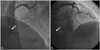
A 57-year old male patient with history of a failed PCI of a RCA CTO lesion since 6 months ago was readmitted to our center with a chief complaint of effort chest pain and background history of hypertension, dyslipidemia and diabetes mellitus. The transthoracic echocardiogram showed preserved systolic function (LVEF 54%), and akinesia of the basal to mid inferior and basal septum and mid posterior wall. The bilateral angiography, done through both femoral artery approaches demonstrated total occlusion of the mid RCA and visualization of grade 2 collateral flow from left anterior descending artery (Fig. 1). First, therefore, we planned an antegrade RCA CTO intervention. A 7 French Amplatz Left 1 guiding catheter (Med-tronic, Launcher, CA, USA) was used to engage the RCA ostium and 0.014" Runthrough guide wire (Terumo, Tokyo, Japan) was advanced through Corsair microcatheter (Asahi, Tokyo, Japan). Then, the Runthrough guide wire was exchanged with a Fielder XT-A wire (Asahi Intecc, Aichi, Japan), which successfully crossed the CTO segment. Then, the Fielder XT-A wire was exchanged to Runthrough wire for balloon dilation. Sequ-ential predilation using Laxa balloon 1.3×10 mm (Goodman, Nagoya, Japan) and Genoss 2.0×15 mm (Genoss, Suwon, Korea) was done for lesion pretreatment. OCT examination of RCA revealed that reference lumen diameter was 2.89 mm and lesion length 41 mm.
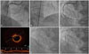
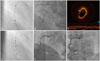
First BVS (Abbott Vascular, Santa Clara, CA, USA) 3.0×18 mm was delivered into the distal RCA lesion and deployed by slow inflation (2 atm, every 5 seconds) up to nominal pressure. After acquisition of the angiographic and stent boost images during second BVS positioning, multiple analyses and observations were done to understand the relationship between the BVS marker beads and balloon markers of distal and proximal BVSs. After clear identification of marker-to-marker and market-to-bead relationship, the second BVS was successfully overlapped and deployed. The distal markers of the second scaffold was over the proximal markers of the first scaffold, resulting in 1–2 mm of overlap. By minimal overlapping of two scaffolds, 3.0×18 mm and 3.0×28 mm BVSs were implanted from mid to distal RCA without complications (Fig. 2). Stent boost imaging showed that the overlapping was safely achieved (Fig. 3A). For further stent optimization, more aggressive adjuvant post-dilation was done with a 3.0×8.0 mm Pantera LEO (Biotronik, AG, Switzerland) balloon, slowly inflated at high pressure (18 atm) (Fig. 3B and D). A final assessment using OCT showed a fully expanded BVS without malapposition, dissection, and intramural hematoma (Fig. 3C). The patient was stabilized and safely discharged 3 days post-procedure. Patient was clinically followed, and 6-month routine angiographic finding showed good patency without significant restenosis (Fig. 3E and F).
DISCUSSION
Even newer generation drug-eluting stent (DES) has better clinical outcomes than bare metal stents or first-generation DESs, nevertheless, but their progress to thrombosis and restenosis limit long-term safety and efficacy.2 The BVS has been restricted to simple lesions, but CTO ABSORB pilot study showed excellent safety and long-term patency of the BVS for CTO PCI without increasing major adverse cardiovascular events.3
Previous study showed that the enhanced stent visualization system (ESV) is effective, particularly for overlapping BVS implantation, and the Poznan CTO-Absorb registry showed that the CTO stenting with BVS was associated with good performance and midterm clinical outcomes.5 Biscaglia, et al.6 evaluated whether ESV system-guided implantation for overlapping BVS is superior to angiography alone-guided implantation in the significant reduction of overlap length. In this WOLFIE study, 30 patients were treated with at least two overlapping BVS. In the ESV-guided group, overlap length was lower than that in angiography-guided group [0.9 (0.6–1.8) mm vs. 2.2 (1.3–3.2) mm, p=0.02]. Thus, they concluded that ESV-guided implantation of overlapping BVS is safe and effective in both shorter overlap length and number of stacked struts. The UNDERDOGS study also showed overlapping BVS for CTO lesions.7 This study included 162 consecutive patients who received overlapping BVS implantation, and a propensity-score was applied with 162 patients who received second generation DES in overlap to compare a device-oriented endpoint. Similarly, ESV system, as well as intravascular ultrasound (IVUS) and OCT, were employed significantly more in the BVS group than in the DES group (both groups, p<0.0001) with the “marker-to-marker” was the most applied overlap technique in the BVS group. Due to radiolucency of the BVS on the fluoroscopy and very tiny radiopaque markers, there are greater chances of geographic miss in case of overlapping stenting, and optimal BVS expansion is not easy without high resolution intravascular imaging including OCT or IVUS. Finally, they reported two cases of OCT evaluation of overlapping everolimus-eluting BVS implantation guided by ESV system, and the result showed that ESV system helped intentional achievement of minimum BVS overlap and reduced the number of overlapped struts.8
For the optimal clinical outcomes by immediate successful procedure, we attempted OCT and stent boost imaging guided PCI for this RCA CTO lesion. In addition to the benefit of stent boost imaging system for BVS minimal overlapping, we reported the usefulness of stent boost imaging.
In our present case, after a successful implantation of two overlapping BVS implantation by combined OCT and stent boost imaging guidance in relatively complex long RCA CTO lesion, immediate optimal angiographic and OCT findings were achieved without post-procedural BVS malapposition, fracture, thrombosis and acute recoil.
 XML Download