

 PDF
PDF ePub
ePub Citation
 Print
Print


INTRODUCTION
Ureteral stone is one of the most common urologic diseases. Affected patients can experience extreme pain. There are many options for ureteral stone treatment [e.g., shock wave lithotripsy (SWL), and ureteroscopic, laparoscopic, or open surgery]. Since the 1980s, SWL has been recommended as a non-invasive, effective, first-line treatment for small-sized radio-opaque stones (≤2 cm diameter).1 The overall stone-free rate after treatment using SWL is 80–90%.23 A patient's age, sex, and stone characteristics (stone site, size, and density) are factors associated with stone-free rates. The number of stones, a history of urolithiasis, presence of renal colic, degree of hydronephrosis, and presence of a double J stent are additional factors associated with stone-free rates after treatment using SWL.456
Ureteral stone is the most common cause of ipsilateral hydronephrosis. The presence of one or more ureteral stones warrants urgent intervention to resolve the patient's symptoms and to prevent damage to renal function. The relationship between the degree of preoperative hydronephrosis and stone free rate after SWL has been studied. However, unanswered questions remain, and a complete understanding would aid clinical decision-making during treatment selection.5678 We aimed to investigate the predictors of one-session success rates after treatment using SWL; we specifically focused on the significance of pretreatment hydronephrosis in patients with a ureteral stone.
MATERIALS AND METHODS
Patient cohort
Medical records were obtained from a database of patients (n=1824) who underwent an initial session of SWL between November 2005 and December 2014 at Severance Hospital, Seoul, Korea. The study inclusion criteria were a single, 4–20 mm, radiopaque calculus located within the ureter on plain-film X-rays, presentation within 1 month prior to SWL treatment, and no evidence of stone migration. Patients with bilateral ureteral stones, urinary tract congenital anomalies, single kidney, concomitant medical expulsive treatment, or a history of stone surgery were excluded from the analysis. Data from a total of 700 patients were eligible for the analysis.
Good clinical practice protocols
The study was performed in accordance with all applicable laws and regulations, good clinical practices, and the ethical principles described in the Declaration of Helsinki. The Institutional Review Board of Severance Hospital approved the study protocol (4-2016-0791). The study was exempt from the need for written informed consent, because of its retrospective design and because the patients' records and information were anonymized and de-identified before analysis.
Shock wave lithotripsy
SWL was performed using an electro-conductive lithotripter (EDAP Sonolith Praktis, Technomed, Lyon, France) until December 2011. On January 2012, this lithotripter was replaced by an electromagnetic generative lithotripter (Dornier Compact Delta II lithotripter, Dornier Medtech, Wessling, Germany). All SWL procedures were performed under fluoroscopic guidance. The total numbers of shocks ranged from 2500 to 4000 in each session, at a rate of 60–90 shock waves per minute and at a launch intensity ranging from 16 to 55 MPa.
Demographic data and non-contrast computed tomography stone characteristics
A detailed medical history that included the number of past stone events was obtained for each patient. Stone characteristics, such as location, maximal stone length (MSL), stone heterogeneity index (SHI), skin-to-stone distance (SSD), and mean stone density (MSD), were evaluated. SSD was measured in the axial plane, 45° from the vertical axis.9 MSL was the longest stone length measured in three dimensions on non-contrast computed tomography (NCCT) images. We used the GE Centricity system (GE Healthcare Bio-Sciences Corp., Piscataway, NJ, USA) for the measuring procedures. MSD was measured using bone windows on the magnified axial NCCT image of the stone in the maximal diameter; the elliptical region of interest incorporated the largest cross-sectional area of the stone without including adjacent soft tissue.10 The SHI was defined as the standard deviation of Hounsfield units (HU) in the same way used by Lee, et al.11 Successful SWL treatment of the ureteral stone was defined as for patients who were rendered stone-free or had asymptomatic residual fragments ≤3 mm in largest stone diameter on simple X-ray at 2 weeks after a single SWL treatment and who did not require additional treatment within the 3-month follow-up period.12
Hydronephrosis grading system
Hydronephrosis was graded according to the degrees of change in the upper collecting system and defined using the Society of Fetal Ultrasound Grade system.13 Grade 0 hydronephrosis was defined as no dilatation of the renal pelvis, with calyceal walls opposed to each other. Grade 1 was defined as dilatation of the renal pelvis without dilatation of the calyces (could also be present in the extra renal pelvis), and no parenchymal atrophy. Grade 2 hydronephrosis was defined as dilatation of the renal pelvis (mild) and calyces (the pelvicalyceal pattern was retained), and no parenchymal atrophy. Grade 3 was defined as moderate dilatation of the renal pelvis and calyces, blunting of the fornicies and flattening of the papillae, and possible mild cortical thinning. Grade 4 hydronephrosis was defined as gross dilatation of the renal pelvis and calyces with a ballooned appearance, loss of the borders between the renal pelvis, calyces, and renal atrophy indicated by the presence of cortical thinning.
Statistical analyses
The statistical comparisons of the continuous variables from the patients' demographic information were performed using Student's or Welch's two-sample t-tests or the Wilcoxon rank sum test. One-way analysis of variance was used for subgroup analysis. Categorical variables were compared using Pearson's chi-square tests. Univariate and binomial multivariate logistic regression analyses were performed to identify factors that predicted post-SWL outcomes. To exclude potential multicollinearity between variables, variance inflation factors for these variables were also analyzed. The statistical analyses were performed using R software (version 3.0.3, R Foundation for Statistical Computing, Vienna, Austria; http://www.r-project.org) and SPSS 23 software (IBM Co., Armonk, NY, USA).
RESULTS
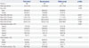
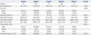
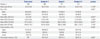
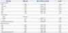
Table 1 presents the results for the baseline characteristics of the 700 patients who underwent an initial SWL treatment for single ureteral calculus. The mean patient age for the cohort was 52.5±13.8 years. The mean MSL and SSD values were 9.2±3.9 and 110.8±18.9 mm, respectively. The mean MSD and SHI values were 707.0±272.1 and 244.9±110.1 HU, respectively. The results for pretreatment hydronephrosis grade in patients with ureteral stone were 76 (10.9%) cases of grade 0, 383 (46.9%) cases of grade 1, 177 (25.3%) cases of grade 2, 100 (14.3%) cases of grade 3, and 19 (2.7%) cases of grade 4 hydronephrosis. There were 573 (81.9%) cases of upper ureteral stone, 48 (6.9%) cases of mid-ureteral stone, and 79 (11.3%) cases of lower ureteral stone. The overall one-session success rate was 69.6%. The data from the study population were divided into two groups (treatment success or failure). The results indicated that there were statistical differences between the two groups in age (51.8±13.8 years in success group vs. 54.1±13.8 years in failure group; p=0.042), MSL (8.3±3.2 vs. 11.2±4.5, respectively; p<0.001), MSD (642.6±243.8 vs. 854.3±276.4, respectively; p<0.001), SHI (252.0±114.0 vs. 228.5±98.9, respectively; p=0.006), and grade of hydronephrosis (p<0.001). The one-session success rates were 68.4, 75.0, 75.1, 54.0, and 10.5% for patients with hydronephrosis grades 0, 1, 2, 3, and 4, respectively (Table 2). The results indicated a statistically significant association between hydronephrosis grade and the values for MSL and MSD (p<0.001) (Table 2). The associations between hydronephrosis grade and one-session success rate and stone location were also significantly different (p<0.001). Among three groups with hydronephrosis grade 0–1, grade 2, and grades 3–4, there were statistically significant differences in sex (p<0.001), MSL (p<0.001), MSD (p<0.001), stone location (p<0.022), and one-session success rate (p<0.001) (Table 3). The univariate logistic regression models revealed that the following predictive factors were associated with SWL outcome after ureteral stone treatment: age [odds ratio (OR): 0.988, 95% confidence intervals (CI): 0.976–0.999; p=0.043], MSL (OR: 0.822, 95% CI: 0.782–0.861; p<0.001), MSD (OR: 0.997, 95% CI: 0.996–0.998; p<0.001), higher SHI (OR: 1.002, 95% CI: 1.001–1.004; p=0.010), and hydronephrosis grade (OR: 0.309, 95% CI: 0.206–0.463; p<0.001). Multivariate analyses revealed that shorter MSL, lower MSD, and higher SHI were independent predictors and that hydronephrosis grades 3 and 4 were negative predictors of one-session success after SWL treatment for ureteral stone (Table 4).
DISCUSSION
SWL has been established as the preferred treatment for urinary stones due to its noninvasiveness, few absolute contraindications, and favorable clinical outcomes.1415 However, if a satisfactory outcome for SWL is not expected for a specific clinical situation, then the other benefits (e.g., non-invasiveness) are no longer available to the patient. Therefore, it is essential to accurately predict individual treatment outcomes in terms of proper treatment selection for patients who are candidates for SWL.
Previous studies of SWL success rates have revealed that age, sex, and stone characteristics (stone site, size, and density) are factors associated with post-treatment stone-free rates.58 Number of stones, a history of urolithiasis, the presence of renal colic, the degree of hydronephrosis, and use of a ureteral stent are other factors that affect success rate.6 MSL is a potentially useful independent predictor of SWL outcome.56 Patients with larger stones are more likely to experience failure of treatment and require further intervention.16 MSD is also an independent factor associated with SWL outcome.17 MSD is the mean value of the HU of each pixel in a specific stone area and can be easily determined from NCCT images using a picture archiving and communication system.18 SSD has been extensively investigated as a predictor of SWL success, but its use remains controversial for patients with ureter stones: SSD has been deemed a significant factor in one-half of all published studies.9 Recently, SHI was introduced as a novel independent predictor of SWL success in patients with ureteral stone and as a useful clinical parameter for stone fragility.11 Other studies of stone sites have found that treatment of proximal and distal ureteral stones results in better outcomes, compared with treatment of mid-ureteral stones. However, the results of recent studies suggest that there are no differences between groups in regards to stone site and treatment results.619 A history of urolithiasis has been reported as a negative factor affecting SWL success.2021 Ureteral stenting is a significant factor affecting stone-free rates.19 Our study revealed that MSL, MSD, SHI, and severe hydronephrosis were significant predictors of one-session success rate after SWL treatment of single ureteral stone.
Several studies have evaluated the effects of preoperative hydronephrosis on the success rate of SWL; their findings have been inconsistent.5678 El-Assmy, et al.7 divided a total of 215 patients with single distal ureteral stone into two groups according to the absence or presence of hydronephrosis. There were no significant differences between the degree of stone-induced hydronephrosis and SWL outcome (83.2% in the non-hydronephrotic group vs. 74.2% in hydronephrotic group; p=0.27). They performed a similar study of 284 patients with proximal ureteral stone and found that the stone-free rate was 80.3% in the hydronephrotic group, compared with 89.1% in the patients without hydronephrosis (p=0.12).7 The results of Wang, et al.'s6 multivariate analysis also suggested that hydronephrosis was not a significant factor for SWL treatment success rate (OR: 1.272, 95% CI: 0.471–3.433; p=0.635). In contrast, Kageyama, et al.8 found that mid to lower ureteral stone and moderate-to-severe hydronephrosis were negative predictive factors of SWL treatment success rate. Delakas, et al.5 also found that the likelihood of SWL treatment failure increases as the severity of the obstruction increases; hydronephrosis was associated with poorer results after SWL treatment (borderline significance; OR: 1.93, 95% CI: 0.99–3.77; p=0.053). The results of our multivariate analyses indicated that severe hydronephrosis (grades 3 and 4) is an independent predictor of a poor outcome after SWL treatment for ureteral stone (Table 4).
Generally, ureteral stones cause sudden ureteral obstruction that results in the development of hydronephrosis; continuous obstruction results in deterioration of renal function. Hydronephrosis has adverse effects on renal function and decreases ureteral peristalsis and hydraulic pressure. These changes may adversely affect the expulsion of ureteral stones. This relationship may help explain our results. Severe hydronephrosis can also be linked to ureteral stone impaction into the ureteral mucosa. Impacted stones are frequently associated with ureteral polyps or strictures. A chronically impacted stone may cause inflammation and edema of the ureteral wall; these changes may also involve the surrounding tissues. The impacted stone can thus cause a more complete ureteral obstruction that results in severe hydronephrosis. Impacted ureteral stones are more difficult to fragment using SWL, because of the lack of a natural expansion space for the targeted stones. There are currently two clinical definitions used for impacted stones.22 The first commonly used definition of impaction is the inability to pass a wire or catheter beyond the stone at the initial attempt.23 The second definition of impaction is that the stone has remained at the same location in the ureter for more than 2 months.24
Most urologists know that impacted stones are much more resistant to treatment using SWL. However, if currently available definitions for stone impaction are used, it is almost impossible to determine whether a specific ureteral stone is impacted at initial diagnosis. For these reasons, severe hydronephrosis can be used as ancillary clinical evidence of ureteral stone impaction in patients with ureteral stone when SWL is being considered as a primary treatment. European Association of Urology (EAU) guidelines recommend laparoscopic ureterolithotomy for treatment of large impacted stones when endoscopic lithotripsy or SWL has failed.25 However, our study revealed one-session success rates of 54.0% and 10.5% in patients with hydronephrosis grades 3 and 4, respectively (Table 2). Therefore, even though SWL is less invasive, compared with surgical lithotripsy; we are not sure whether physicians should offer SWL as a first-line therapy for ureteral stone patients with concomitant grade 4 hydronephrosis. Physicians and patients should discuss this low SWL performance rate before selection of SWL as a treatment for ureteral stones with accompanying grade 4 hydronephrosis.
This study had some inherent limitations, because the use of a retrospective study design may have introduced sampling bias. However, we used a relatively large cohort of patients who underwent SWL for treatment of single ureteral stone. The presence of renal stones and anatomical considerations, including the location of calyx and renal pelvic stones or stones in the infundibulopelvic angle, were additional possible sources of bias. To overcome this type of limitation and more clearly elucidate the effects of various stone-related factors on SWL outcomes, we limited the study population to patients who only had ureteral stones. Unlike previous studies, we also classified hydronephrosis grade into five groups using a current grading system. This approach resulted in a better characterization of the contribution of hydronephrosis to treatment outcome. Prospective studies that use large sample sizes are needed to confirm our results regarding the relationships between pretreatment hydronephrosis and stone clearance.
In conclusion, the presence of grade 3 or 4 hydronephrosis before SWL was a negative predictive factor for one-session success in patients treated for a single ureteral stone. Severe hydronephrosis can be used as an indicator of possible ureteral stone impaction. In the presence of severe hydronephrosis, especially grade 4 hydronephrosis, physicians should be cautious when choosing and offering SWL as the primary treatment for ureteral stone.
 XML Download