

 PDF
PDF ePub
ePub Citation
 Print
Print


INTRODUCTION
Children with cerebral palsy (CP) are at risk for respiratory dysfunction due to various causes, such as pneumonia, atelectasis, bronchiectasis, sleep apnea, and chronic obstructive lung disease. Restrictive lung disease is also common in patients with CP.12 In addition, poor nutritional status, drooling, aspiration, gastroesophageal reflux, impairment of airway clearance due to muscular weakness or incoordination, and poor pulmonary reserve increase the risk of morbidity and mortality as a result of respiratory infection.345678 According to a recently reported study, children with spastic CP have lower pulmonary function than normal, healthy children.9 Poor chest mobility, trunk extensibility, and weak respiratory muscle strength are related to poor respiratory function in children with CP.31011
Incentive spirometer exercise (ISE) is widely used in chest physiotherapy, and it encourages the patient to perform slow and deep inspiration through visual feedback, allowing for the stretching and opening of collapsed airways. ISE is useful as it is inexpensive and simple to use with no known side effects, and also it does not require supervision once the child is trained in its use. Furthermore, achievement of the visual target encourages the children to try their best and thus promotes patient compliance.12 Previous reports demonstrated some beneficial effects of ISE on pulmonary function and arterial blood gases in patients with chronic obstructive pulmonary disease,131415 as well as in patients with ankylosing spondylitis.12
According to a recently published study, feedback respiratory training (FRT) in children with CP leads to improvement in pulmonary function.16 However, there is still a paucity of data regarding the effectiveness of respiratory training on pulmonary function in children with CP.
Maximum phonation time (MPT) represents the ability to maximally sustain a vowel sound after having taken a maximal inspiration. It is widely used to evaluate the efficiency of respiratory mechanisms during phonation, because it is a quick, non-invasive, and inexpensive method. According to a previous report, MPT has been used to objectively assess the degree of severity of dysphonia and to determine the effects of voice therapy. 17 Additionally, it has proven to be a highly reliable measure in voice assessment.18 To the best of our knowledge, the effects of respiratory training on MPT have not yet been investigated in children with CP.
Therefore, the aims of this study were to evaluate the effect of ISE on pulmonary function tests (PFT) and MPT in children with spastic CP.
MATERIALS AND METHODS
This was a prospective, randomized, case-control study (registration number NCT02406404). Ethical approval was granted by the Institutional Review Board and ethics committee of Severance Hospital. Since all of the children in this study were younger than 18 years, informed consent was obtained from the parents of the children who agreed to participate in this study.
Participants
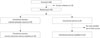
This study was conducted in a rehabilitation hospital affiliated with a university between May 2013 and April 2015. Among the children who were admitted to our hospital for intensive therapy, those who met the following inclusion and exclusion criteria were selected for this study: the inclusion criteria included 1) children with spastic CP between the ages of 8 and 15 years, 2) level I to IV on the Gross Motor Function Classification System (GMFCS), who have the ability to maintain antigravity head and trunk postures, 3) cognitive and cooperative function allowing for pulmonary function measurements, and 4) no history of psychiatric or neurological disorders other than CP. The exclusion criteria included 1) any uncontrolled, clinically significant medical condition, such as coexistent cardiac disease or respiratory disease, 2) children with cognitive impairment who are unable to comply with the protocol-required procedure, 3) children with the presence or history of tracheostomy, 4) children who are taking medications that can affect respiratory function, and 5) children with bone deformities of the spine, such as kyphosis or scoliosis. As a result, 60 children met the criteria, and 10 eligible participants declined to participate in the study. Thus, 50 children participated in this study. A computerized random generator was used to assign each participant to the experimental group (n=25) or the control group (n=25) (Fig. 1).
Study design
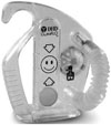
Participants in the experimental group were treated using a flow-oriented incentive spirometer (DHD CliniFLO®, Smiths medical ASD, Inc., Rockland, MA, USA) (Fig. 2), which has a chamber-containing ball. The ball is raised in its chamber upon generation of different inspiratory flow rates (six flow settings from 100 mL/sec to 600 mL/sec) by participants breathing through a mouthpiece. The children in the ISE group were instructed on how to use the incentive spirometer. After a quiet expiration, they were encouraged to close their lips tightly around the mouthpiece and then inhale slowly and deeply until the ball in the spirometer lifted. They were then instructed to hold their breath for as long as possible, or for at least 5 seconds, and then to breathe out slowly. Corresponding to the inspiratory flow, the balls lifted and remained suspended by the sustained inspiratory flow, which served as visible feedback. The flow rate progressively increased at 100 mL/sec intervals from 100 mL/sec to 600 mL/sec, if the participants could maintain the balls lifted at least 5 seconds.
The participants were encouraged to use the device for 10–15 breaths per session. Ten training sessions were performed daily for 4 weeks. They trained themselves with checklist in a hospital room with regular supervision twice a week. Children in both groups received conventional physical therapy and occupational therapy that focused on gross and fine motor tasks five times per week for a period of four weeks.
Outcome measures
The Gross Motor Function Measure (GMFM)-66 was used to assess improvement in gross motor function. GMFM-66 has been shown to be a reliable and valid measure of gross motor function and useful for measuring the effect of intervention programs.19
The PFT was done in the resting period at least 1 hour after physical and occupational therapy and ISE. The PFT was performed before training and at the end of the 4-week training period by the same investigator. PFT was performed using a portable spirometer (Micro Spirometer MS01; Micro Medical Ltd., Kent, UK) assessing forced expiratory volume at one second (FEV1) and forced vital capacity (FVC). A peak flow meter (ASSESS® Respironics International Inc., Murrysville, PA, USA) was also used to measure peak expiratory flow (PEF). The children were seated on a chair with the head and trunk straight and the hip and knee joints flexed to 90° with use of external supporting pad. For the children at GMFCS level IV, the test was performed in a chair sitting with external supporting pad and also the shoulder supported by their parent's hands. We used various chairs in consideration of each child's size. The children were told to inhale as deeply as possible and to blow their entire lung volume through the spirometer. This process was repeated at least three times, and the highest value was selected. PFT data were normalized for age, gender, and height, and the predicted FEV1 and FVC values (%) were calculated using the equations provided by Yoon, et al.20 based on values acquired from healthy Korean children. These calculations were used to obtain percent predicted values for FVC (FVC, %) and FEV1 (FEV1, %).
Maximal phonation time (MPT) testing was conducted in a quiet room with children seated on a chair in the same body position with measurement of PFT. The children were asked to inhale deeply and then produce a sustained phonation of the vowel /a:/ at a comfortable pitch and loudness in the seated position for as long as possible. The children were allowed three trials in a row with a 30-second break in between. Each phonation was video recorded (Handycam DCR-SR67; Sony Co., Tokyo, Japan), and the longest value was used for analysis.
Statistical analysis
The baseline characteristics of the two groups were compared using the Student's t-test or Mann-Whitney test for continuous variables and the chi-square test for categorical variables. Comparisons of pulmonary function before and after training within each group were made using the paired samples t-test or Wilcoxon signed rank test. Differences between the experimental group and the control group were assessed using an independent samples t test or Mann-Whitney test. Statistical software SPSS 20.0 (SPSS Inc., Chicago, IL, USA) was used for statistical analysis. The level of significance was chosen to be 0.05.
RESULTS
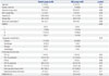
Although 50 children with CP were recruited for the study, two children in the control group did not complete the study protocol due to early discharge. As a result, 25 children in experimental group and 23 children in control group were included in this study. Their mean age was 11.6 years old (±2.3 years). The demographic and baseline characteristics of the study population are shown in Table 1. The two groups were not significantly different in gestational age, birth weight, height, or body weight. The PFT parameters and GMFM scores at baseline were not significantly different between the two groups.
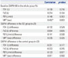
FEV1, FVC, PEF, and MPT increased significantly after ISE in the experimental group, compared with baseline data. The improvements in FEV1, FVC, and MPT were significantly higher in the experimental group than in the control group (Table 2).
Compared to baseline GMFM-66 scores, GMFM scores significantly increased after 4 weeks of training in both groups, although improvements in the GMFM scores were not significantly different between the two groups.
There were no significant relationships between GMFM-66 scores and PFT parameters, such as FEV1, FVC and PEF, but significant correlation with MPT (Table 3); and also the improvements in GMFM-66 scores did not significantly relate to changes in pulmonary function or MPT.
DISCUSSION
The FEV1, FVC, and FEV1/FVC ratio are useful parameters for assessing restrictive lung disease. On the other hand, PEF is a measure of the maximal or peak flow produced during exhalation with maximal effort, and it assesses maximal expiratory effort as a surrogate measure for expiratory muscle strength and is useful for assessing obstructive lung disease, such as asthma. These parameters are considered to be potentially very useful in integrated measures of respiratory function in Duchenne muscular dystrophy patients.21 In addition, previous studies have shown that pulmonary function in the children with CP had characteristics of both obstructive and restrictive lung disease,222232425 and thus, FEV1, FVC, and PEF were assessed as outcome measures in this study.
Shallow breathing, paradoxical breathing, and low breathing volume are commonly noted in children with CP, particularly low functioning CP, and these findings may lead to the development of widespread microatelectasis and a decrease in lung distensibility.2627 Morbidity and mortality associated with CP is mainly related to respiratory compromise, especially in low functioning CP. Therefore, interventions for enhancing respiratory function should be included as part of the comprehensive management for children with CP.
Some previous studies, report positive effects of various exercise programs on vital capacity (VC).112427 However, the effect of respiratory training in children with CP has rarely been reported. In a previous randomized controlled study by Lee, et al.,16 FRT led to significant gains in FEV1 and FVC, but not in PEF. In that study, the FRT group consisted of only nine children, and thus, the improvement in PEF after FRT did not reach a statistically significant level. On the other hand, our randomized controlled study revealed significant improvements in FEV1, FVC, and PEF after ISE training. FRT consists of repetitive, continuous performance of both maximal inspiration and expiration training, while ISE is designed to achieve and sustain maximal inspiration for a prolonged period using slow inspiration and deep breaths.162829 Although there are some differences between FRT and ISE, both ISE and FRT are effective in enhancing pulmonary function. The ISE, which increases transpulmonary pressure, inspiratory volumes, and inspiratory muscle performance, by encouraging the patient to take long, slow, deep breaths,30 led to improvement in these parameters. The results of our study suggest that ISE is useful for enhancing both inspiratory and expiratory muscle strengthening.
According to another previous study by Kwon and Lee,9 the children at GMFCS level III had a higher gain in FVC after FRT than the children at GMFCS level I or II.31 They suggested that children at GMFCS level I or II may have already reached the upper level of their respiratory function capacity, and thus, their improvement is not significant after FRT. The normal values of FVC and FEV1 can vary depending on various factors such as height, weight, and sex, and thus, predicted values (%) for FVC or FEV1 are preferred to compare the results across studies. In Kwon and Lee9 and Lee, et al.'s16 previous studies, predicted values for FVC and FEV1 were not assessed. In addition, the responses of respiratory training on pulmonary function in children with CP may differ between unilateral CP and bilateral CP. Further studies are needed to determine the best candidates for respirator training.
Respiration is an essential physiologic component for maintaining vital function and for performing physical activity in daily life.32 Respiration is also one of the key elements of physical fitness, along with muscular function and cardiovascular function.33 A recent study pointed out that respiratory muscle strength in children with CP is positively correlated with the activities of daily living, self-care, and social function.10 From this perspective, it is interesting to note whether improvement in respiratory function can lead to better exercise endurance, perception of dyspnea, and quality of life. Further studies to address this issue may be helpful for understanding the role of respiratory exercise in children with CP.
According to a previous study, MPT increases in normal children as they grew older, and MPT of normal children in the United States at the age of 8 to 15 years is between 17.1 and 20.7 sec.34 On the other hand, MPT of normal Korean children aged from 8 to15 years reportedly range from 12.6 to 15.8 sec.35 Compared to those values, the MPTs of our children are shorter. In a previous study, children with spastic CP had a shorter MPT than the normal control group.36 Also, a significant positive correlation between MPT and peak VO2 and also between MPT and the severity of chronic heart failure (CHF) were reported in patients with CHF.3738 On the other hand, there was no significant relationship between MPT and VC in adults with normal speech and voice.39 The proportion of VC used by individual speakers for the MPT task can vary, leading to no significant relationship between VC and MPT.404142 Also, in our study, there were no significant relationships between MPT and the parameters of PFT. ISE is a breathing technique in which deep breathing exercises are performed through a device that offers visual feedback in response to inspired flow and volume. These exercises allow patients to maximally inflate their lungs and to sustain that inflation. Thus, through this exercise, improvement in breath control appears to result in the observed increase in MPT. Breathy speech, weak breath control, and short utterances are major speech and voice concerns in children with CP.43 The results of our study suggest that ISE may be helpful for enhancing breath control for speech production in children with CP. Further studies are needed to delineate the clinical relevance of the improvement in MPT in terms of speech and voice quality in children with CP.
There are some limitations of our study. Most of children of our study were at level II or III, and thus, the effects of ISE according to GMFCS level were not investigated in our study. Further studies are needed to outline optimal candidates for ISE in terms of GMFCS level. Another limitation comes from the duration of respiratory training. According to a previous report, 16 significant improvements in pulmonary function were obtained with 4 weeks of respiratory training in children with CP. Four weeks is the maximal duration of stay in our hospital in general, and thus, 4 weeks of respiratory training was selected for our study. However, the optimal duration of training in order to maximize pulmonary function needs to be clarified in children with CP through further studies.
In conclusion, this randomized controlled study demonstrated significant benefits of ISE in terms of pulmonary function and MPT in children with spastic CP. ISE is a simple and inexpensive tool for children to use, and therefore, wider use of ISE may be helpful for children at risk of pulmonary dysfunction and poor breath control in speech production.

 XML Download