Journal List > Yonsei Med J > v.57(3) > 1031992

Ko, Kim, Sol, Kim, Yoon, Kim, and Kim: High-Sensitivity C-Reactive Protein Can Reflect Small Airway Obstruction in Childhood Asthma

Abstract

Purpose

High-sensitivity assays enabled the identification of C-reactive protein (hs-CRP) at levels that were previously undetectable. We aimed to determine if hs-CRP could reflect airway inflammation in children, by comparing hs-CRP with spirometry and impulse oscillometry (IOS) parameters and symptomatic severities.

Materials and Methods

A total of 276 asthmatic children who visited Severance Children's Hospital from 2012–2014 were enrolled. Serum hs-CRP and pulmonary function tests were performed on the same day. Patients were divided into hs-CRP positive and negative groups (cut-off value, 3.0 mg/L).

Results

Of the 276 asthmatic children [median age 7.5 (5.9/10.1) years, 171 boys (62%)], 39 were hs-CRP positive and 237 were negative. Regarding spirometry parameters, we observed significant differences in maximum mid-expiratory flow, % predicted (FEF25–75) (p=0.010) between hs-CRP positive and negative groups, and a negative correlation between FEF25–75 and hs-CRP. There were significant differences in the reactance area (AX) (p=0.046), difference between resistance at 5 Hz and 20 Hz (R5–R20) (p=0.027), resistance at 5 Hz, % predicted (R5) (p=0.027), and reactance at 5 Hz, % predicted (X5) (p=0.041) between hs-CRP positive and negative groups. There were significant positive correlations between hs-CRP and R5 (r=0.163, p=0.008), and X5 (r=0.164, p=0.007). Spirometry and IOS parameters had more relevance in patients with higher blood neutrophil levels in comparison to hs-CRP.

Conclusion

Hs-CRP showed significant correlation with FEF25–75, R5, and X5. It can reflect small airway obstruction in childhood asthma, and it is more prominent in neutrophil dominant inflammation.

INTRODUCTION

Asthma is the most common chronic airway inflammatory disease in childhood. Approximately 40% of all young children experience at least one episode of asthmatic symptoms such as coughing, wheezing, and dyspnea.12 Asthma is characterized by bronchial hyper-responsiveness and chronic airway inflammation. Various cells, cytokines, and mediators participate in the process.3 In addition to local inflammation, systemic inflammation is also present in asthma. This is indicated by the increase in plasma fibrinogen and serum amyloid A levels in asthmatic patients.45 However, its pathogenesis remains unclear.
C-reactive protein (CRP), named after its capacity to precipitate the somatic C-polysaccharide of Streptococcus pneumoniae, was the first acute-phase reactant to be described. It is widely used as a marker for inflammation and tissue injury.67 Assays for CRP, although simple and cost-effective, lacked the sensitivity using conventional methods. Standard assays of CRP, with its lower detection limit of 3–8 mg/L, could not detect low-grade inflammation.89 However, recent advancements have made high-sensitivity assays for CRP available in clinical laboratories. High-sensitivity assays for CRP, reported to have 100-fold higher sensitivity than standard assays, can detect CRP at extremely low concentrations with its lower detection limit of 0.1–0.2 mg/L.910 CRP measured using high-sensitivity assays is referred to as high-sensitivity CRP (hs-CRP). Application of hs-CRP revealed the presence of low-grade inflammation in several disorders, such as cardiovascular disorders and diabetes mellitus.11
With its ability to detect even low-grade inflammation, it is now thought that hs-CRP can reflect systemic inflammation in asthma.245 It has been reported that asthmatic patients have higher serum hs-CRP levels than healthy controls.245910121314 Negative relationships between serum hs-CRP levels and spirometry indices were reported, reflecting positive associations between increased serum hs-CRP levels and respiratory impairment. 9151617 Positive relationships between serum hs-CRP level and asthma severity have also been reported.213141819 A study with pediatric subjects showed that serum hs-CRP levels were higher in asthmatic patients than in healthy controls, and that there were positive associations between serum hs-CRP levels and asthma severity.219
In this context, we designed a study to check if there are any significant relationships between symptomatic severities of asthma, pulmonary function test (PFT)–spirometry and impulse oscillometry (IOS)–parameters, and hs-CRP in asthmatic children.

MATERIALS AND METHODS

Subjects

This study included 276 asthmatic children who visited the Pulmonology and Allergy Department of Severance Children's Hospital from January 2012 to April 2014. Blood sampling, PFT, and histories to determine symptomatic severities were conducted on the day of the clinic visit. Exclusion criteria included fever, acute respiratory infections, and signs of other acute infections seven days prior to blood sampling. Patients with other comorbidities that can influence the levels of hs-CRP, such as diabetes mellitus, cancer, recent history of surgery, and chronic inflammatory conditions like systemic lupus erythematosus, rheumatic arthritis, and inflammatory bowel disease were also excluded.
Patients were classified as asthmatic patients if they showed any typical asthma symptoms–such as wheezing, cough, or dyspnea–together with reversibility in airflow obstruction or bronchial hyper-responsiveness. Reversibility in airflow obstruction was defined as an increase in the forced expiratory volume in 1 second (ΔFEV1) ≥12% from baseline after inhalation of a short-acting beta-2 agonist. Bronchial hyper-responsiveness was defined as a positive result in bronchoprovocation by the methacholine challenge test (MCT). This meant that provocation concentration resulted in a 20% fall in FEV1 (PC20) ≤16 mg/mL.20
All patients were then divided into two groups based on whether they had positive hs-CRP values or negative hs-CRP values. The reference ranges of CRP and hs-CRP are <10 mg/L and <3 mg/L, respectively.2122 Therefore, the cut-off value of hs-CRP was set to 3.0 mg/L, making hs-CRP ≥3.0 mg/L positive, and hs-CRP <3.0 mg/L negative.
Patients were also divided into two groups according to their neutrophil counts: the high neutrophil group with neutrophil counts ≥median of neutrophil counts of all patients (3665/µL) and the low neutrophil group with neutrophil counts <median. Patients were likewise divided according to eosinophil counts into a high eosinophil group with eosinophil counts ≥median of eosinophil counts of all patients (350/µL) and a low eosinophil group with eosinophil counts <median. Within each group, patients were further divided into positive and negative hs-CRP subgroups, and their spirometry and IOS parameters were compared separately.
This study was approved by the Institutional Review Board of Severance Hospital (Seoul, Korea, IRB No. 4-2004-0036).

Laboratory tests and allergy screening tests

From the sampled blood, a complete blood count (CBC), including white blood cell (WBC), neutrophil, and eosinophil counts, as well as total and specific immunoglobulin E (IgE) and hs-CRP levels were measured. The CBC was measured using the ADVIA 2120i hematology system with autoslide (Siemens Healthcare Diagnostics Inc., Deerfield, IL, USA), hs-CRP using the Hitachi 7600 P module (Hitachi High-Technologies Corporation, Tokyo, Japan), and total and specific IgE using the Pharmacia CAP assay (Uppsala, Sweden).
Skin prick tests were done for 12 common aeroallergens, which included two types of dust mites, cat and dog epithelia, as well as mold and pollen allergens such as Alternaria, Aspergillus, birch, oak, mugwort, Japanese hop, ragweed, and Bermuda grass. Negative control (using saline solution) and positive control (using 0.5% histamine hydrochloric acid solution) were conducted simultaneously. A positive reaction was defined as a wheal of diameter >3 mm after 15 minutes.
Atopy was defined as specific IgE >0.7 kU/L to more than 1 allergen, total IgE >150 IU/mL, or one or more positive skin reactions to 12 common aeroallergens.

Spirometry and impulse oscillometry

The Jaeger MasterScreen PFT system (Jaeger Co., Wurzberg, Germany) was used for spirometry and the MCT. Flow-volume curves were acquired, according to the American Thoracic Society guidelines, before and after inhalation of a short-acting beta 2 agonist. For the MCT, increasing concentrations of methacholine (0.075, 0.15, 0.31, 0.62, 1.25, 2.5, 5, 10, 25, and 50), nebulized by the dosimeter (MB3; Mefar; Bresica, Italy), were inhaled by each patient until FEV1 was reduced by 20% from a post-nebulized saline solution value. Provocation concentration causing a 20% fall in FEV1 (PC20) was calculated by linear interpolation of the log dose-response curve.
The Jaeger MasterScreen IOS system (Jaeger Co., Wurzberg, Germany) was used for IOS. IOS was performed prior to spirometry, both before and after bronchodilator inhalation, to avoid any possible influence by forced breathing during spirometry. Calibration of the system was performed through a single volume of air (3 L) at different flow rates using a reference device (0.2 kPa/L/s). The machine was also calibrated to the air temperature and pressure of the saturated gas. The impulse generator produced pressure pulses at intervals of 0.2 seconds. From the measurement over 60 seconds, the mean resistance (R) values were calculated at frequencies of 5 Hz (R5) and 10 Hz (R10), and the reactance (X) values were calculated at 5 Hz (X5). The reactance area (AX), which is an integrated response index for reactance developed by Goldman,23 was also computed. It demonstrates the integral of the negative values of reactance from 5 Hz to the resonant frequency. The difference between R5 and R20 (R5–R20) was also calculated as the parameter of small airway obstruction, since R5 demonstrates obstruction in total airways, while R20 demonstrates obstruction in only the large airways. X5 and AX also demonstrate obstruction of peripheral airways.24
During IOS, patients sat upright with their heads placed against the back of the chair. While wearing nose clips, they were instructed to breathe quietly through a mouthpiece. In order to minimize shunt compliance of the cheeks and to observe for any artifacts produced by coughing, breath holding, swallowing, or vocalization, an investigator supported the patients' cheeks and chins from behind. Three correct measurements without any artifacts were averaged using an acceptable coherence value of ≥0.8 at 10 Hz.24

Statistical analysis

The values that follow parametric distributions are expressed as mean±standard deviation, while the values that follow non-parametric distributions are expressed as median with interquartile ranges. Comparisons of two groups were performed using t tests or Fisher's exact tests if the data were parametric and continuous, or using Kruskal-Wallis test and Mann-Whitney test if the data were non-parametric and continuous. If the data were categorical variables, the chi-square test was used. Correlations between data were analyzed using Spearman's rank correlation test.
A p value of <0.05 was considered significant. The Statistical Package for the Social Sciences (version 18.0, SPSS Inc., Chicago, IL, USA) was used for all analyses.

RESULTS

Clinical characteristics, asthma severity, and hs-CRP

The median age of the 276 patients was 7.5 (5.9/10.1) years, with 171 (62%) patients being boys. Among them, 39 patients had positive hs-CRP values, while 237 patients had negative hs-CRP values. Patients were divided into two groups by hs-CRP levels; however, there were no significant differences in age, gender, atopy (defined based on their specific IgE, total IgE, and results of the skin prick tests), WBC count, neutrophilcount, eosinophil count, total IgE level, or body mass index between the groups (Table 1). The patients were categorized by asthma severity into three groups according to the National Heart, Lung, and Blood Institute (NHLBI) guidelines: 146 patients were intermittent, 74 were mild persistent, and 56 were moderate to severe persistent.20 Median hs-CRP values were as follows: intermittent group, 0.5 (0.4/1.2), mild persistent group, 0.6 (0.4/1.2), and the moderate to severe persistent group, 0.6 (0.4/1.7). There was no significant difference in hs-CRP values among the three asthma severity groups (p=0.706). There was also no significant difference in asthma severity between the positive hs-CRP group and the negative hs-CRP group (Table 1).

Spirometry parameters and hs-CRP

We compared the forced vital capacity (FVC), % predicted FEV1, FEV1/FVC ratio in percentage, and FEF25–75, with hs-CRP. Out of these, FEV1 and FEF25–75 showed significant differences between positive and negative hs-CRP groups (Fig. 1). Among spirometry, only FEF25–75 had a significant negative correlation with hs-CRP (Table 2).

IOS parameters and hs-CRP

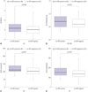
Among the IOS parameters, AX, R5–R20, R5, R10, and X5 were compared between the positive and negative hs-CRP groups. Out of these, AX, R5–R20, R5, and X5 showed significant differences between the positive and negative hs-CRP groups (Fig. 2). Among IOS parameters, only R5 and X5 showed significant positive correlation with hs-CRP values (Table 2).

Subgroup analysis according to blood neutrophil/eosinophil levels

The high and low neutrophil groups were each comprised of 138 patients. In the high neutrophil group, there were significant differences in the FVC1, FEV1, FEF25–75, and X5 between the positive and negative hs-CRP subgroups. There were also significant correlations between hs-CRP and FEF25–75, AX, R5, and X5, individually. Meanwhile, in the low neutrophil group, there was no significant association between the PFT parameters and hs-CRP (Table 3).
Similarly, all 276 patients were divided into high eosinophil (n=140) and low eosinophil (n=136) groups. In the high eosinophil group, there was no significant association between the PFT parameters and hs-CRP. Meanwhile, in the low eosinophil group, there were significant differences in the FEV1, FEF25–75, AX, R5–R20, R5, and X5 between the positive and negative hs-CRP subgroups, and hs-CRP had significant correlations with the FEV1, FEF25–75, R5–R20, R5, and X5 (Table 4).

DISCUSSION

This study is of significance because it is, to our best knowledge, the first study that compared hs-CRP with IOS parameters in asthmatic patients. In the present study, we found there were significant differences in the FEV1, FEF25–75, AX, R5–R20, R5, and X5 between the hs-CRP positive and negative groups. Hs-CRP also showed a significant negative correlation with FEF25–75, and significant positive correlations with R5 and X5. The associations between hs-CRP and the PFT indices were stronger in patients with higher blood neutrophil counts than in patients with lower counts. However, hs-CRP showed no significant relationship with symptomatic asthma severities.
CRP is an inflammatory marker synthesized mainly by hepatocytes and Browicz-Kupffer cells, but also by monocytes and lymphocytes. Its main roles are recognition and elimination of bacteria and damaged cells through complement and activation of phagocytic cells.1325 CRP has been widely used as an inflammatory marker because of its simple and cost-effective measurement methods; however, routine assays lacked the sensitivity to detect the presence of low concentrations of serum CRP in chronic low inflammatory diseases. With the development of high-sensitivity assays of CRP, the detection of subtle elevations of serum hs-CRP in conditions associated with chronic low-grade inflammation became possible. Indeed, hs-CRP has been demonstrated to be a strong independent predictor of future myocardial infarction, stroke, and peripheral arterial diseases in healthy people, as well as recurrent events in patients with coronary diseases.71226
Chronic airway inflammation is characteristic to asthma, but it has been proven that systemic inflammation is also present.245 Systemic inflammation in asthma is supported by the increase in circulating pro-inflammatory cytokines, such as interleukin (IL)-6 and tumor necrosis factor (TNF)-α, a consequent increase in acute-phase reactants such as CRP, and the presence of immune cells, such as neutrophils and natural killer cells in asthmatic patients.27 Accordingly, several studies report associations between hs-CRP, a sensitive marker of systemic inflammation, and asthma. Serum hs-CRP levels in asthmatic patients were reportedly higher than in healthy controls.5910 This was the same in pediatric populations.2 Associations, such as a significant negative correlation between hs-CRP and FEV1 1517 or FEV1/FVC916 in asthmatic patients, between hs-CRP and spirometry indices have also been reported. However, the pathogenesis of systemic inflammation in asthmatic patients remains unclear. It may be because of an overspill effect of local airway inflammation, tissue hypoxia, subclinical respiratory tract inflammation, or other environmental factors.28293031
In the present study, we found that FEV1 and FEF25–75 among spirometry parameters were significantly lower in the hs-CRP positive group than in the hs-CRP negative group, and that only FEF25–75 had a statistically significant negative correlation with hs-CRP. FEF25–75 reflects small airway patency and is reduced in asthmatic patients with a history of wheezing.3233 Hs-CRP, therefore, may have a greater association with small airway obstruction or inflammation.
IOS is a noninvasive technique to measure pulmonary impedance, which comprises pulmonary resistance and reactance.34 Since it is measured during normal tidal breathing, it is now popular for its applicability to younger children who are unable to conduct forced breathing, which is required to measure spirometry parameters. It is also highlighted as a marker for small airway obstruction. In the present study, AX, R5–R20, R5, and X5 were significantly higher in the hs-CRP positive group than in the hs-CRP negative group, and R5 and X5 showed statistically significant positive correlations with hs-CRP. A decrease in FEF25–75 and an increase in R5 and X5 are all characteristics of small airway obstruction.24 Accordingly, serum hs-CRP can reflect small airway inflammation.
CRP is a systemic marker for neutrophilic inflammation. In this context, Wood, et al.35 reported that hs-CRP was higher in patients with neutrophilic asthma. Some studies showed that hs-CRP had no association with markers of eosinophilic inflammation, history of atopy, or allergic sensitization,9313536 while other studies reported that hs-CRP was increased in non-atopic asthmatic patients but not in atopic asthmatic patients.121837 In the present study, when the subjects were subdivided according to their neutrophil and eosinophil levels, only patients in the high neutrophil and low eosinophil groups showed any statistically significant associations between serum hs-CRP and PFT indices. This result shows that hs-CRP can reflect small airway obstruction in asthma with neutrophil dominant inflammation.
As mentioned above, the reason for the presence of systemic inflammation, especially neutrophilic inflammation, in asthma is still unknown. Hypotheses that cytokines from airway 'spill over' to systemic circulation have been proposed in chronic obstructive pulmonary disease, but not for asthma.38 However, Wood, et al.35 demonstrated that receptors α for IL-8 (IL8RA), which are highly selective for neutrophil chemotaxis, were elevated in neutrophilic asthma. Meanwhile, Fu, et al.39 showed that systemic inflammation in asthma was associated with a body of upregulated genes that were involved in IL-1, TNF-α/nuclear factor-κB, and Kit receptor pathways, which were associated with innate immune response, defense and inflammatory responses, and particularly neutrophilic inflammation.
Navratil, et al.19 reported that serum hs-CRP was higher in uncontrolled asthmatic patients than in controlled asthmatic patients. Kilic, et al.7 reported that there is a negative association between hs-CRP and the asthma control test (ACT). However, in the present study, we could not prove a statistically significant relationship between hs-CRP and asthma severity according to NHLBI guidelines.20 The discrepancies between the results might be due to the fact that while ACT reflects the present condition, NHLBI severities reflect conditions of the past four weeks. As CRP is an acute-phase reactant, it may have less relevance with past symptoms than with present conditions.
The present study has limitations, in that the findings were not compared with those of normal healthy controls. However, several studies already reported that hs-CRP levels were more increased in asthmatic patients than to healthy control groups.245910121314 There was also only a small number of patients with positive hs-CRP (39 out of 276 patients, 14.1%). Additionally, neutrophil and eosinophil counts from induced sputum were unavailable, and relationships between hs-CRP and airway neutrophilia could not be investigated.
In conclusion, the present study showed that serum hs-CRP has significant associations with indices of small airway obstruction. This was more prominent in patients with higher blood neutrophil counts. We, therefore, propose the possibility of hs-CRP as a marker for small airway inflammation.

Figures and Tables

Fig. 1

Comparisons of forced expiratory volume in 1 second, % predicted (FEV1) and maximum midexpiratory flow, % predicted (FEF25–75) between high-sensitivity C-reactive protein (hs-CRP) positive and negative groups. FEV1 (p=0.046) and FEF25–75 (p=0.010) were significantly lower in the hs-CRP positive group than in the negative group.

ymj-57-690-g001
Fig. 2

Comparisons of impulse oscillometry (IOS) parameters between high-sensitivity (hs-CRP) positive and negative groups. (A) Reactance area (AX) was significantly higher in the hs-CRP positive group than in the negative group (p=0.046). (B) Difference between resistance at 5 Hz and 20 Hz (R5–R20) was significantly higher in the hs-CRP positive group than in the negative group (p=0.027). (C) Resistance at 5 Hz, % predicted (R5) was significantly higher in the hs-CRP positive group than in the negative group (p=0.027). (D) Reactance at 5 Hz, % predicted (X5) was significantly higher in the hs-CRP positive group than in the negative group (p=0.041).

ymj-57-690-g002
Table 1

Comparison of Clinical Characteristics between Patients in Positive and Negative hs-CRP Groups (n=276)

ymj-57-690-i001
Positive hs-CRP (n=39) Negative hs-CRP (n=237)
Age, yrs 6.5 (5.4/4.9) 7.6 (6.1/10.4)
Gender, male/female 25/14 146/91
Atopy 31 (79.5%) 172 (72.6%)
WBC, /µL 8000 (6380/11150) 7970 (6820/9555)
Neutrophil count, /µL 4210 (2810/6450) 3660 (2850/4650)
Eosinophil count, /µL 230 (160/470) 380 (180/560)
Total IgE, kU/L 297.0 (121.0/577.0) 194.5 (71.7/512.3)
BMI, kg/m2 16.8 (15.1/20.2) 16.7 (15.4/19.0)
hs-CRP, mg/L 6.7 (3.6/10.7) 0.5 (0.4/0.8)*
Severity according to NHLBI guidelines
 Intermittent (n=146) 20 (51.3%) 126 (53.2%)
 Mild persistent (n=74) 10 (25.6%) 64 (27.0%)
 Moderate to severe persistent (n=56) 9 (23.1%) 47 (19.8%)

hs-CRP, high-sensitivity C-reactive protein; WBC, white blood cell; BMI, body mass index; NHLBI, national heart, lung, and blood institute.

Data are given as median (interquartile range) or number (%).

*p<0.05 vs. positive hs-CRP.

Table 2

Correlations of hs-CRP and PFT Parameters

ymj-57-690-i002
Total (n=276) r p value
Spirometry parameters
 FVC, % predicted 101.23±14.78 -0.034 0.574
 FEV1, % predited 102.18±17.02 -0.094 0.118
 FEV1/FVC (%) 87.17 (81.34/92.42) -0.087 0.149
 FEF25-75, % predicted 84.31±26.13 -0.178 0.003
IOS parameters
 AX, kPa/L 3.16 (2.00/4.20) 0.083 0.180
 R5-R20, kPa/(L/s) 0.62 (0.51/0.79) 0.105 0.088
 R5, % predicted 104.50 (86.40/126.00) 0.163 0.008
 R10, % predicted 82.70 (73.30/95.90) 0.078 0.204
 X5, % predicted 139.30 (111.60/175.90) 0.164 0.007

hs-CRP, high-sensitivity C-reactive protein; PFT, pulmonary function test; FVC, forced vital capacity; FEV1, forced expiratory volume in 1 second; FEF25–75, maximum midexpiratory flow; IOS, impulse oscillometry; AX, reactance area; R5–R20, difference between resistance at 5 Hz and 20 Hz; R5, resistance at 5 Hz; R10, resistance at 10 Hz; X5, reactance at 5 Hz.

Data are given as mean±standard deviation, median (interquartile range), or number. r, Spearman's correlation coefficient.

Table 3

Comparison of Spirometry and IOS Parameters between Positive and Negative hs-CRP Groups, and Correlation between Spirometry and IOS Parameters and hs-CRP, within Separate High and Low Neutrophil Groups (n=276)

ymj-57-690-i003
High neutrophil group (n=138) Low neutrophil group (n=138)
Positive hs-CRP (n=21) Negative hs-CRP (n=117) Correlation with hs-CRP Positive hs-CRP (n=18) Negative hs-CRP (n=120) Correlation with hs-CRP
FVC, % predicted 97 (81/107) 102 (93/110)* r=-0.105 101 (81/117) 101 (92/110) r=0.868
FEV1, % predicted 93 (78/109) 105 (94/114)* r=-0.153 101 (82/117) 103 (91/110) r=-0.052
FEV1/FVC, % 86 (76/92) 87 (82/92) r=-0.091 89 (80/94) 87 (81/93) r=-0.082
FEF25-75, % predicted 63 (44/95) 86 (70/105)* r=-0.197 73 (60/110) 83 (64/101) r=0.063
AX, kPa/L 3.56 (2.56/4.83) 3.18 (2.00/4.19) r=0.178 3.44 (2.19/4.65) 3.05 (1.99/3.91) r=-0.036
R5-R20, kPa/(L/s) 0.72 (0.55/0.85) 0.64 (0.51/0.79) r=0.138 0.74 (0.52/0.92) 0.60 (0.50/0.74) r=0.036
R5, % predicted 116 (103/136) 105 (84/131) r=0.231 109 (90/137) 100 (86/119) r=0.048
X5, % predicted 176 (126/220) 142 (107/171)* r=0.282 141 (120/171) 134 (109/173) r=0.025

IOS, impulse oscillometry; hs-CRP, high-sensitivity C-reactive protein; FVC, forced vital capacity; FEV1, forced expiratory volume in 1 second; FEF25–75, maximum midexpiratory flow; AX, reactance area; R5–R20, difference between resistance at 5 Hz and 20 Hz; R5, resistance at 5 Hz; X5, reactance at 5 Hz.

Data are expressed as median (interquartile range). r, Spearman's correlation coefficient.

*p<0.05 vs. positive hs-CRP, p<0.05 in correlation with hs-CRP by Spearman's rank correlation test.

Table 4

Comparison of Spirometry and IOS Parameters between Positive and Negative hs-CRP Groups, and Correlation between Spirometry and IOS Parameters and hs-CRP, within Separate High and Low Eosinophil Groups (n=276)

ymj-57-690-i004
High eosinophil group (n=140) Low eosinophil group (n=136)
Positive hs-CRP (n=21) Negative hs-CRP (n=117) Correlation with hs-CRP Positive hs-CRP (n=18) Negative hs-CRP (n=120) Correlation with hs-CRP
FVC, % predicted 96 (78/111) 101 (94/109) r=-0.044 99 (83/116) 101 (92/112) r=-0.031
FEV1, % predicted 100 (76/112) 104 (92/111) r=-0.002 93 (79/113) 104 (95/114)* r=-0.181
FEV1/FVC, % 89 (78/94) 86 (82/92) r=-0.022 87 (77/92) 88 (82/93) r=-0.160
FEF25-75, % predicted 74 (60/101) 81 (65/99) r=-0.600 70 (45/89) 91 (68/105)* r=-0.301
AX, kPa/L 3.98 (2.46/4.79) 3.24 (2.12/4.26) r=0.028 3.51 (2.48/4.81) 2.89 (1.60/3.77)* r=0.154
R5-R20, kPa/(L/s) 0.70 (0.54/0.94) 0.66 (0.54/0.79) r=0.037 0.74 (0.55/0.88) 0.60 (0.46/0.68)* r=0.199
R5, % predicted 108 (86/133) 103 (86/126) r=0.092 110 (102/142) 102 (84/118)* r=0.236
X5, % predicted 135 (117/168) 133 (107/178) r=0.097 169 (123/210) 140 (114/166)* r=0.240

IOS, impulse oscillometry; hs-CRP, high-sensitivity C-reactive protein; FVC, forced vital capacity; FEV1, forced expiratory volume in 1 second; FEF25–75, maximum midexpiratory flow; AX, reactance area; R5–R20, difference between resistance at 5 Hz and 20 Hz; R5, resistance at 5 Hz; X5, reactance at 5 Hz.

Data are expressed as median (interquartile range). r, Spearman's correlation coefficient.

*p<0.05 vs. positive hs-CRP, p<0.05 in correlation with hs-CRP by Spearman's rank correlation test.

ACKNOWLEDGEMENTS

This research was supported by Basic Science Research Program through the National Research Foundation of Korea (NRF) funded by the Ministry of Education (2007-0056092 and 2011-0024036).

Notes

The authors have no financial conflicts of interest.

References

1. van de Kant KD, Klaassen EM, Jöbsis Q, Nijhuis AJ, van Schayck OC, Dompeling E. Early diagnosis of asthma in young children by using non-invasive biomarkers of airway inflammation and early lung function measurements: study protocol of a case-control study. BMC Public Health. 2009; 9:210.
crossref
2. Deraz TE, Kamel TB, El-Kerdany TA, El-Ghazoly HM. High-sensitivity C reactive protein as a biomarker for grading of childhood asthma in relation to clinical classification, induced sputum cellularity, and spirometry. Pediatr Pulmonol. 2012; 47:220–225.
crossref
3. Busse WW, Lemanske RF Jr. Asthma. N Engl J Med. 2001; 344:350–362.
crossref
4. Jousilahti P, Salomaa V, Hakala K, Rasi V, Vahtera E, Palosuo T. The association of sensitive systemic inflammation markers with bronchial asthma. Ann Allergy Asthma Immunol. 2002; 89:381–385.
crossref
5. Hoshino M, Ohtawa J, Akitsu K. Increased C-reactive protein is associated with airway wall thickness in steroid-naive asthma. Ann Allergy Asthma Immunol. 2014; 113:37–41.
crossref
6. Ramirez D, Patel P, Casillas A, Cotelingam J, Boggs P, Bahna SL. Assessment of high-sensitivity C-reactive protein as a marker of airway inflammation in asthma. Ann Allergy Asthma Immunol. 2010; 104:485–489.
crossref
7. Kilic H, Karalezli A, Hasanoglu HC, Erel O, Ates C. The relationship between hs-CRP and asthma control test in asthmatic patients. Allergol Immunopathol (Madr). 2012; 40:362–367.
crossref
8. Ridker PM. High-sensitivity C-reactive protein: potential adjunct for global risk assessment in the primary prevention of cardiovascular disease. Circulation. 2001; 103:1813–1818.
crossref
9. Takemura M, Matsumoto H, Niimi A, Ueda T, Matsuoka H, Yamaguchi M, et al. High sensitivity C-reactive protein in asthma. Eur Respir J. 2006; 27:908–912.
crossref
10. Shimoda T, Obase Y, Kishikawa R, Iwanaga T. Serum high-sensitivity C-reactive protein can be an airway inflammation predictor in bronchial asthma. Allergy Asthma Proc. 2015; 36:e23–e28.
crossref
11. Rifai N, Tracy RP, Ridker PM. Clinical efficacy of an automated highsensitivity C-reactive protein assay. Clin Chem. 1999; 45:2136–2141.
crossref
12. Sahoo RC, Acharya PR, Noushad TH, Anand R, Acharya VK, Sahu KR. A study of high-sensitivity C-reactive protein in bronchial asthma. Indian J Chest Dis Allied Sci. 2009; 51:213–216.
13. Zietkowski Z, Tomasiak-Lozowska MM, Skiepko R, Mroczko B, Szmitkowski M, Bodzenta-Lukaszyk A. High-sensitivity C-reactive protein in the exhaled breath condensate and serum in stable and unstable asthma. Respir Med. 2009; 103:379–385.
crossref
14. Sävykoski T, Harju T, Paldanius M, Kuitunen H, Bloigu A, Wahlström E, et al. Chlamydia pneumoniae infection and inflammation in adults with asthma. Respiration. 2004; 71:120–125.
crossref
15. Kony S, Zureik M, Driss F, Neukirch C, Leynaert B, Neukirch F. Association of bronchial hyperresponsiveness and lung function with C-reactive protein (CRP): a population based study. Thorax. 2004; 59:892–896.
crossref
16. Fujita M, Ueki S, Ito W, Chiba T, Takeda M, Saito N, et al. C-reactive protein levels in the serum of asthmatic patients. Ann Allergy Asthma Immunol. 2007; 99:48–53.
crossref
17. Shaaban R, Kony S, Driss F, Leynaert B, Soussan D, Pin I, et al. Change in C-reactive protein levels and FEV1 decline: a longitudinal population-based study. Respir Med. 2006; 100:2112–2120.
crossref
18. Olafsdottir IS, Gislason T, Thjodleifsson B, Olafsson I, Gislason D, Jögi R, et al. C reactive protein levels are increased in non-allergic but not allergic asthma: a multicentre epidemiological study. Thorax. 2005; 60:451–454.
crossref
19. Navratil M, Plavec D, Dodig S, Jelcic Z, Nogalo B, Erceg D, et al. Markers of systemic and lung inflammation in childhood asthma. J Asthma. 2009; 46:822–828.
crossref
20. Crapo RO, Casaburi R, Coates AL, Enright PL, Hankinson JL, Irvin CG, et al. Guidelines for methacholine and exercise challenge testing-1999. This official statement of the American Thoracic Society was adopted by the ATS Board of Directors, July 1999. Am J Respir Crit Care Med. 2000; 161:309–329.
21. Pearson TA, Mensah GA, Alexander RW, Anderson JL, Cannon RO 3rd, Criqui M, et al. Markers of inflammation and cardiovascular disease: application to clinical and public health practice: a statement for healthcare professionals from the Centers for Disease Control and Prevention and the American Heart Association. Circulation. 2003; 107:499–511.
22. Greenland P, Alpert JS, Beller GA, Benjamin EJ, Budoff MJ, Fayad ZA, et al. 2010 ACCF/AHA guideline for assessment of cardiovascular risk in asymptomatic adults: a report of the American College of Cardiology Foundation/American Heart Association Task Force on Practice Guidelines. Circulation. 2010; 122:e584–e636.
23. Goldman MD. Clinical application of forced oscillation. Pulm Pharmacol Ther. 2001; 14:341–350.
crossref
24. Komarow HD, Myles IA, Uzzaman A, Metcalfe DD. Impulse oscillometry in the evaluation of diseases of the airways in children. Ann Allergy Asthma Immunol. 2011; 106:191–199.
crossref
25. Pepys MB. C-reactive protein fifty years on. Lancet. 1981; 1:653–657.
crossref
26. Pepys MB, Baltz ML. Acute phase proteins with special reference to C-reactive protein and related proteins (pentaxins) and serum amyloid A protein. Adv Immunol. 1983; 34:141–212.
crossref
27. Gabay C, Kushner I. Acute-phase proteins and other systemic responses to inflammation. N Engl J Med. 1999; 340:448–454.
crossref
28. Wouters EF, Reynaert NL, Dentener MA, Vernooy JH. Systemic and local inflammation in asthma and chronic obstructive pulmonary disease: is there a connection? Proc Am Thorac Soc. 2009; 6:638–647.
crossref
29. Wilkinson TM, Donaldson GC, Johnston SL, Openshaw PJ, Wedzicha JA. Respiratory syncytial virus, airway inflammation, and FEV1 decline in patients with chronic obstructive pulmonary disease. Am J Respir Crit Care Med. 2006; 173:871–876.
crossref
30. Maclay JD, McAllister DA, Macnee W. Cardiovascular risk in chronic obstructive pulmonary disease. Respirology. 2007; 12:634–641.
crossref
31. Livnat G, Yoseph RB, Nir V, Hakim F, Yigla M, Bentur L. Evaluation of high-sensitivity serum CRP levels compared to markers of airway inflammation and allergy as predictors of methacholine bronchial hyper-responsiveness in children. Lung. 2015; 193:39–45.
crossref
32. Weiss ST, Tosteson TD, Segal MR, Tager IB, Redline S, Speizer FE. Effects of asthma on pulmonary function in children. A longitudinal population-based study. Am Rev Respir Dis. 1992; 145:58–64.
crossref
33. Rao DR, Gaffin JM, Baxi SN, Sheehan WJ, Hoffman EB, Phipatanakul W. The utility of forced expiratory flow between 25% and 75% of vital capacity in predicting childhood asthma morbidity and severity. J Asthma. 2012; 49:586–592.
crossref
34. Beydon N, Davis SD, Lombardi E, Allen JL, Arets HG, Aurora P, et al. An official American Thoracic Society/European Respiratory Society statement: pulmonary function testing in preschool children. Am J Respir Crit Care Med. 2007; 175:1304–1345.
crossref
35. Wood LG, Baines KJ, Fu J, Scott HA, Gibson PG. The neutrophilic inflammatory phenotype is associated with systemic inflammation in asthma. Chest. 2012; 142:86–93.
crossref
36. Halvani A, Tahghighi F, Nadooshan HH. Evaluation of correlation between airway and serum inflammatory markers in asthmatic patients. Lung India. 2012; 29:143–146.
crossref
37. Butland BK, Strachan DP, Rudnicka AR. C-reactive protein, obesity, atopy and asthma symptoms in middle-aged adults. Eur Respir J. 2008; 32:77–84.
crossref
38. Barnes PJ, Celli BR. Systemic manifestations and comorbidities of COPD. Eur Respir J. 2009; 33:1165–1185.
crossref
39. Fu JJ, Baines KJ, Wood LG, Gibson PG. Systemic inflammation is associated with differential gene expression and airway neutrophilia in asthma. OMICS. 2013; 17:187–199.
crossref
TOOLS
Similar articles