

 PDF
PDF ePub
ePub Citation
 Print
Print


INTRODUCTION
Acute aortic disease consists mainly of acute aortic dissection (and its variants, intramural hematoma and penetrating aortic ulcers) and thoracic or abdominal aortic aneurysm with or without rupture. Acute aortic disease is uncommon, yet associated with life-threatening vascular conditions that can result in high mortality if misdiagnosed and untreated.12 Timely diagnosis is especially challenging for emergency physicians, as clinical features vary and are often atypical.3456 Along with early diagnosis, the keys to successful treatment for patients with acute aortic disease are rapid provision of medical therapy and emergency intervention (endovascular or surgical) and transfer to the intensive care unit (ICU).789 In ascending aortic dissection, mortality rates have been reported to increase by 1–2% per hour after symptom onset.10 For Stanford type A aortic dissection without surgical treatment, the mortality rate is 20% by 24 hours, 30% by 48 hours, and 40% at 1 week.2
To ensure prompt treatment, cooperation among multidisciplinary departments is crucial. Clinical pathways (CPs) that provide the ideal treatment sequence in a timely manner have been developed for several urgent disease entities, such as ST-elevation myocardial infarction, acute cerebral infarction, severe sepsis, and major trauma.111213 However, a CP is not currently applied to patients with acute aortic disease. One study reported the effects of protocol-based management of type A aortic dissections,14 while another study reported the effects of the initiation of an acute aortic treatment center and the subsequent implementation of a treatment pathway.8
In the present study, we compared outcomes before and after the implementation of a CP for patients with acute aortic disease at the emergency department (ED). We hypothesized that we could reduce mortality among patients with acute aortic disease by shortening the time to diagnosis and emergency intervention via this CP.
MATERIALS AND METHODS
Study design and setting
This was a retrospective, observational cohort study, conducted at an urban tertiary care hospital with approximately 80000 visits per year to the ED. This study was approved by the hospital's Institutional Review Board committee. The first meeting for the CP was held in January 2012, and a protocol for patients with acute aortic disease was created and implemented in March 2012. Taking the education required and the adaptation period of three months into consideration, we compared patients treated during pre-CP implementation (January 1, 2010 to December 31, 2011) with those treated during post-CP implementation (July 1, 2012 to June 30, 2014). We screened patients who were admitted to the ED during the study period and discharged from the hospital with a diagnosis of aortic disease (International Classification of Disease codes I71.0–I71.9, aortic aneurysm and dissection). Data for all patients with aortic disease were investigated, and acute aortic diseases, such as symptomatic aortic aneurysms, acute aortic dissections (Stanford type A and type B), and intramural hematomas, were included. We excluded patients who did not have acute disease, such as those with asymptomatic uncomplicated aneurysm without the need of intervention, asymptomatic Stanford type B aortic dissection, intramural hematoma without the need of hypertensive control, and aortic disease without interval change from previous state. If diagnosis was not confirmed at the ED and CP was not activated, the patients were not included in analysis.
CP protocol
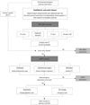
This CP was composed of two phases named PRE-AORTA and AORTA (Fig. 1).
PRE-AORTA
The aim of PRE-AORTA was timely diagnosis of acute aortic disease. Indications for PRE-AORTA activation were patients with a possibility of acute aortic disease based on clinical manifestations (mainly chest pain, abdominal pain, and back pain) and physical examination (including bedside ultrasonography) who had not yet been diagnosed with computerized tomography (CT). Even in cases with atypical symptoms and negative ultrasonography results, an emergency physician could activate PRE-AORTA with any suspicion of acute aortic disease. When PRE-AORTA was activated by clicking the CP button in the order communication system, an ED nurse, diagnostic laboratory, CT room, and transport staff were immediately facilitated. To easily recognize a CP activated patient, the color of the patients line on the emergency patient list screen of the electronic health record (EHR) system changed to sky-blue. An ED nurse obtained a blood sample and secured an intravenous line before treating other patients. The blood sample was then delivered to the diagnostic laboratory using an air shooter, specifically designed for CPs. In the diagnostic laboratory, a blood test including creatinine point-of-care testing was conducted, and the result was reported as soon as possible. Staff in the CT room arranged for an aorta CT, giving top priority to the PRE-AORTA-activated patient, and transport staffs were on standby for the timely transport of the patient to the CT room. If a diagnosis of acute aortic disease was ruled out as a result of the aorta CT, the emergency physician deactivated PRE-AORTA.
AORTA
The aim of AORTA was timely emergency intervention and rapid transfer to the ICU. If the CT results indicate the patient with new acute aortic disease or aggravation of previous aortic disease, the emergency physician changed the CP from PRE-AORTA to AORTA. Some patients were also transferred from other hospitals after diagnosis of acute aortic disease by CT imaging. In this case, AORTA was activated without first activating PRE-AORTA. When AORTA was activated, the color of the patient's line on the EHR changed to blue, and a short message service (SMS) notification was sent automatically to cardiologist, cardiovascular surgeon, radiologist, anesthetist, and ICU manager. Additionally, emergency physicians directly contacted the resident on duty of cardiology and cardiovascular surgery. The resident examined the CP-activated patient before other patients and notified his/her faculty. The cardiologist and cardiovascular surgeon then determined the appropriate emergency intervention (operation or endovascular repair) with assistance of a radiologist. They also decided whether the patient needed intensive care or not. Anesthetist and ICU manager then assigned the operation room and ICU bed with preference to AORTA-activated patients. If the patient did not need emergency intervention or ICU care, AORTA was deactivated.
Data collection
Patient data were collected retrospectively from medical records. Patient demographics, underlying disease, chief complaint, emergency resuscitations (e.g., central catheterization and endotracheal intubation at the ED), diagnosis, emergency intervention (operation or endovascular repair), and mortality were investigated. As the chief complaint could influence the timing of suspicion of acute aortic disease, typical chief complaints were classified as chest pain, abdominal pain, and back pain, and compared between the two periods. Considering the different severities and treatment options according to the diagnosis, we classified diagnosis, based on the interpretation of radiologists, into aortic dissection Stanford type A, aortic dissection Stanford type B, intramural hematoma, unruptured aortic aneurysm, and ruptured aortic aneurysm. Emergency intervention was confined to cases in which patients were transferred directly from the ED to the operation or procedure room to receive operation or endovascular repair. Mortality was assessed at the time of hospital discharge.
The timings of the patient's arrival at the ED, acquisition of CT images, emergency intervention, departure from the ED, departure from the ICU, and hospital discharge were evaluated. The timing of emergency intervention was defined as when the patient arrived at the operation or procedure room. The door-to-CT time (interval from ED arrival to CT acquisition), CT-to-intervention time (from CT acquisition to emergency intervention), ED length of stay (LOS), and ICU/hospital LOS were compared between the two groups.
Statistical analysis
The patient characteristics and treatment outcomes between the two groups were compared using Student's t-test for continuous variables and the chi-squared test or Fisher's exact test for dichotomous variables. We also used the Mann-Whitney U test for the time factors due to their positively skewed distribution. To analyse whether differences in patient characteristics between the two groups influenced the hospital mortality, multivariable logistic regression was performed by using the stepwise methods (variables were selected using the entry and exit criteria of p<0.1). Statistical analyses were performed using SAS 9.3 (SAS Institute Inc., Cary, NC, USA). A p-value of <0.05 was regarded as statistically significant.
RESULTS
Acute aortic disease was diagnosed in 94 patients treated during the pre-period and in 104 patients treated during the post-period. Thirteen patients were not in acute state and excluded: six aortic aneurysms and three intramural hematomas were asymptomatic and did not need intervention or hypertensive control, and two aortic aneurysms and two aortic dissections (Stanford type B) were the same as the previous state.
Patient characteristics
Characteristics of patients diagnosed with acute aortic disease were compared between the two periods (Table 1). More patients treated during the pre-period had underlying hypertension and coronary artery disease. The proportion of typical chief complaints was similar between the two groups. Thirty (31.9%) pre-period patients and 32 (30.8%) post-period patients had atypical chief complaints [pre-period: dyspnea (8), abdominal mass (3), syncope (3), mental change (2), etc.; post-period: dyspnea (10), flank pain (7), abdominal mass (2), mental change (2), etc.]. There was no significant difference in the number of patients transferred from other hospitals between the two groups. Among transferred patients, there were 11 atypical chief complaints (23.4%) during the pre-period and 18 (29.5%) during the post-period. More patients received endotracheal intubation at the ED during the pre-period; however, there was no statistical significance [14 (14.9%) vs. 8 (7.7%); p=0.107]. There were no significant differences in initial vital signs and the proportion of diagnoses between the two periods.
Outcome of PRE-AORTA phase
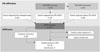
After the CP was implemented, PRE-AORTA was activated in 76 patients (Fig. 2). Among them, acute aortic disease was diagnosed in 19 patients in PRE-AORTA, and CP was deactivated in 57 patients. There were 30 patients who were not suspected of having acute aortic disease at first, yet diagnosed incidentally because of abdomen or chest CT images examined for other purposes. Of 61 patients who were transferred from another hospital after CT, 6 went through the PRE-AORTA phase, as the previous hospital's CT images were lost or insufficient for diagnosis.
The door-to-CT time was compared between the two periods (Table 2). Patients who were diagnosed at a previous hospital were not included in analysis. The median door-to-CT time was 119.0 (61.0, 192.0) during the pre-period and 82.0 (59.0, 161.5) during the post-period (p=0.414). The door-to-CT time of typical patients was reduced by 66.0 min during the post-period, although there was no statistical significance [139.0 (49.0, 204.8) vs. 73.0 (55.0, 128.5); p=0.161]. During the post-period, the door-to-CT time of PRE-AORTA-activated patients was shorter than non-activated patients by 42.0 min [71.0 (61.0, 115.0) vs. 113.0 (56.0, 170.5); p=0.026]. The proportion of typical chief complaints was similar between PRE-AORTA-activated patients and non-activated patients (14 of 19 vs. 19 of 30; p=0.541).
Outcome of AORTA phase
After CP implementation, AORTA was activated in 104 patients, of whom 38 (36.5%) received emergency intervention and 88 (84.6%) were admitted to the ICU (Fig. 2). Treatment results of patients were compared between the two periods (Table 3). During the post-period, more patients were admitted to the ICU [66 (70.2%) vs. 88 (84.6%); p=0.015] and received emergency intervention [21 (22.3%) vs. 38 (36.5%); p=0.029] than during the pre-period. Emergency intervention for aortic dissection Stanford type A and ruptured aortic aneurysm was performed more frequently during the post-period. The number of patients who died at the ED without receiving emergency intervention declined from 11 (11.7%) in the pre-period to 4 (3.8%) in the post-period (p=0.037). Most importantly, death from ruptured aortic aneurysm decreased by the largest margin after CP implementation [7 of 18 (38.9%) vs. 2 of 14 (14.3%)]. During the pre-period, three patients with aortic dissection Stanford type A were transferred to another hospital for emergency operation as there was no available ICU bed or operation rooms, and two patients (unruptured aortic aneurysm and aortic dissection Stanford type B) opted out from the ED against medical advice. During the post-period, one patient with intramural hematoma was transferred to a hospital near his home at his request. Hospital mortality decreased from 26.6% to 14.4% (p=0.033). Death of patients with aortic aneurysm (ruptured or unruptured) was reduced most in the post-period. In multivariable logistic analysis adjusting for previous aortic disease, and endotracheal intubation, and central catheterization in the ED, the post-period showed a negative association with hospital mortality, but this was not statistically significant [odds ratio (95% confidence interval)=0.416 (0.150, 1.156); p=0.093]. Hospital mortality was not influenced by hypertension (p=0.163) and coronary disease (p=0.319).
Table 4 shows that the median ED LOS decreased by 63.0 min in the post-period [236.0 (128.8, 353.8) min vs. 173.0 (104.5, 267.3) min; p=0.016]. We separately analyzed patients who were diagnosed after ED arrival and those who were diagnosed at a previous hospital. In patients diagnosed at the ED, ED LOS was reduced from 294.0 (211.0, 485.0) min to 207.0 (150.5, 305.0) min (p=0.041). The door-to-intervention time of these patients was reduced by 178.0 min in the post-period [378.0 (302.0, 489.0) min vs. 200.0 (170.0, 299.0) min; p=0.001]. The difference in door-to-CT time was not statistically significant; however, the CT to intervention time was reduced from 198.0 (180.0, 247.5) min to 138.0 (95.0, 183.0) min (p=0.013). On the other hand, there was no efficacy of time reduction in patients who were diagnosed with aortic disease before the transfer. ICU LOS was 3.5 (2.0, 11.0) days during the pre-period and 3.0 (2.0, 7.0) days during the post-period (p=0.214). The total hospital LOS was similar [pre-period: 9.5 (2.0, 17.3) days; post-period: 9.0 (6.0, 14.8) days; p=0.690].
DISCUSSION
We developed a CP composed of two phases: PRE-AORTA for diagnosis and AORTA for treatment. Unfortunately, our CP was unable to reach a productive outcome in the PRE-AORTA phase. When PRE-AORTA was activated, the time until acquisition of CT images was effectively reduced due to the CP protocol. However, we were able to diagnose acute aortic disease via PRE-AORTA only in 19 of 49 patients. The most important problem was the absence of a detailed screening protocol that took each aortic disease into consideration. Although this CP included various aortic diseases, our screening protocol merely included typical symptoms as chest pain, abdominal pain, and back pain. Many patients presented with chest pain or abdominal pain at the ED; however, the decision to activate the CP depended on each emergency physician, as our CP protocol did not specify the typical presentation of each disease. For this reason, 30 patients who presented with typical chief complaints were diagnosed with acute aortic disease without PRE-AORTA activation.
One other problem was that a fair number of patients with acute aortic disease did not show typical disease presentation.561516 In our study, atypical chief complaints accounted for 31.3% of cases. Previous studies have reported that timely diagnosis of acute aortic disease was challenging, and the rate of missed diagnoses was reported to be 38.2% for acute aortic dissection14 and 42.0% for ruptured aortic aneurysm.17 When preparing for CP implementation, we anticipated that we would be able to reduce the delay in diagnosis by applying bedside-echocardiography as a screening tool in a broader range of patients. However, we did not clarify candidates for echocardiography and entrusted the decision to individual emergency physicians, which did not appear to be effective. To prevent the omission of patients who potentially have acute aortic disease, more obvious criteria for screening need to be introduced. In 2010, the American College of Cardiology Foundation and the American Heart Association published guidelines for the diagnosis and management of thoracic aortic disease.18 In these guidelines, atypical symptoms defined as syncope and symptoms consistent with perfusion deficit such as cerebral, myocardial, mesenteric, or limb ischemia are included as screening symptoms. Rogers, et al.19 reported that when applying this screening tool to patients with acute aortic dissection who were enrolled in the International Registry of Acute Aortic Dissection, it had a sensitivity of 95.7%. Biomarkers such as D-dimer could also potentially be used in screening.202122
After CP implementation, fewer patients died at the ED and more patients received emergency intervention within a shorter time after diagnosis, resulting in improved hospital mortality in the AORTA phase. This effect was obvious only in patients who were diagnosed after ED arrival. A study by Grau, et al.14 is the only previous study to report the performance of a type A aortic dissection management protocol during a 7-year period. Consistent with our findings, they reported that after implementation of the protocol, patients were transported to the operation room faster, improving hospital mortality.14 Providing definite care on time is critical to reduce mortality in patients who need an emergency operation. This goes beyond resuscitation in the emergency room, and a well-prepared and organized hospital process involving the cooperation of a multidisciplinary team is essential for timely emergency operations. It has been reported that designation of a trauma center decreased the time from the ED to the operating room and overall mortality in patients with ruptured abdominal aortic aneurysms.92324 Although the infrastructure of a trauma center allows for the immediate mobilization of physical and human resources to prepare for major trauma patients, patients with other diseases requiring immediate operation also benefit from an improved hospital system that is designed to cope with emergency situations. Our achievement in developing this AORTA protocol, which mainly involved the cooperation of other department staff and the prioritization of hospital resources, can be understood in the same context. One major challenge is that too many patients are concentrated in large tertiary hospitals in Korea, and there is almost always no available operation room or ICU bed even for emergency patients. In cases of transferred patients, preparation for operation or an ICU bed was performed before his/her arrival regardless of the CP; thus, the process at the ED for these patients did not change greatly after CP implementation. However, the AORTA protocol shone with unannounced visits of patients with acute aortic disease.
In our study, another factor that reduced the time until emergency intervention was the improvement of the notification system of the consulted department. In most tertiary teaching hospitals in Korea, resident physicians at each department are primarily in charge of the care of consulted patients at the ED. As they do not have sufficient experience in managing critically ill patients, it can take a significant amount of time to notify senior residents or faculty and to make a decision as to whether or not to enforce emergency intervention. After implementing the CP, we were able to reduce the delay related to the notification system by automatically transmitting an SMS notification to staff at each department.
This study had several limitations. First, our CP protocol embraced all acute aortic diseases, which have a broad spectrum of urgency and treatment options. Due to the low incidence of each disease, we did not confine the inclusion criteria to one disease entity, considering that some degree of frequency of CP activation is needed to educate related physicians and staff and to maintain the CP. Therefore, specific treatment guidelines were not included in the protocol, and treatment for each patient was based on the decision of the attending physician. This physician factor might have affected the rates of emergency intervention and hospital mortality. However, the attending physicians in charge of aortic disease did not change during the study period. Second, advances in treatment methods such as endovascular repair might have contributed to the reduced hospital mortality observed in the post-period. Finally, there might have been some patients who died before diagnosis and were not included in the analysis, thus being unable to assess the missed diagnoses.
In conclusion, after the implementation of a CP for patients with acute aortic disease, emergency intervention was performed in more patients within a shorter time, and hospital mortality improved. However, our screening protocol should be revised to include more precise and detailed indication to facilitate timely diagnosis.


 XML Download