

 PDF
PDF ePub
ePub Citation
 Print
Print


INTRODUCTION
An estimated 1.8 to 2.5 million venomous snakebites occur worldwide each year and result in at least 100000 to 125000 deaths.12 As in other parts of the world, snakebites are common in Korea. Generally, in the treatment of venomous snakebite, focus of treatment is coagulation abnormalities, neuromuscular paralysis and respiratory failure in the elapid bites.3 However, of the various possible complications, cardiac involvement is also an infrequently recognized manifestation of ven-omous snakebites, and is seen mainly associated with viperine bites.4
However, little is known of the adverse cardiovascular events (ACVEs) arising as a result of snakebites in Korea, except one case report on the rare development of acute myocardial infarction after snakebite.5 In particular, there has been no report on myocardial injury in Korea as determined using serum high sensitivity troponin I (hs-TnI) resulting from venomous snakebites.
Accordingly, we studied the prevalence of ACVEs associated with venomous snakebites in Korea and compared the clinical features of patients with and without ACVEs.
MATERIALS AND METHODS
Study design and data
This retrospective, observational study was conducted on consecutive patients who presented with a venomous snakebite at the emergency department (ED) of Wonju Severance Christian Hospital, Wonju College of Medicine, Yonsei University, between May 2011 and October 2014. The study exclusion criteria applied were; an age of <18 years, end-stage renal disease, no admittance for treatment at our hospital, ED arrival >48 hrs after being bitten, and the absence of a measurable serum TnI level within 48 hrs of arrival at the ED.
Diagnostic criteria of venomous snakebite included the presence of one or more of the following: 1) a confirmed two fang wound; 2) a triangular head; 3) a venomous snake confirmed by patient's experience; and 4) abnormal physical and laboratory findings such as shock, tachypnea, bleeding, thrombocytopenia, disseminated intravascular coagulation (DIC), or diplopia despite insufficient other findings.6 When patients had systemic signs and symptoms or severe local symptoms associated with snakebite, antivenin was administered and, if symptoms persisted, antivenin was repeated.
The following information was obtained from medical records: age, gender, site of the bite, ED arrival times, underlying diseases, severity at ED & admission, initial systolic blood pressure, and initial symptoms and signs. A traditional snakebite severity grading scale was used (Table 1).7 Complications including hematologic (anemia,8 thrombocytopenia, prothrombin time/partial thromboplastin time prolongation, DIC,9 and bleeding) and neurologic complications (blurred vision or diplopia) occurring during hospitalization, and mortality were investigated.
Cardiac biochemical markers including hs-TnI were investigated in the ED. A hs-TnI (Siemens Healthcare Diagnostics Inc., Newark, DE, USA) was used to determine blood TnI levels (reference range: <0.046 ng/mL) and myocardial injury was defined as an elevation of hs-TnI within 48 hrs of arrival at the ED.
ACVEs were defined as the occurrence of at least one of the followings: 1) myocardial injury [based on hs-TnI elevation within 48 hrs of presentation or electrocardiogram (ECG) evidence of ischemic change, such as ST elevation, ST depression, or T wave inversion]; 2) shock (defined as hypotension requiring vasopressors); 3) ventricular dysrhythmia (ventricular tachycardia, ventricular fibrillation, torsades des pointes); and 4) cardiac arrest. We investigated ACVEs that developed within 48 hrs of treatment commencement because we wanted to minimize the possibility of ACVEs secondary to malfunctions of other organs or systems, such as, multiple organ failure.
This study was approved by the Institutional Review Board of Wonju College of Medicine, Yonsei University.
Statistical analysis
Categorical variables are presented as frequencies and percentages, and continuous variables are presented as mean and standard deviation or median and interquartile range after assessments for normality using the Shapiro-Wilk test. The chi-square test or Fisher's exact test was used to compare nominal variables, and the two-sample t-test or the Mann-Whitney U test was used to compare continuous variables. p values of <0.05 were considered statistically significant. The analysis was performed using IBM SPSS 20 Ver. (IBM, Armonk, NY, USA).
RESULTS
General and laboratory characteristics of patients with venomous snakebite
A total of 65 consecutive patients were found eligible for this study while 33 patients were excluded from the study due to the following exclusion criteria: age less than 18 years (7 patients), end-stage renal disease (1 patient), patients had not been admitted for treatment in our hospital or transfer out (4 patients), ED arrival more than 48 hrs after snakebite (4 patients), and the absence of serum hs-TnI level within 48 hrs of arrival at the ED (15 patients), and insufficient data (2 patients).
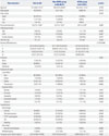
General characteristics are shown in Table 2. Forty six of the patients were male (70.8%) and the ages of the 65 study subjects ranged from 20 to 90 years with a median of 57 years. All patients were bitten in upper and lower extremities. There were 5 patients (7.7%) with cardiac diseases including coronary artery disease (unstable angina 1 patient and myocardial infarction 1 patient), valvular heart disease (aortic regurgitation 1 patient), hypertensive heart disease (hypertensive heart failure 1 patient), and arrhythmia (atrial fibrillation 1 patient). Most common grading of severity was grade II. The common symptoms and signs at initial presentation were pain (65 patients, 100%), edema (53 patients, 81.5%), discoloration (18 patients, 27.7%), and dizziness (9 patients, 13.8%). The most common hematologic complication during hospitalization was anemia (39 patients, 60%), and bleeding developed in 6 patients at puncture site (2 patients), gastrointestinal tract (3 patients), and brain (1 patient). DIC developed in 46.2% of patients. Out of neurologic complications, blurred vision was developed in 3.0%. Other complication was a rhabdomyolysis (13 patients, 20%) and there was no mortality (Table 2).
ACVEs occurred in 9 patients (13.8%); myocardial injury (9 patients, 13.8%) included hs-TnI elevation (7 patients, 10.8%) or ECG determined ischemic change (2 patients, 3.1%), and shock (2 patient, 3.1%). Neither ventricular dysrhythmia nor cardiac arrest was observed. Cardiac biochemical markers analysis revealed median initial levels of hs-TnI of 0.015 ng/mL. ECG showed a T wave inversion in 4 patients (6.2%). QT prolongation was observed in 25 patients (38.5%) (Table 2 and 3).
General characteristics and laboratory findings according to the presence of ACVEs
Underlying cardiac diseases were more common in the ACVEs group than in the non-myocardial injury group (p=0.017). Ho-wever, age, severity, and initial symptoms & signs were not significantly different between the two groups (Table 2).
Regarding complications during hospitalization, 3 patients (5.4%) in the non-ACVEs group and 3 patients (33.3%) in the ACVEs group developed bleeding (p=0.031). Blurred vision or diplopia developed in 2 patients in non-ACVEs group (Table 2).
DISCUSSION
Cardiac involvement is known mainly to be associated with viperine bites.4 There are four types of venomous snakes in Korea. Three belong to the family Viperidae; that is, Agkistrodon brevicaudus, Agkistrodon ussuriensis, and Agkistrodon saxatilis. The other Rhabdophis tigrinus belongs to the family Cloubridae.10 In this study, the incidence of ACVEs development within 48 hrs following treatment commencement in a venomous snakebite cases was greater than expected (13.8%). To the best of our knowledge, this is the first report to address ACVEs after a venomous snakebite in Korea. In this study, there was no mortality, irrespective of ACVEs. In the present study, therefore, we cannot predict the prognosis of patients with ACVEs. Nevertheless, previous studies showed that the most severe complications and fatalities are unrelated to cardiac abnormalities.1112
Serum TnI is used to detect myocardial injury, because it is expressed as a cardio-specific isoforms.13 For this reason therefore, the occurrence of myocardial injury was checked in the present study by hs-TnI assay and ECG. In fact, all 9 patients who experienced ACVEs exhibited myocardial injury. Furthermore, 7 patients showed TnI elevation, but none showed TnI elevation with ECG determined ischemic change. The elevated hs-TnI levels observed in the present study (median: 0.063 ng/mL, maximum: 3.000 ng/mL) were lower than those of other myocardial injuries reported, such as myocardial infarction and myocarditis.14
In a study conducted in northeastern Nigeria involving 108 patients with a viperine snakebite, including carpet viper (93%), burrowing asp, and puff adder (4.6%) bites, more than 60% of patients were shown to have electrocardiographic abnormalities, but cardiac troponin T was elevated in only 2% of patients.11 The only other systematic study, conducted in Papua New Guinea, using ECG and cardiac troponin T levels, also concluded that myocardial damage is uncommon following elapid sna-kebites.15
In the ACVEs group, two patients had shock requiring vasopressors. We thought that shock might be caused by anaphylaxis because all two patients had skin rash and subsequently recovered after treatment of intravenous epinephrine.
Although the mechanism of myocardial damage from snakebite remains unclear,16,17,18,19,20,21,22 four possible mechanisms have been proposed for the development of acute myocardial infarction after a snakebite: 1) hypovolemic shock due to increased vascular permeability; 2) coronary thrombosis resulting from hypercoagulability; 3) direct cardio-toxicity; and 4) vasospasm caused by snake venom. In this study, shock occurred only in 2 patients and bleeding tendency was rather higher than hypercoagulability tendency in the ACVEs group. A well designed prospective study is needed to determine the pathogenesis of myocardial injury.
Coagulopathy is a feature of viperin snakebites.3 In Korea, the prevalence of coagulopathy varies from 2.1–82.4% in snakebite victims.23242526 In the present study, the prevalence of DIC was high (46.2%) in total 65 patients. Although no significant intergroup difference was observed, except for bleeding, hematologic complications were more severe in the ACVEs group. ACVEs group had more cardiac disease history, which resulted in higher chance of taking antiplatelet or anticoagulant such as aspirin or warfarin. Therefore, it is likely that ACVEs group had more bleeding complication.
This study has some limitations that bear consideration. First, the study was conducted using a retrospective design at one hospital, and thus, some relevant parameters were not included. Second, we could not conclude for cardiac function in patients with ACVEs because we performed echocardiography on only 2 out of 9 patients with ACVEs. Two patients had normal heart function. Third, our study is limited by lack of serial ECG and serum hs-TnI testing, which would have confirmed any reversal of abnormalities. Fourth, a bias might be caused by exclusions. Therefore, we propose that further prospective studies are needed to investigate serial echocardiography and prognosis of cardiac injury in patients with venomous snakebite.
In conclusion, significant proportion of the patients with venomous snakebite is associated with occurrence of ACVEs. Patients with ACVEs have more underlying cardiac disease and bleeding complication.
 XML Download