Journal List > Yonsei Med J > v.57(2) > 1031955

Koo, Kim, and Yoon: Expression of Autophagy and Reactive Oxygen Species-Related Proteins in Lacrimal Gland Adenoid Cystic Carcinoma

Abstract

Purpose

To investigate the difference of expression of autophagy and reactive oxygen species (ROS) related proteins in adenoid cystic carcinoma (ACC) of lacrimal gland in comparison with ACC of salivary gland.

Materials and Methods

Formalin-fixed, paraffin-embedded tissue samples from patients pathologically diagnosed as lacrimal gland ACC (n=11) and salivary gland ACC (n=64) were used. Immunochemistry was used to measure expression of autophagy related proteins [beclin-1, light chain (LC) 3A, LC3B, p62, and BCL2/adenovirus E1B 19 kDa protein-interacting protein 3 (BNIP3)] and ROS related proteins [catalase, thioredoxinreductase, glutathione S-transferasepi (GSTpi), thioredoxin interacting protein, and manganese superoxide dismutase (MnSOD)]. The prognostic factors related to disease-free and overall survival (OS) in lacrimal gland ACC by log-rank tests, were determined.

Results

GSTpi in stromal cells was more highly expressed in lacrimal gland ACC (p=0.006), however, MnSOD in epithelial cells was expressed more in salivary gland ACC (p=0.046). LC3B positivity and BNIP3 positivity in epithelial component were associated with shorter disease-free survival (both p=0.002), and LC3A positivity in stromal component was the factor related to shorter OS (p=0.005).

Conclusion

This is the first study to demonstrate the expression of autophagy and ROS related proteins in lacrimal gland ACC in comparison with the salivary gland ACC, which would provide a basis for further study of autophagy and ROS mechanism as novel therapeutic targets in lacrimal gland ACC.

INTRODUCTION

Adenoid cystic carcinoma (ACC) is a malignancy of secretory glands, including the lacrimal and salivary gland, which are highly aggressive and prone to local recurrence, and spread to adjacent tissues.1 ACC is characterized by a slow but persistent progression, with multiple local recurrence, and metastasis to the lung, bone, and brain, occurring in approximately 50% of patients.23 There are only a few studies of lacrimal gland ACC, in comparison with salivary gland ACC, due to rarity of the tumor. Advanced stage, solid architecture, high histologic grade, perineural invasion, and positive surgical resection margin are known as factors related to poor prognosis of salivary gland ACC.456 Lacrimal gland ACC showed clinical features of younger age and worse prognosis compared to salivary gland ACC.7 The causes of worse prognosis of lacrimal gland ACC were the high rate of incomplete excision rate due to complex orbital anatomy, invasion to nearby structures, and subsequent metastases.18
Primary treatments of lacrimal gland ACC are en bloc surgical excision and postoperative radiation, as needed.9 Recently, neoadjuvant intra-arterial cytoreductive chemotherapy has been introduced to improve overall survival (OS) and decrease recurrence in 19 lacrimal gland ACCs, although controversy exists.1011 Neutron radiation therapy achieved 80% of 5 year local control in 11 cases, although late recurrence and distant metastasis remained as challenges.12 No effective treatment is available in cases of recurrence or metastasis of lacrimal gland ACC. New treatments targeting this rare and life-threatening cancer are needed.
Recent advances have highlighted that alterations in both reactive oxygen species (ROS) and autophagy regulation are associated with cancer initiation and progression. It is now clear that these processes are mutually linked and play a crucial role in cancer progression and in response to cancer therapeutics.13141516 Autophagy, a self-digestion process that facilitates cellular survival by maintaining energy homeostasis and macromolecular synthesis during cellular stress and nutrient deprivation, which can also induce ROS generation.13151617181920 Recently, it was demonstrated that ROS can induce autophagy through several distinct mechanisms involving Atg4, catalase, and the mitochondrial electron transport chain, and some of the ROS stimulator, such as 2-methoxyestrodial and arsenic trioxide which are under clinical investigation as cancer treatments.1921 Autophagy can lead to cell-survival as well as cell-death responses and could be selective toward cancer cells. Cancer cell is able to survive in such an environment of hypoxia and nutrient deprivation, through angiogenesis and/or aerobic glycolysis. In highly aggressive malignant tumor requiring high metabolic demand, alternative metabolic pathway such as autophagy can provide cellular energy by recycle of cytoplasmic component, acting as a cytoprotective mechanism that help cancer cells resist anti-cancer treatments.722 In the case of salivary gland ACC, ongoing investigations are taking place to better understand autophagy related proteins such as beclin-1 and YM155 and to develop chemotherapeutic agents targeting these markers.23242526 However, no study has yet been conducted to investigate autophagy and ROS status in lacrimal gland ACC. The aim of this study was to investigate the expression and its implications of autophagy and ROS-related proteins in lacrimal gland ACC, compared to salivary gland ACC.

MATERIALS AND METHODS

Patient selection and clinicopathologic evaluation

Formalin-fixed, paraffin-embedded tissue samples of lacrimal gland ACC, collected from January 1997 to December 2012, at Severance Hospital, Yonsei University College of Medicine, were used for analyses. The study was approved by the Institutional Review Board of Severance Hospital. Clinical informations such as age at surgery, gender, tumor side, symptoms, and visual acuity were obtained from medical chart recordings. Tumor stage classifications followed the 7th American Joint Committee on Cancer staging system, and histologic features of hematoxylin and eosin slides were reviewed by a specialized pathologist (JSK). Histologic grading of tumors followed the indications established by Szanto, et al.27 as follows: grade I, no solid component; grade II, ACC with less than 30% solid component; and grade III, ACC with more than 30% solid component. Histologic type was determined by predominant morphological growth patterns and divided into cribriform, tubular, and solid patterns. Perineural invasion, tumor margin (expanding, infiltrative), and tumor involvement in the surgical resection margin were evaluated. As a control group, 64 cases of salivary gland ACC in the same time periods of tissue collection were included.

Immunohistochemistry

The antibodies used for immunohistochemistry (IHC) in this study are listed in Supplementary Table 1 (only online). IHC was performed on formalin-fixed, paraffin-embedded tissue sections. After sectioning the tissue at a thickness of 3 µm, the samples were deparaffinized and rehydrated using xylene and alcohol solutions. IHC was performed using the Ventana Discovery XT automated staining system (Ventana Medical System, Tucson, AZ, USA). CC1 buffer (Cell Conditioning 1; citrate buffer, pH 6.0; Ventana Medical System) was used to wash samples for antigen exposure. IHC included the appropriate positive and negative controls.
IHC was performed to measure expression of proteins related to autophagy including beclin-1, light chain (LC) 3A, LC3B, p62 and BCL2/adenovirus E1B 19 kDa protein-interacting protein 3 (BNIP3), and ROS including catalase, thioredoxinreductase, glutathione S-transferasepi (GSTpi), thioredoxin interacting protein, and manganese superoxide dismutase (MnSOD) in 11 cases of lacrimal gland ACC and 64 cases of salivary gland ACC.

Interpretation of immunohistochemical results

Results of IHC were defined as the proportion of stained cells×immunostaining intensity. The proportion of stained cell was defined as follows: 0 as negative, 1 as less than 30% positivity, and 2 as 30% or more positivity. Immunostaining intensity was defined as follows: 0 as negative, 1 as weak, 2 as moderate, and 3 as strong. The proportion of stained cells×immunostaining intensity was defined as follows: 0–1 was negative, 2–6 was positive.28

Statistical analysis

Data were statistically processed using SPSS for Windows version 12.0 (SPSS Inc., Chicago, IL, USA). The Student's t-test and Fisher's exact test were used for continuous and categorical variables, respectively. Statistical significance was defined as p<0.05. Kaplan-Meier survival curves and log-rank statistics were employed to evaluate survival time and time to tumor metastasis, respectively. Multivariate regression analysis was performed using the Cox proportional hazards model.

RESULTS

Basal characteristics of lacrimal gland ACC

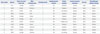
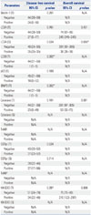
This study included 11 cases of lacrimal gland ACC (Table 1). Clinical characteristics were 21–72 years of age, and 3 were female and 8 were male. The size of the tumor was 2.5 to 4.0 cm, and the main histologic type was cribriform type (n=5). The most common histologic grade was grade 1 (n=5). Lymphovascular invasion was observed in 2 cases, local recurrence in 5 cases, and distant metastases in 7 cases [brain (n=5) and lung (n=2)]. Three patients died of disease.

Expression of autophagy and ROS-related proteins in lacrimal gland ACC: comparison with salivary gland ACC

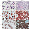
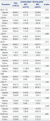
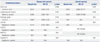
Autophagy and ROS-related proteins expression was compared between lacrimal gland ACC and salivary gland ACC (Table 2). Compared to salivary gland ACC, lacrimal gland ACC showed higher expression of GSTpi in stromal component (p=0.006), and lower expression of MnSOD in epithelial component (p=0.046) (Fig. 1).

Impact of expression of autophagy and ROS-related proteins on patient prognosis in lacrimal gland ACC

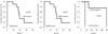
In lacrimal gland ACC, the effect of expression of autophagy and ROS-related proteins on the prognosis was evaluated using univariate analysis (Fig. 2, Table 3). Factors associated with a shorter disease-free survival (DFS) were LC3B and BNIP3 positivity in epithelial component (both p=0.002). The factor associated with shorter OS was LC3A positivity in stromal component (p=0.005), however, no independent influencing factors on prognosis were found by multivariate Cox analyses (Table 4).

DISCUSSION

In this study, expression of autophagy and ROS-related proteins was examined in lacrimal gland ACC, in comparison to salivary gland ACC, and effects of prognostic variables effects on DFS and OS in lacrimal gland ACC were explored using the log-rank test. First, GSTpi isoenzyme protein expression level was higher in lacrimal gland ACC than salivary gland ACC. Until now, there has been no study on the expression of GSTpi in ACC. GSTpi isoenzyme is known to suppress toxin-induced DNA damage by catalyzing the conjugation of electrophilic molecules with glutathione.2930 High GSTpi expression is consistently found in tumor cells, and seems to be directly related to the development of chemotherapeutic resistance in several types of cancer, especially in breast cancer by detoxifying chemotherapeutic drugs inside neoplastic cells.11123132 GSTpi expression in stromal cells in breast tumor microenvironment, namely cancer-associated fibroblast, is also recognized to have major roles in cancer progression.32 Likewise, higher level of GSTpi in stromal cells of lacrimal gland ACC could be related to chemoresistance, although the mechanism is unclear and requires a further investigation.
The major antioxidant enzyme that scavenges superoxide anion radical in mitochondria is MnSOD.33 In our study, the expression level of MnSOD was lower in lacrimal gland ACC than salivary gland ACC. MnSOD was reported to be expressed lower in tumor tissue than in normal tissue, playing a role as a tumor suppressor.10 MnSOD have been demonstrated to play a critical role in the development and progression of cancer.33 Many human cancer cells such as neuroblastoma, lung cancer, hepatoma, esophageal cancer, and colorectal cancer harbor low levels of MnSOD proteins and enzymatic activity.3435363738 Enzymatic activity of MnSOD rapidly declined in stage IV colon cancer tissue, suggesting that a decrease of in MnSOD in cancer tissue could be related to aggressiveness of tumor.39 However, some cancer cells possess high levels of MnSOD proteins and enzymatic activity,40 suggesting that differential regulation of MnSOD exists in cancer cells, depending on the type and stage of cancer development. Lower expression of MnSOD proteins in lacrimal gland ACC could be associated with poorer prognosis of lacrimal gland ACC than salivary gland ACC.
Cancer is one of the first diseases found to genetically be linked to autophagy malfunction.1841 A study has reported that beclin-1, an autophagy related protein, was correlated with OS in salivary gland ACC.42 In this study, there was no statistical difference in beclin-1 expression level between lacrimal gland and salivary gland. Also, LC3B and BNIP3 were closely associated with shorter DFS in lacrimal gland. Expression of LC3B in breast cancer and BNIP3 in lung cancer, larynx cancer, and breast cancer were related to poor prognosis, compatible to our results.43444546 In addition, LC3A expression in stromal component was associated with shorter OS in our study, which was also compatible to the previous reports that the expression of LC3A was a poor prognostic factor in other cancers including stomach cancer, ovary cancer, and lung cancer.474849 Current cancer therapies, including chemotherapy and radiation, are known to induce autophagy within tumor cells.50 Recently, autophagy related to ROS, pathway is thoroughly discussed as a target of anticancer treatment.1921 ROS produced endogenously, by deranged metabolism of cancer cells, or exogenously, by ROS-generating drugs, have been shown to promote macroautophagy, a lysosomal pathway of self-degradation with essential prosurvival functions.16 Furthermore, there are safe, clinically available drugs known to both inhibit and stimulate autophagy, however, there are conflicting positive and negative effects of autophagy reported and no current consensus on how to manipulate autophagyto improve clinical outcomes.
In conclusion, lacrimal ACC showed different expression of ROS related protein from salivary gland ACC. Lacrimal gland ACC was shown to express higher level of GSTpi in stromal component and lower level of MnSOD in epithelial component than salivary gland ACC. Also, autophagy related proteins such as LC3A, LC3B, and BNIP3 were associated with poor prognosis in lacrimal gland ACC. We found that some of autophagy and ROS related proteins were expressed in both cell and stromal component of lacrimal gland ACC. Further studies are mandatory to understand the role of autophagy in the pathogenesis, and to confirm association between autophagy and ROS pathways, in order to find out whether inhibition or stimulation of autophagy and/or ROS is beneficial in the treatment of lacrimal gland ACC. Our data would provide a basis for further study of investigation of autophagy and ROS pathway as targets for possible anticancer treatment.

Figures and Tables

Fig. 1

Immunohistochemical expression of GSTpi and MnSOD in lacrimal and salivary gland adenoid cystic carcinoma (ACC). Expression of GST in stromal component was higher and MnSOD in cell component was lower in lacrimal gland ACC than in salivary gland ACC (×100). GSTpi, glutathione S-transferasepi; MnSOD, manganese superoxide dismutase; H&E, hematoxylin and eosin.

ymj-57-482-g001
Fig. 2

Kaplan-Meier curves for disease-free survival and overall survival of lacrimal gland ACC, correlated with expression of the autophagy related proteins such as LC3A, LC3B, and BNIP3. BNIP3, BCL2/adenovirus E1B 19 kDa protein-interacting protein 3; ACC, adenoid cystic carcinoma; LC, light chain.

ymj-57-482-g002
Table 1

Clinicopathologic Characteristics of Lacrimal Gland Adenoid Cystic Carcinoma

ymj-57-482-i001
Case number Age/sex Tumor size (cm)/tumor side Main histologic type Histologic grade Lymphovascular invasion Ocular symptom Local recurrence/metastasis Survival Chemotherapy/radiation therapy
1 41/F 3.0/right Cribriform 2 No Proptosis Yes/yes Dead No/yes
2 21/M 2.6/left Cribriform 1 No Proptosis No/yes Alive No/yes
3 63/F 4.0/left Solid 3 No Lid swelling Yes/yes Dead No/no
4 54/M 3.5/right Solid 3 No Proptosis No/yes Dead No/yes
5 28/M 3.5/right Tubular 1 No Proptosis No/no Alive Yes/yes
6 72/M 3.0/right Solid 3 No Proptosis Yes/no Alive Yes/yes
7 57/M 2.5/left Cribriform 1 Yes Palpable mass No/no Alive No/yes
8 51/M 3.6/right Tubular 2 No Proptosis No/yes Alive Yes/yes
9 35/M 3.0/right Cribriform 1 Yes Proptosis No/no Alive Yes/yes
10 61/F 3.5/left Tubular 2 No Proptosis Yes/yes Alive Yes/yes
11 43/M 2.5/right Cribriform 1 No Lid swelling Yes/yes Alive Yes/yes
Table 2

Comparison to the Expression of Autophagy and Redox-Related Proteins between Lacrimal Gland ACC and Salivary Gland ACC

ymj-57-482-i002
Parameters Total, n=75 (%) Lacrimal gland ACC, n=11 (%) Salivary gland ACC, n=64 (%) p value
Beclin-1 (T) 0.742
 Negative 50 (66.7) 8 (72.7) 42 (65.6)
 Positive 25 (33.3) 3 (27.3) 22 (34.4)
LC3A (T) 0.333
 Negative 67 (89.3) 9 (81.8) 58 (90.6)
 Positive 8 (10.7) 2 (18.2) 6 (9.4)
LC3A (S) 0.719
 Negative 56 (74.7) 9 (81.8) 47 (73.4)
 Positive 19 (25.3) 2 (18.2) 17 (26.6)
LC3B (T) 0.269
 Negative 55 (73.3) 10 (90.9) 45 (70.3)
 Positive 20 (26.7) 1 (9.1) 19 (29.7)
p62 (T) 0.272
 Negative 68 (90.7) 9 (81.8) 59 (92.2)
 Positive 7 (9.3) 2 (18.2) 5 (7.8)
BNIP3 (T) 0.477
 Negative 71 (94.7) 10 (90.9) 61 (95.3)
 Positive 4 (5.3) 1 (9.1) 3 (4.7)
Catalase (T) 0.742
 Negative 50 (66.7) 8 (72.7) 42 (65.6)
 Positive 25 (33.3) 3 (27.3) 22 (34.4)
Catalase (S) 1.000
 Negative 70 (93.3) 11 (100.0) 59 (92.2)
 Positive 5 (6.7) 0 (0.0) 5 (7.8)
TxNR 0.341
 Negative 65 (86.7) 11 (100.0) 54 (84.4)
 Positive 10 (13.3) 0 (0.0) 10 (15.6)
GSTpi (T) 1.000
 Negative 16 (21.3) 2 (18.2) 14 (21.9)
 Positive 59 (78.7) 9 (81.8) 50 (78.1)
GSTpi (S) 0.006*
 Negative 60 (80.0) 5 (45.5) 55 (85.9)
 Positive 15 (20.0) 6 (54.5) 9 (14.1)
TxNIP 1.000
 Negative 72 (96.0) 11 (100.0) 61 (95.3)
 Positive 3 (4.0) 0 (0.0) 3 (4.7)
MnSOD (T) 0.046*
 Negative 12 (16.0) 4 (36.4) 8 (12.5)
 Positive 63 (84.0) 7 (63.6) 56 (87.5)
MnSOD (S) 0.681
 Negative 63 (84.0) 10 (90.9) 53 (82.8)
 Positive 12 (16.0) 1 (9.1) 11 (17.2)

S, stromal; T, tumor; BNIP3, BCL2/adenovirus E1B 19 kDa protein-interacting protein 3; TxNR, thioredoxinreductase; GSTpi, glutathione S-transferasepi; TxNIP, thioredoxin interacting protein; MnSOD, manganese superoxide dismutase.

Data are presented as the number of cases with positive expression (%).

*Characters refer to statistical significance (p<0.05).

Table 3

Univariate Analysis by Log-Rank Test of the Impact of Autophagy and Redox-Related Proteins Expression in Lacrimal Gland ACC on Disease-Free Survival and Overall Survival Times

ymj-57-482-i003
Parameters Disease-free survival Overall-survival
95% CI p value 95% CI p value
Beclin-1 (T) 0.201 N/A
 Negative 44 (30–58) N/A
 Positive 24 (0–50) N/A
LC3A (T) 0.761 0.431
 Negative 44 (30–59) 74 (61–86)
 Positive 27 (0–77) 245 (245–245)
LC3A (S) 0.634 0.005*
 Negative 40 (24–55) 201 (97–305)
 Positive 35 (35–35) 38 (38–38)
LC3B (T) 0.002* N/A
 Negative 44 (31–56) N/A
 Positive 1 (1–1) N/A
p62 (T) 0.100 N/A
 Negative 45 (31–58) N/A
 Positive 18 (0–52) N/A
BNIP3 (T) 0.002* N/A
 Negative 44 (31–56) N/A
 Positive 1 (1–1) N/A
Catalase (T) 0.101 0.061
 Negative 44 (31–58) 201 (97–305)
 Positive 23 (0–49) 53 (32–73)
Catalase (S) N/A N/A
 Negative N/A N/A
 Positive N/A N/A
TxNR N/A N/A
 Negative N/A N/A
 Positive N/A N/A
GSTpi (T) 0.534 N/A
 Negative 43 (33–52) N/A
 Positive 37 (23–52) N/A
GSTpi (S) 0.714 N/A
 Negative 39 (32–46) N/A
 Positive 37 (17–56) N/A
TxNIP N/A N/A
 Negative N/A N/A
 Positive N/A N/A
MnSOD (T) 0.397 0.808
 Negative 51 (24–78) 75 (70–80)
 Positive 34 (22–46) 210 (123–297)
MnSOD (S) N/A N/A
 Negative N/A N/A
 Positive N/A N/A

ACC, adenoid cystic carcinoma; CI, confidence interval; LC, light chain; T, tumor; S, stromal; BNIP3, BCL2/adenovirus E1B 19 kDa protein-interacting protein 3; TxNR, thioredoxinreductase; GSTpi, glutathione S-transferasepi; TxNIP, thioredoxin interacting protein; MnSOD, manganese superoxide dismutase.

*Characters refer to statistical significance (p<0.05).

Table 4

Multivariate Analysis of Disease-Free Survival and Overall-Survival in Lacrimal Gland ACC

ymj-57-482-i004
Included parameters Disease-free survival Overall-survival
Hazard ratio 95% CI p value Hazard ratio 95% CI p value
Tumor size 0.456 0.632
 ≤3 cm vs. >3 cm 0.419 0.042–4.132 0.012 0.000–815171
Lymphovascular invasion 0.287 0.635
 Absent vs. present 0.003 0.000–116.7 0.012 0.000–977358
Histologic grade 0.415 N/A
 1/2 vs. 3 0.444 0.063–3.128 N/A N/A
LC3A (S) 0.289 N/A
 Negative vs. positive 5.210 0.247–109.9 N/A N/A
LC3B (T) N/A Not included
 Negative vs. positive N/A N/A
BNIP3 (T) N/A Not included
 Negative vs. positive N/A N/A
Catalase (T) N/A N/A
 Negative vs. positive N/A N/A N/A N/A

ACC, adenoid cystic carcinoma; CI, confidence interval; LC, light chain; T, tumor; S, stromal; BNIP3, BCL2/adenovirus E1B 19 kDa protein-interacting protein 3.

ACKNOWLEDGEMENTS

This research was supported by the Basic Science Research Program through the National Research Foundation of Korea (NRF), funded by the Ministry of Education, Science and Technology (2012R1A1A1002886). The funding organization had no role in the design or conduct of this research.

Notes

The authors have no financial conflicts of interest.

References

1. Esmaeli B, Ahmadi MA, Youssef A, Diba R, Amato M, Myers JN, et al. Outcomes in patients with adenoid cystic carcinoma of the lacrimal gland. Ophthal Plast Reconstr Surg. 2004; 20:22–26.
crossref
2. Florentine BD, Fink T, Avidan S, Braslavsky D, Raza A, Cobb CJ. Extra-salivary gland presentations of adenoid cystic carcinoma: a report of three cases. Diagn Cytopathol. 2006; 34:491–494.
crossref
3. Matsuba HM, Simpson JR, Mauney M, Thawley SE. Adenoid cystic salivary gland carcinoma: a clinicopathologic correlation. Head Neck Surg. 1986; 8:200–204.
crossref
4. Khan AJ, DiGiovanna MP, Ross DA, Sasaki CT, Carter D, Son YH, et al. Adenoid cystic carcinoma: a retrospective clinical review. Int J Cancer. 2001; 96:149–158.
5. Spiro RH, Huvos AG. Stage means more than grade in adenoid cystic carcinoma. Am J Surg. 1992; 164:623–628.
crossref
6. Prokopakis EP, Snyderman CH, Hanna EY, Carrau RL, Johnson JT, D'Amico F. Risk factors for local recurrence of adenoid cystic carcinoma: the role of postoperative radiation therapy. Am J Otolaryngol. 1999; 20:281–286.
crossref
7. Li N, Xu L, Zhao H, El-Naggar AK, Sturgis EM. A comparison of the demographics, clinical features, and survival of patients with adenoid cystic carcinoma of major and minor salivary glands versus less common sites within the Surveillance, Epidemiology, and End Results registry. Cancer. 2012; 118:3945–3953.
crossref
8. Polito E, Leccisotti A. Epithelial malignancies of the lacrimal gland: survival rates after extensive and conservative therapy. Ann Ophthalmol. 1993; 25:422–426.
9. Garden AS, Weber RS, Morrison WH, Ang KK, Peters LJ. The influence of positive margins and nerve invasion in adenoid cystic carcinoma of the head and neck treated with surgery and radiation. Int J Radiat Oncol Biol Phys. 1995; 32:619–626.
crossref
10. Oberley LW, Buettner GR. Role of superoxide dismutase in cancer: a review. Cancer Res. 1979; 39:1141–1149.
11. Su F, Hu X, Jia W, Gong C, Song E, Hamar P. Glutathion S transferase pi indicates chemotherapy resistance in breast cancer. J Surg Res. 2003; 113:102–108.
crossref
12. Huang J, Tan PH, Thiyagarajan J, Bay BH. Prognostic significance of glutathione S-transferase-pi in invasive breast cancer. Mod Pathol. 2003; 16:558–565.
crossref
13. Garg AD, Dudek AM, Ferreira GB, Verfaillie T, Vandenabeele P, Krysko DV, et al. ROS-induced autophagy in cancer cells assists in evasion from determinants of immunogenic cell death. Autophagy. 2013; 9:1292–1307.
crossref
14. Gibson SB. A matter of balance between life and death: targeting reactive oxygen species (ROS)-induced autophagy for cancer therapy. Autophagy. 2010; 6:835–837.
crossref
15. Dewaele M, Maes H, Agostinis P. ROS-mediated mechanisms of autophagy stimulation and their relevance in cancer therapy. Autophagy. 2010; 6:838–854.
crossref
16. Azad MB, Chen Y, Gibson SB. Regulation of autophagy by reactive oxygen species (ROS): implications for cancer progression and treatment. Antioxid Redox Signal. 2009; 11:777–790.
crossref
17. Bellot GL, Liu D, Pervaiz S. ROS, autophagy, mitochondria and cancer: Ras, the hidden master? Mitochondrion. 2013; 13:155–162.
crossref
18. Levine B, Kroemer G. Autophagy in the pathogenesis of disease. Cell. 2008; 132:27–42.
crossref
19. Li L, Ishdorj G, Gibson SB. Reactive oxygen species regulation of autophagy in cancer: implications for cancer treatment. Free Radic Biol Med. 2012; 53:1399–1410.
crossref
20. Mathew R, Karantza-Wadsworth V, White E. Role of autophagy in cancer. Nat Rev Cancer. 2007; 7:961–967.
crossref
21. Janku F, McConkey DJ, Hong DS, Kurzrock R. Autophagy as a target for anticancer therapy. Nat Rev Clin Oncol. 2011; 8:528–539.
crossref
22. Roy S, Debnath J. Autophagy and tumorigenesis. Semin Immunopathol. 2010; 32:383–396.
crossref
23. Jiang L, Huang S, Zhang D, Zhang B, Li K, Li W, et al. Inhibition of autophagy augments chemotherapy in human salivary adenoid cystic carcinoma. J Oral Pathol Med. 2014; 43:265–272.
crossref
24. Jiang LC, Huang SY, Zhang DS, Zhang SH, Li WG, Zheng PH, et al. Expression of beclin 1 in primary salivary adenoid cystic carcinoma and its relation to Bcl-2 and p53 and prognosis. Braz J Med Biol Res. 2014; 47:252–258.
crossref
25. Wang YF, Zhang W, He KF, Liu B, Zhang L, Zhang WF, et al. Induction of autophagy-dependent cell death by the survivin suppressant YM155 in salivary adenoid cystic carcinoma. Apoptosis. 2014; 19:748–758.
crossref
26. Chen G, Hu X, Zhang W, Xu N, Wang FQ, Jia J, et al. Mammalian target of rapamycin regulates isoliquiritigenin-induced autophagic and apoptotic cell death in adenoid cystic carcinoma cells. Apoptosis. 2012; 17:90–101.
crossref
27. Szanto PA, Luna MA, Tortoledo ME, White RA. Histologic grading of adenoid cystic carcinoma of the salivary glands. Cancer. 1984; 54:1062–1069.
crossref
28. Won KY, Kim GY, Kim YW, Song JY, Lim SJ. Clinicopathologic correlation of beclin-1 and bcl-2 expression in human breast cancer. Hum Pathol. 2010; 41:107–112.
crossref
29. Brockstedt U, Krajinovic M, Richer C, Mathonnet G, Sinnett D, Pfau W, et al. Analyses of bulky DNA adduct levels in human breast tissue and genetic polymorphisms of cytochromes P450 (CYPs), myeloperoxidase (MPO), quinone oxidoreductase (NQO1), and glutathione S-transferases (GSTs). Mutat Res. 2002; 516:41–47.
crossref
30. Van Emburgh BO, Hu JJ, Levine EA, Mosley LJ, Case LD, Lin HY, et al. Polymorphisms in drug metabolism genes, smoking, and p53 mutations in breast cancer. Mol Carcinog. 2008; 47:88–99.
crossref
31. Chekhun VF, Zhylchuk VE, Lukyanova NY, Vorontsova AL, Kudryavets YI. Expression of drug resistance proteins in triple-receptor-negative tumors as the basis of individualized therapy of the breast cancer patients. Exp Oncol. 2009; 31:123–124.
32. Chaiwun B, Sukhamwang N, Trakultivakorn H, Saha B, Young L, Tsao-Wei D, et al. GSTPi-positive tumour microenvironment-associated fibroblasts are significantly associated with GSTPi-negative cancer cells in paired cases of primary invasive breast cancer and axillary lymph node metastases. Br J Cancer. 2011; 105:1224–1229.
crossref
33. Dhar SK, St Clair DK. Manganese superoxide dismutase regulation and cancer. Free Radic Biol Med. 2012; 52:2209–2222.
crossref
34. Oberley LW, Bize IB, Sahu SK, Leuthauser SW, Gruber HE. Superoxide dismutase activity of normal murine liver, regenerating liver, and H6 hepatoma. J Natl Cancer Inst. 1978; 61:375–379.
35. Coursin DB, Cihla HP, Sempf J, Oberley TD, Oberley LW. An immunohistochemical analysis of antioxidant and glutathione S-transferase enzyme levels in normal and neoplastic human lung. Histol Histopathol. 1996; 11:851–860.
36. Van Driel BE, Lyon H, Hoogenraad DC, Anten S, Hansen U, Van Noorden CJ. Expression of CuZn- and Mn-superoxide dismutase in human colorectal neoplasms. Free Radic Biol Med. 1997; 23:435–444.
crossref
37. Czeczot H, Skrzycki M, Podsiad M, Gawryszewska E, Nyckowski P, Porembska Z. [Antioxidant status of patients with primary colorectal cancer and liver metastases of colorectal cancer]. Pol Merkur Lekarski. 2005; 18:58–61.
38. Sun GG, Wang YD, Chen LQ, Wang SJ, Liu GL, Yu XR, et al. Novel cancer suppressor gene for esophageal cancer: manganese superoxide dismutase. Dis Esophagus. 2011; 24:346–353.
crossref
39. Kocot J, Kiełczykowska M, Da¸browski W, Piłat J, Rudzki S, Musik I. Total antioxidant status value and superoxide dismutase activity in human colorectal cancer tissue depending on the stage of the disease: a pilot study. Adv Clin Exp Med. 2013; 22:431–437.
40. Tamura M, Matsui H, Tomita T, Sadakata H, Indo HP, Majima HJ, et al. Mitochondrial reactive oxygen species accelerate gastric cancer cell invasion. J Clin Biochem Nutr. 2014; 54:12–17.
crossref
41. Abd El-Rehim DM, Ball G, Pinder SE, Rakha E, Paish C, Robertson JF, et al. High-throughput protein expression analysis using tissue microarray technology of a large well-characterised series identifies biologically distinct classes of breast cancer confirming recent cDNA expression analyses. Int J Cancer. 2005; 116:340–350.
crossref
42. Liang LZ, Ma B, Liang YJ, Liu HC, Zheng GS, Zhang TH, et al. High expression of the autophagy gene Beclin-1 is associated with favorable prognosis for salivary gland adenoid cystic carcinoma. J Oral Pathol Med. 2012; 41:621–629.
crossref
43. Zhao H, Yang M, Zhao J, Wang J, Zhang Y, Zhang Q. High expression of LC3B is associated with progression and poor outcome in triple-negative breast cancer. Med Oncol. 2013; 30:475.
crossref
44. Karpathiou G, Sivridis E, Koukourakis M, Mikroulis D, Bouros D, Froudarakis M, et al. Autophagy and Bcl-2/BNIP3 death regulatory pathway in non-small cell lung carcinomas. APMIS. 2013; 121:592–604.
crossref
45. Jin T, Lin HX, Lin H, Guo LB, Ge N, Cai XY, et al. Expression TGM2 and BNIP3 have prognostic significance in laryngeal cancer patients receiving surgery and postoperative radiotherapy: a retrospective study. J Transl Med. 2012; 10:64.
crossref
46. Koop EA, van Laar T, van Wichen DF, de Weger RA, Wall Ev, van Diest PJ. Expression of BNIP3 in invasive breast cancer: correlations with the hypoxic response and clinicopathological features. BMC Cancer. 2009; 9:175.
crossref
47. Liao W, Sun L, Wang C, Huang H, Liu J, Liao W, et al. LC3A-positive "stone-like" structures predict an adverse prognosis of gastric cancer. Anat Rec (Hoboken). 2014; 297:653–662.
crossref
48. Spowart JE, Townsend KN, Huwait H, Eshragh S, West NR, Ries JN, et al. The autophagy protein LC3A correlates with hypoxia and is a prognostic marker of patient survival in clear cell ovarian cancer. J Pathol. 2012; 228:437–447.
crossref
49. Karpathiou G, Sivridis E, Koukourakis MI, Mikroulis D, Bouros D, Froudarakis ME, et al. Light-chain 3A autophagic activity and prognostic significance in non-small cell lung carcinomas. Chest. 2011; 140:127–134.
crossref
50. Levy JM, Thorburn A. Targeting autophagy during cancer therapy to improve clinical outcomes. Pharmacol Ther. 2011; 131:130–141.
crossref

Supplementary Material

Supplementary Table 1

Clone, Dilution, and Source of Antibodies Used
TOOLS
Similar articles