

 PDF
PDF ePub
ePub Citation
 Print
Print


INTRODUCTION
The risk of deep vein thrombosis (DVT) is high in hospitalized elderly patients,1234 and major surgery, cancer, acute medical illness, cardio-pulmonary diseases, and immobilization increase the risk of DVT formation.5678910 The longer the period of immobility, the higher the risk of developing DVT.11 In the subacute to chronic phase of diseases, careful prophylactic treatment for DVT should be taken into consideration. However, in patients in acute phase, causative diseases should be treated with priority, although the risk of developing DVT is high in such patients.21213 Early initiation of heparin treatment for elderly patients is recommended for prophylaxis of DVT,3 nevertheless, it is still debatable, especially in hemorrhagic diseases.1415 To assess the necessity of early initiation of heparin treatment, the incidences of DVT and pulmonary embolism (PE) were analyzed in comatose elderly patients in the acute phase of illness or in one week after surgery, by using D-dimer measurement, lower-extremity ultrasonography, and chest computed tomography (CT).
MATERIALS AND METHODS
Teikyo University Ethics Committee approved this study and protocols (Teirin 14-149). Study details were explained to each patient's family, and informed consent was obtained in written form with a signature.
Patients
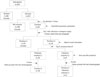
From January to December 2014, 323 patients were admitted to the Department of Neurosurgery in Teikyo University Mizonokuchi Hospital. Among them, 43 comatose patients aged ≥60 years old were present. These patients were selected in the current study, as shown in Fig. 1. Intermittent pneumatic compression was applied to all 43 patients on admission. A Glasgow Coma Scale (GCS) score of ≤11 (E≤3, V≤3, and M≤5) was defined as comatose in this study.
D-dimer measurement and lower-extremity vein ultrasonography
D-dimer is a reliable marker for the diagnosis of DVT,1617181920 and DVT can be ruled out in a patient whose D-dimer value is within the normal range.16 Laboratory analysis of D-dimer was performed for all 43 patients on admission, and a second D-dimer measurement and lower-extremity vein ultrasonography examination were performed in 7 days after admission or emergent surgery. The presence of DVT was evaluated by ultrasonography from the common femoral to the tibial vein through the popliteal vein. The correlation between increased D-dimer and existence of DVT in the lower extremities was analyzed. When DVT was identified, D-dimer measurement and ultrasonography study were repeated every week until DVT became negative on ultrasonography.
Chest CT with contrast enhancement
When DVT was positive in the lower extremities of the patients, chest CT with contrast medium was performed for PE evaluation. If a PE was identified, vena cava filter protection was applied to the patients regardless of being symptomatic or asymptomatic.
Treatment with heparin sodium (Novo-heparin) and rivaroxaban (Xarelto)
All patients diagnosed as DVT positive underwent intravenous heparin sodium (150 units/kg body weight in 24 hours) treatment2122 for 2 weeks, and were switched to oral rivaroxaban,2324 administered through a naso-gastric tube. The treatment continued for one month after the DVT became negative on ultrasonography.
Statistical analysis
Statistical analysis was conducted using Microsoft Excel 2010 (Microsoft Corp., Redmond, WA, USA). Chi-squared test and Student's t-test were used to identify statistical differences in patients' age, gender, diseases, and the D-dimer value between DVT-negative and positive groups. A value of p less than 0.05 (p<0.05) was defined as statistically significant.
RESULTS
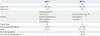
Incidence of DVT and PE, and contributing factors (Table 1 and 2)
Among the 43 comatose elderly patients, DVT was identified in 19 (44.2%) patients and PE was detected in 4 (9.3%) in one week after admission. The mean age of all 43 patients was 77.3±9.3 years, and that of DVT-positive and negative groups was 78.4±9.3 years (range, 65–94 years) and 76.4±9.5 years (range, 61–92 years), respectively. There was no significant difference in age between the two groups (p>0.10). All four PE-positive patients were aged >80 years old with a mean age of 86.3±0.96 years, which was significantly higher than that of PE-negative patients (76.3±9.4) (p<0.01). These results suggested that age is not a contributing factor for DVT occurrence as long as the patients are older than 60; however, a risk of developing to PE is significantly high in the patients older than 80 years. Among the four cases with PE, three were asymptomatic, but one patient demonstrated sudden-onset of dyspnea with a rapid decrease of arterial oxygen saturation (SaO2) to 75% in room-air on the 7th day of admission, and oxygen was administered. The patient recovered spontaneously from dyspnea 1 hour after the onset and regained 100% SaO2. Both DVT and PE were identified on radiological examination, and heparin treatment was then, initiated immediately and vena cava filter was inserted in the patient.
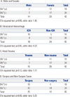
The number of female patients was greater than that of male in this study. However, gender was not a risk factor for DVT, and the number of incidence of DVT between male and female was not significantly different (p>0.05) (Table 2A). There were 15 cases of intracerebral hemorrhage (ICH), 15 head traumas with brain contusion, 9 cerebral infarctions, 2 status epileptics, 1 case of hydrocephalus, and 1 case of encephalitis (Table 1). DVT was not found in any of cerebral infarction patients. On the other hand, DVT was identified in 10 ICH (66.7%) and 8 head trauma (53.3%) patients. Occurrence of DVT was significantly higher in the patients with ICH than in those without ICH (p<0.05) (Table 2B), leading to the conclusion that ICH is a definite risk factor for DVT. Head trauma did not strongly affect the occurrence of DVT and there was no statistical difference in incidence of DVT between head trauma and non-trauma cases (p>0.10) (Table 2C). In 25 patients who underwent surgery, DVT was identified in 15 patients (60.0%) in one week after surgery, whereas DVT was confirmed in 4 of 18 (22.2%) in non-surgical patients. The risk of DVT was significantly higher in surgical cases than with non-surgical cases (p<0.05) with an odds ratio of 5.25 (Table 2D).
D-dimer value and DVT (Table 1)
The normal range of D-dimer in our testing was ≤1.0 µg/mL. On admission and in 1 week after admission or surgery, D-dimer was higher than normal range in both the DVT-negative and positive groups. In the DVT-negative group, D-dimer increased from 1.8±0.7 µg/mL to 4.1±4.2 µg/mL, whereas the value increased from 3.1±2.8 µg/mL to 20.1±14.5 µg/mL in the DVT-positive group. D-dimer values were not significantly different between DVP-positive and negative groups on admission (p>0.05); however the value significantly different at day 7 (p<0.01). The high value of D-dimer on admission was not always associated with high incidence of DVT. When DVT turned to be negative on ultrasonography examination after treatment, D-dimer values decreased to 3.5±2.3 µg/mL. Moreover, D-dimer values decreased further to 1.6±0.8 µg/mL after one month of continuous treatment. D-dimer was an effective follow-up marker for DVT.
Period of treatment (Table 1)
Thrombosis was alleviated by treatment and no recurrence of DVT was observed in any of the 19 patients during the mean follow-up period of 68.1±9.5 days. The mean duration from initiating treatment for DVT to the absence of thrombosis on ultrasonography was 22.3±14.8 days (from 8 to 56 days) in 19 patients; 24.2±16.6 days in the PE-negative group, and 17.8±9.8 days in the PE-positive group. There was no significant difference between the two data (p>0.10). The treatment never caused hemorrhagic diseases in the study.
DISCUSSION
DVT can occur not only in prolonged immobilized patients but also in immobilized acute stroke patients within 7 days after the onset.25 Our study was carried out in an emergency institute, and the authors investigated the incidence of DVT in 7 days after admission and evaluated the necessity of early heparin treatment in comatose patients with severe acute neurological diseases, especially in hemorrhagic diseases.
All DVT-positive patients in our study were aged ≥65 years, in agreement with the finding that the risk of DVT is higher in patients aged 65 years and over.2627 In addition, the incidence of venous embolism is considered extremely high in patients older than 80 years,2628 and all patients with PE in the current study were aged over 80 years.
In ischemic disease, no cases with DVT were identified, and administration of anti-platelet or anti-coagulant drugs to the patients might have played a role of prophylaxis for DVT. In contrast, the incidence of DVT was significantly high in patients with ICH (Table 2B). Hemostatic drugs such as carbazochrome and tranexamic acid were routinely administered for 3 days to the ICH patients. However, these drugs were not correlated with the DVT formation in immobilized older patients, because the incidence of DVT was not statistically high in head trauma patients who were administered hemostatic drugs (Table 2C). Surgery is a major risk factor for DVT, especially in elderly patients, 29 and the risk of DVT was significantly higher in surgical patients than in non-surgical patients (Table 2D). In this study, there was no case of surgery in ischemic patients and this is also the reason that the incidence of DVT was extremely low in the patients with ischemic diseases. Head trauma causing brain contusion itself was not a risk factor of DVT; however, the risk of DVT turned significantly high if surgery was performed to the traumatic patients.
D-dimer is a reliable marker for DVT or PE detection.1617181920 However, D-dimer increases not only in thrombotic disorders, but also in a variety of conditions including recent major surgery, hemorrhage, trauma, infection, and cancer.20 In addition, there is no set value of D-dimer that indicates the existence of DVT. In the present study, the mean values of D-dimer on admission were greater in both DVT-negative and positive patients, than the normal upper limit, and the value increased at day 7. However, the D-dimer value on admission were not statistically higher in DVT-positive patients (p>0.05), although their value at day 7 was significantly higher (p<0.01). This result suggested that high value of D-dimer on admission does not imply the occurrence of DVT, and examination of D-dimer value at day 7 is important in old comatose patients whose D-dimer value was within normal range on admission.
It has been reported that 90% of cases of distal DVT resolved spontaneously without treatment.30 However, DVT is a potentially life-threatening disease, and the mortality rate is extremely high when symptomatic PE occurs.2730 In our institute, intermittent pneumatic compression is applied to all comatose older patients on admission and continue for more than 1 week, but DVT was identified in 44.2% of our patients, suggesting that mechanical treatment alone is insufficient for prophylaxis of DVT. Heparin is considered to be an effective treatment for DVT.314 In geriatric patients, aggressive pharmacologic prophylaxis for DVT is recommended, and should be initiated either at admission or shortly after surgery.3 However, heparin carries a risk of ICH, and even low-molecular-weight heparin is associated with an increased risk for ICH.20 Moreover, recent studies have questioned the effectiveness of heparin as a prophylaxis treatment.15 In this study, the mortality rate of DVT was fortunately 0% and no patients had recurrence of either DVT or PE after treatment during 2 months of admission.
The elderly comatose patients, who suffer from ICH or who undergo emergent surgery, possess extremely high risk of DVT; however, such patients are also the patients who should avoid anticoagulant therapy at an acute period. High value of D-dimer on admission does not indicate the necessity of early initiation of heparin treatment in those patients. Rather, D-dimer measurement and lower-extremity vein ultrasonography examination at day 7 after admission or surgery are important to assess the necessity of heparin treatment. Our present study proved that initiation of heparin treatment 7 days after admission or surgery is safe and effective for prophylaxis of DVT in comatose elderly patients with acute neurological diseases.
In this study, patients with malignancy, cardio-pulmonary diseases, hip fracture, or other diseases were not included, and further studies with a larger sample size are required to validate the findings reported herein.
In conclusion, the risk of developing DVT and PE is extremely high in comatose elderly patients who suffer from ICH or undergo surgery. And initiation of heparin treatment 7 days after admission or surgery is safe and effective for prophylaxis in such patients.
 XML Download