

 PDF
PDF ePub
ePub Citation
 Print
Print


INTRODUCTION
Prosthesis-patient mismatch (PPM) can be considered after insertion into the patient when the effective orifice area (EOA) is of the prosthesis valve and is less than that of a normal human valve, which results in an increased postoperative transvalvular gradient.12
Previous studies reported that PPM after aortic valve replacement is associated with worse hemodynamics, decreased regression of left ventricular (LV) hypertrophy, more cardiac events and higher mortality.3456 However, PPM after mitral valve replacement (MVR) has not been widely investigated, and its incidence has been reported to vary.78910 In addition, studies on the clinical impact of PPM following MVR on survival have shown conflicting results, although two recent trials showed that PPM in the mitral position independently affects longterm survival.78 The most important reason for the discrepancy among the previous studies might be that the methods used to define PPM were different. Three different techniques have been used to calculate EOA, including the continuity equation (CE),7 pressure half time (PHT) method,1011 and reference EOA (EOAR).812131415 However, differences among the methods of calculating PPM after MVR have not yet been clarified.
Dumesnil and Pibarot16 have addressed that indexed EOA derived from in vivo postoperative measures is the only parameter that can consistently be correlated with postoperative gradients as well as clinical outcomes in defining PPM. Therefore, it is emphasized that the indexed EOA, not geometric specifications of the prosthesis, should be used to define PPM.17 However, data showing correlation between indexed EOA from postoperative echocardiography and clinical outcomes have been investigated mainly in patients with aortic prosthesis, and clinical evidences supporting the use of calculated EOA using CE for defining PPM in mitral prosthesis are scarce.
We hypothesized that EOA derived from in vivo postoperative measure using CE would be most valid compared with other parameters including PHT method and reference EOA for the evaluation of mitral PPM. Therefore, we investigated the incidence of PPM in the mitral position using different methods of EOA determination, including calculation by CE, calculation by PHT and use of reference EOA, and compared them with various echocardiographic variables in patients with mitral stenosis after MVR.
MATERIALS AND METHODS
Patient population
We retrospectively analyzed the data of patients who underwent isolated MVR due to rheumatic mitral stenosis at Severance Cardiovascular Hospital from January 2004 to December 2012. Patients with >2+ mitral valve regurgitation, >1+ aortic valve regurgitation and/or >mild aortic stenosis on preoperative and postoperative echocardiography, and patients with cardiomyopathy or coronary artery disease requiring concurrent bypass surgery were excluded. This study was approved by the Institutional Review Board of Yonsei University, Severance Hospital, Seoul, Korea.
Echocardiographic measurement
Clinical and echocardiographic assessment was performed prior to MVR and 12-60 months after operation. The echocardiographic images of the included patients were reanalyzed by 2 experienced echocardiographers who were unaware of the patient's clinical data. LV internal diameter, septal thickness, and LV posterior wall thickness were measured at end-diastole. LV mass was calculated using the formula developed by Devereux, et al.,18 and LV mass was indexed for the body surface area. The left atrial volume was calculated from the parasternal long-axis view and apical four-chamber view using the prolate ellipse method19 and was indexed for the body surface area. The severity of tricuspid regurgitation (TR) was assessed using color flow imaging and regurgitant jet area.20 The calculated systolic pulmonary artery (PA) pressure was defined as: 4×(maximum velocity of TR jet)2+right atrial pressure. Right atrial pressure was estimated by the inferior vena cava diameter and its response to inspiration.1011 Doppler color flow mapping was used to assess the competency of the prosthetic valves.
EOA calculation and definition of PPM
Mitral valve EOA, calculated by CE (EOACE), was determined using the stroke volume measured in the LV outflow tract divided by the integral of the mitral valve trans-prosthetic velocity during diastole. Mitral valve EOA, calculated by PHT (EOAPHT), was calculated using the formula 220/PHT.21 Three cardiac cycles for patients in sinus rhythm and five for patients in atrial fibrillation were recorded, and the results were averaged for every patient. EOAR was determined from the literature or values offered by the manufacturer. EOACE, EOAPHT, and EOAR were indexed for the body surface area (EOAICE, EOAIPHT, and EOAIR, respectively). Indexed EOA was used to define PPM as not significant if >1.2 cm2/m2, moderate if >0.9 cm2/m2 and ≤1.2 cm2/m2, and severe if ≤0.9 cm2/m2.7
Statistical analysis
The distributions of all relevant variables are reported as percentages or as mean±SD. The groups were compared using chi-square statistics for categorical variables and Student's t-test for continuous variables. Correlation between the variables was assessed with the Pearson correlation test. Intraclass correlation coefficient (ICC) for a consistency was used to measure and compare agreement between EOACE and EOAPHT and between EOACE and EOAR. Good correlation was defined as an ICC >0.8. To determine independent predictors of postoperative systolic PA pressure, linear relationships were checked with univariate linear regression analysis. Variables that had statistical significance in univariate analysis and EOA values were entered in the multiple linear regression model. A p value <0.05 was considered statistically significant.
RESULTS
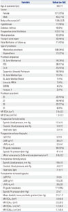
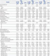
Among the eligible 185 patients who underwent MVR due to mitral stenosis, 167 patients received postoperative echocardiography between 12 and 60 months after MVR. After excluding one patient who showed paravalvular leakage on postoperative echocardiography, the remaining 166 patients (age 56±11 years) with a median follow-up time of 16 months comprised the study population. Preoperative and postoperative variables of the studied populations are shown in Table 1. Of the 166 patients included in this study, there were 45 men and 121 women with a mean age of 56±11 years. Prevalence of hypertension and diabetes were 10% and 9%, respectively. Maze procedure and TV repair were performed in 42 (25%) and 66 (40%) patients, respectively, and atrial fibrillation during postoperative echocardiography was shown in 71 patients (43%).
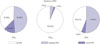
Fig. 1 shows the prevalence of mitral valve PPM according to the methods used to calculate EOA. Prevalence ranged from 7% in PHT method to 49% in referred EOA method to 62% in CE method. There were 63 patients without PPM (38%), 80 patients with moderate PPM (48%) and 23 patients with severe PPM (14%), when EOAICE was used to define PPM. In contrast, the prevalence of moderate and severe PPM was relatively very low [8 patients (5%) and 3 patients (2%), respectively] according to EOAIPHT. There were 85 patients without PPM (51%), 73 patients with moderate PPM (44%) and 8 patients with severe PPM (5%) according to EOAIR.
Comparison of characteristics according to the method of PPM calculation is shown in Table 2. When PPM was defined by EOAICE, the prevalence of males was higher, body surface area was greater and trans-prosthetic pressure and postoperative systolic PA pressure were higher in the PPM group. When PPM was defined by EOAIPHT, age at operation and prevalence of atrial fibrillation at follow-up were higher and left atrial volume index (LAVI) was larger in the PPM group. When we used EOAIR, prevalence of atrial fibrillation at follow-up and postoperative LV ejection fraction was higher in the PPM group. There were no significant differences in prevalence of moderate or greater TR at preoperative echocardiography between the groups in all three methods. However, there was a tendency of high occurrence of moderate or greater TR at postoperative echocardiography in PPM group, only when PPM was defined according to CE (p=0.069).
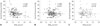
Correlations between EOAICE and EOAIPHT, between EOAICE and EOAIR, and between EOAIPHT and EOAIR are demonstrated in Fig. 2. All the agreements between EOAICE and EOAIPHT, between EOAICE and EOAIR, and between EOAIPHT and EOAIR were poor [0.430, 95% confidence interval (CI) 0.226-0.580; 0.320, 95% CI 0.077-0.500; and 0.362, 95% CI 0.133-0.530, respectively]. When the patients were divided into two groups according to the presence of PPM using various methods, the results of correlation were similar. CE methods poorly correlated with referred method (ICC=0.249) and PHT method (ICC=0.228) in defining PPM. Correlation between referred and PHT method was also poor (ICC=0.306).
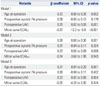
Fig. 3 shows the correlation between indexed EOAs and postoperative systolic PA pressure. Among the indexed EOAs, EOAICE showed significant correlation with postoperative systolic PA pressure (r=-0.384, p<0.001). In contrast, there were no significant correlations between EOAIPHT and postoperative systolic PA pressure (r=-0.089, p=0.254) or between EOAIR and postoperative systolic PA pressure (r=-0.110, p=0.158). Linear regression analysis was performed to identify the predictors of postoperative systolic PA pressure as a dependent variable (Table 2). On multivariate regression, age, mitral valve EOAICE, and postoperative LAVI were found to be independent predictor of postoperative systolic PA pressure (p=0.002, p<0.001, and p=0.001, respectively) (Table 3). On the contrary, EOAIPHT and EOAIR were not predictors for postoperative systolic PA pressure.
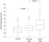
Fig. 4 compares of postoperative systolic PA pressure among the patients without PPM (n=63), with moderate PPM (n=80) and with severe PPM (n=23) according to EOAICE. Systolic PA pressure was statistically different between the patients without PPM and with moderate PPM (25±6 mm Hg vs. 28±6 mm Hg, p=0.007), as well as between patients with moderate and severe PPM (28±6 mm Hg vs. 33±11 mm Hg, p=0.037).
DISCUSSION
The principal findings of the present study are that 1) the incidence of PPM after MVR in patients with mitral stenosis was variable according to the different methods of determining EOA, and 2) among the EOAs assessed by 3 different methods, including the CE method, PHT method and use of reference values, only the EOAICE was found to be independently associated with the postoperative systolic PA pressure.
Conflicting data on PPM after MVR
Since first described in 1978,1 PPM after MVR has been suggested to correlate with poor clinical outcomes including persistent pulmonary hypertension and late functional TR.49 However, some authors have insisted that PPM did not affect survival after MVR,1015 although several recent trials suggested that mitral PPM independently affects long-term survival.78 In addition, the overall incidence of PPM (<1.3 to <1.2 cm2/m2) after MVR has been shown to vary, ranging from 3.7% to 85.9%.78101112131415 The incidence of moderate PPM has also been reported to vary, ranging from 37.4% to 69.5%, and the incidence of severe PPM ranged from 8.7% to 16.4%.22
Explanations for discrepancy
There may be several explanations concerning the conflicting data on the clinical effect of PPM after MVR. First, different valve types and patients groups were employed in the studies. Especially, patients suffering PPM have sometimes additional risk factors, including hypertension and diabetes, which could also affect long-term survival. Second, when evaluating the impact of PPM after MVR, it is also important to consider pathophysiological status of patients. In the current study, minimize those problems, we included only patients with rheumatic mitral stenosis and analyzed postoperative echocardiography 12 to 60 months after MVR, thus avoiding dynamic changes of hemodynamic status at early postoperative period and the late development of prosthetic valve malfunction which might be associated with pannus formation. Consequently, we found that PPM determined by CE showed significant correlation with various postoperative hemodynamic parameters.
Methods to determine effective orifice area
The most important reason for discrepancy among the previous studies might be that the methods used to define PPM were different. In previous studies, three different methods have been used to calculate EOA, including the CE method,7 PHT method,1011 and reference EOA.812131415
We compared the three methods and found that agreement among EOAICE, EOAIPHT, and EOAR was poor, and the prevalence of PPM varied according to the calculation method, ranging from 7% in the PHT method to 49% in the reference EOA method to 62% in the CE method. This finding explains the discrepant prevalence of PPM and conflicting data about the prognosis of PPM in the various studies. Long-term survival data of patients with PPM defined by EOACE has not yet been reported, and further studies with EOACE might be necessary to clarify the clinical impact of PPM.
Interestingly, it has been reported in the previous reports923 that prevalence of PPM was relatively high when CE was used to define PPM , ranging from 42% to 71%. We also found similar results of high prevalence of PPM, especially in moderate PPM. Possible explanation is that in bileaflet valves, the smaller central orifice has a higher velocity than the larger outside orifice, which may lead to underestimation of EOACE.17 Patients who underwent MVR with bileaflet mechanical heart valve were 78% in the current study, and therefore, there is a chance that EOACE might be underestimated in part. Therefore, further research and overall agreement are required to establish the allowable range of EOACE for the definition of PPM, although CE methods might be the most appropriate method to determine EOA. In addition, clinical implication of moderate PPM in mitral position is not clear, since moderate PPM did not show statistically significant result of poor survival, contrast to severe PPM, which showed poor long-term survival.7 Nevertheless, we demonstrated that even patients with moderate PPM showed raised systolic PA pressure compared to those without PPM, suggesting clinical significance of moderate PPM. Therefore, the clinical impact of moderate PPM and EOA range for the diagnosis of PPM might need further investigation.
Among the three methods, mentioned above EOAPHT is not recommended for calculating EOA of the mitral prosthesis because of the influence of chronotropic conditions and atrioventricular compliance.24 Nevertheless, some studies still use PHT method to define EOA.1011 In the current study, as anticipated, EOA calculation via the PHT method was found to be not associated with postoperative hemodynamic parameters and largely overestimated the EOA compared to other methods. Therefore, we should try not to use PHT to calculate EOA in mitral prosthesis.
Pulmonary artery pressure after MVR
A rise in PA pressure can result from elevation of pulmonary blood flow, pulmonary venous pressure and/or vascular resistance,9 and pulmonary hypertension is an important risk factor for morbidity and mortality in patients with various cardiovascular diseases.252627 Pulmonary hypertension is frequently observed in patients with mitral stenosis. Since increased PA pressure is associated with poor functional capacity and a dismal prognosis in patients with severe mitral valve disease, normalization of PA pressure constitutes a crucial goal of MVR.282930
The development of late TR after MVR is an important complication of the surgery, since it is associated with a severe impairment of exercise capacity and a poor symptomatic outcomes. 31 However, the pathogenesis of this condition remains poorly understood, and treatment for the patients with late TR is clinically difficult to decide. Recently, it has been reported that mitral PPM is associated with the persistence of TR and pulmonary hypertension following MVR.23 Our study also demonstrated similar results, showing a tendency of high prevalence in moderate or greater TR in PPM group, defined by CE. In addition, PPM calculated by EOAICE showed higher transprosthetic pressure, and EOAICE was the only parameter which showed independent correlation with postoperative systolic PA pressure. Systolic PA pressure has been known to be influenced by many factors including age, diastolic dysfunction and cardiopulmonary disease,32 as well as trans-prosthetic pressure. Since we enrolled homogenous, relatively young patient population of pure mitral stenosis, we can speculate that systolic PA pressure might be influenced mainly by trans-prosthetic gradient in the studied population. Therefore, the use of EOAICE rather than EOAIPHT and EOAIR may be appropriate for identifying PPM in the mitral position, since hemodynamic variables, including trans-prosthetic pressure and systolic PA pressure, showed significant correlation with EOA, only when it is calculated by using CE.
Unfortunately, however, most previous studies on PPM after MVR used EOAIR to define PPM. EOAICE was used to define PPM in only two studies, and both demonstrated clinical significance of PPM after MVR, including association with persistent pulmonary hypertension and late TR.923 Therefore, further larger studies to define PPM using EOAICE are needed to clarify clinical significance and implications of PPM after MVR.
Limitations
The results of the study were based on retrospective analysis; however, we carefully reviewed patient medical records and echocardiography. As our findings are based upon an observational cohort of patients with rheumatic mitral stenosis, they may not necessarily be generalizable to all patients with mitral regurgitation. We compared indexed EOA to systolic PA pressure, not clinical outcome, since the studied group was at relatively low risk for cardiovascular events, as indicated by the low prevalence of combined co-morbidities such as hypertension and diabetes. In addition, functional data, such as maximum exercise capacity and maximum oxygen consumption, which would be more helpful for identifying the clinical implication of mitral PPM, were not included. Prevalence of pulmonary hypertension (systolic PA pressure >40 mm Hg) was relatively low, and therefore, further investigation into the clinical significance of the results of the current study is needed. Patients with atrial fibrillation were included in the study population, although five cardiac cycles were recorded and these results were averaged. We used linear regression analysis to determine independent predictors of systolic PA pressure, although we cannot conclude that there is no association just because the linear regression is not significant. Nevertheless, the association with EOAIPHT and EOAIR is unlikely, since there were no significant differences in systolic PA pressure between the groups, and the multivariate regression analysis showed that EOAIPHT and EOAIR were not predictors of systolic PA pressure.

 XML Download