

 PDF
PDF ePub
ePub Citation
 Print
Print


INTRODUCTION
Atherosclerosis is the most common cause of cardiovascular diseases, such as unstable angina, myocardial infarction, stroke, and ischemic heart failure, and it is estimated that it will to become the leading cause of death worldwide by the year 2020.1 Atherosclerosis is the result of hyperlipidemia and lipid oxidation and is characterized by inflammatory reactions, endothelial dysfunction, smooth muscle cell proliferation, extracellular matrix alteration, thrombosis and apoptosis.
Rho kinase, one of the effectors of the small GTP-binding protein Rho, has been shown to play an important role in many major cardiovascular diseases, such as hypertension,23 heart failure,4 myocardial infarction, ischemia/reperfusion injury,56 and atherosclerosis.7 Rho kinase consists of an N-terminal located kinase domain, followed by a long coiled-coil domain and a Rho-binding domain, and a C-terminal Pleckstrin-homology domain. Rho kinase activity can be regulated by several distinct mechanisms, such as the Rho/Rho-kinase pathway8 and the Caspase3/Rho-kinase pathway.9 Y-27632 and fasudil are non-isoform-selective Rho kinase inhibitors. Certain lipids, such as arachidonic acid, may effectively stimulate Rho kinase activity independent of RhoA binding.10 The lipids seem to bind to the regulatory C-terminus of Rho kinase, disrupting the autoinhibitory interaction and thus leading to kinase activation.11 Furthermore, other negative regulators have been found that bind to and inhibit Rho kinase activity.
Lipoprotein-associated phospholipase A2 (Lp-PLA2) is a key enzyme responsible for degrading platelet-activating factor and oxidized-LDL (ox-LDL). Many experimental studies have shown that PLA2 is involved in lipid metabolism and immunoinflammatory response and participates in the development of atherosclerosis.1213 Darapladib is an oral, investigational, highly potent, and selective Lp-PLA2 inhibitor.
The principal aim of this study was to examine whether darapladib could reduce the elevated Lp-PLA2 and Rho kinase activity in a dose-dependent manner in a rat atherosclerosis model.
MATERIALS AND METHODS
All procedures were performed in conformity with the Institutional Animal Care and Use Committee and National Institutes of Health guidelines.
Animal model and experimental protocol
Forty-eight male Sprague-Dawley rats (body weight 200–220 g, from Shandong University, Shandong Province, China) were maintained under conditions of standard lighting (alternating 12-h light/dark cycles), temperature (22±0.5℃), and humidity (60±10%) for at least 1 week before the experiments. The rats were then randomly divided into two groups. Twelve rats given a normal diet were designated as a sham group, and the other 36 rats were fed with a high-cholesterol diet for 10 weeks to induce atherosclerosis.1415 The high-cholesterol diet contained 3% cholesterol, 0.5% cholic acid, 0.2% 6-propyl 2-thiouracil, 5% sucrose, and 3% lard. Subsequently, the 36 hyperlipidemic rats were randomly and evenly assigned to three groups: a control group that was administered normal saline, a low-dose darapladib group that received darapladib by gavage (25 mg·kg-1·d-1, reconstituted in normal saline), and a high-dose darapladib group that received darapladib by gavage (50 mg·kg-1·d-1, reconstituted in normal saline). The duration darapladib intervention was 2 weeks. At the end of the experiment, all rats were euthanized via injection of an overdose of sodium pentobarbital solution. The hearts were used for histological and biochemical studies.
Plasma lipid and biological analysis
Fasting blood was taken for laboratory examination before the beginning of the study, after 10 weeks of model production, and after 2 weeks of intervention. Blood samples were collected under chloral hydrate anesthesia and centrifuged at 3500 rpm for 15 minutes using a refrigerated centrifuge (HEMA, Zhuhai, China), and the serum was transferred into a separate vial and stored at 4℃. Serum concentrations of triglycerides (TG), total cholesterol (TC), low-density lipoprotein cholesterol (LDL-C), high-density lipoprotein cholesterol (HDL-C), and high-sensitivity C-reactive protein (hs-CRP) were measured via enzymatic assays using an automated biochemical analyzer (Cobas 8000, Roche, Basel, Switzland).
Plasma level of Lp-PLA2 was measured using a quantitative sandwich enzyme immunoassay (commercial ELISA kits) according to the manufacturer's recommendation (CoWin Bioscience Co., Ltd., Beijing, China). Additionally, serum level of nitric oxide (NO) was evaluated based on the nitrite reductase method using a Total Nitric Oxide Kit (Beyotime, Shanghai, China, S0023).
TUNEL staining
The terminal deoxynucleotidyl transferase-mediated dUTP nick end labeling (TUNEL) technique was used to evaluate apoptotic activity. Myocardial tissues were embedded in paraffin and cut into 5-mm thick slices. The sections were deparaffinized and rehydrated with serial changes of xylene and ethanol. Proteinase K (20 mg/L) was applied to each section for 30 min with the intention of producing optimal proteolysis. The endogenous peroxidase activity was inhibited with 3% hydrogen peroxide for 10 min. A commercial apoptosis detection kit (Roche, Basel, Switzland) was used. The terminal deoxynucleotidyl transferase (TdT) reaction was carried out for 1 h at 37℃ in a humidified chamber, and DAB chromogen was then applied. Hematoxylin was used as a counterstain. TUNEL-positive cells were calculated by randomly counting ten fields of the section and were expressed as a percentage of normal nuclei.16
Assay of protein concentration
The protein concentration of heart tissue was determined based on the method of Lowry with bovine serum albumin as the standard.17
Western blotting
Rho kinase activity was assessed by examining the phosphorylation state of myosin phosphatase targeting subunit 1 (MYPT-1), a well-established Rho kinase specific substrate. MYPT-1 and p-MYPT-1 levels were determined by western blot analysis. Equal amounts of protein (50 µg) were fractionated on 14% sodium dodecyl sulfonate (SDS)-polyacrylamide gels in running buffer [25 mM Tris, 192 mM glycine, 0.1% (wt/vol) SDS, pH 8.3] at 90 V and then electroblotted to nitrocellulose membranes. Membranes were blocked at room temperature with 5% nonfat milk in Tris-buffered saline (25 mM Tris, 137 mM NaCl, and 2.7 mM KCl) containing 0.05% Tween-20 (TBS-T) and then incubated overnight at 4℃ with the following primary antibodies: β-actin (Santa Cruz, TX, USA; dilutions: 1:1000; molecular weight of β-actin: 43 kD), rabbit polyclonal MYPT-1 antibody (Bioworld, MN, USA; dilutions: 1:500; molecular weight of MYPT-1: 130 kD), rabbit polyclonal p-MYPT-1 antibody (Bioworld, MN, USA; dilutions: 1:500; molecular weight of p-MYPT-1: 130 kD). The membranes were then washed three times in TBS-T and incubated with the corresponding secondary antibody (Santa Cruz, TX, USA; dilutions: 1:10000) conjugated to horseradish peroxidase at room temperature. Immunoreactive bands were visualized with the SuperSignal West Pico enhanced chemiluminescence kit (Piece, Rockford, IL, USA) according to the manufacturer's instructions. Band intensities were quantified using a densitometer analysis system (Quantity One; Bio-Rad, Hercules, USA).
RESULTS
Rat parameters
All rats completed the entire study. The body weight of the sham-group rats did not change significantly from week 0 to week 12, while the weight of the atherosclerosis-group rats was 202±11 g at baseline, although this increased to 242±24 g at week 10. There was no significant difference in body weight between week 10 and week 12.
Changes of lipid profile and CRP
As presented in Table 1, the baseline laboratory variables of the different groups were comparable at the outset, and after 10 weeks of high-cholesterol diet administration, the serum levels of TG, TC, and LDL-C in the atherosclerosis-model groups were significantly greater than those in the sham group (p<0.05). Additionally, the serum level of CRP was also profoundly increased in the atherosclerosis-model groups (p<0.05 vs. sham group), indicating that atherosclerosis was significantly associated with systemic inflammation. After two weeks of intervention, the serum levels of TC, LDL-C, and CRP in the low-dose darapladib group and the high-dose darapladib group significantly decreased (p<0.05 vs. control atherosclerosis group). Darapladib therapy produced no significant changes in HDL-C levels. When compared to the low-dose darapladib group, the reduction of TC, LDL-C, and CRP was more prominent in the high-dose darapladib group (p<0.05).
Changes of NO production
As presented in Table 2, after 10 weeks of high-cholesterol diet administration, NO production in the atherosclerosis model groups significantly decreased when compared to the sham group (p<0.05). After 2 weeks of darapladib treatment, NO production increased when compared to the control group (p<0.05). When compared to the low-dose darapladib group, the increase of NO production was more prominent in the high-dose darapladib group (p<0.05).
Changes of Lp-PLA2 activity
As shown in Table 3, after 10 weeks of high-cholesterol diet administration, the serum level of Lp-PLA2 was significantly elevated when compared to the sham group (p<0.05). After 2 weeks of darapladib treatment, the serum level of Lp-PLA2 was lower than the control atherosclerosis group (p<0.05). When compared to the low-dose darapladib group, Lp-PLA2 reduction was more prominent in the high-dose darapladib group (p<0.05).
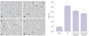
Effect of darapladib on cardiomyocyte apoptosis
Fig. 1 showed the TUNEL positive cells in rat hearts of different groups. TUNEL-positive myocytes were expressed as a percentage of total myocytes. The apoptotic rate was 5.16±0.79% in the control atherosclerosis group. The apoptotic rates were respectively reduced to 4.15±0.67% and 3.63±0.59% in the low-dose darapladib group and the high-dose darapladib group (p<0.05 vs. control atherosclerosis group). When compared to the lowdose darapladib group, cardiomyocyte apoptosis reduction was more prominent in the high-dose darapladib group (p<0.05).
Effect of darapladib on Rho kinase activity
Rho kinase activity was determined by measuring levels of MYPT-1 phosphorylation, a specific Rho kinase substrate. As shown in Fig. 2, MYPT-1 phosphorylation increased during the control atherosclerosis protocol (p<0.05 vs. sham group). Low-dose darapladib therapy resulted in a 23.3% reduction in MYPT-1 phosphorylation (p<0.05 vs. control atherosclerosis group), while high-dose darapladib therapy resulted in a 29.5% reduction (p<0.05 vs. control atherosclerosis group). There was no significant difference between the low-dose darapladib group and the high-dose darapladib group (p>0.05).
DISCUSSION
The results of this study provided experimental evidence that darapladib could reduce elevated Lp-PLA2, Rho kinase activity, and cardiomyocyte apoptosis in atherosclerosis.
Atherosclerosis is a chronic disease with lipids and inflammation playing a crucial role in all stages of the disease from initiation through progression and, ultimately, to thrombotic complications.18 Atherosclerosis is characterized by increased LDL-C and TC levels, and coronary artery disease is its common manifestation. Previous studies have demonstrated that oxidative stress and apoptosis play a fundamental biological role in atherosclerosis, and it is well documented that NO could diminish oxidative stress and reduce ox-LDL production. In the rats from our experiment, a high-cholesterol diet was found to lead to increased blood lipids, as well as atherosclerotic plaque formation. In the past, numerous biomarkers, such as CRP and cytokines, have been found to be related to atherosclerosis.192021 Previous studies have suggested that Lp-PLA2 is a new biomarker for cardiovascular risk, and Lp-PLA2 activity increases during the development of atherosclerosis.22232425 In our study, we also found that serum Lp-PLA2 activity increased in the atherosclerosis-model group after rats were fed a high-cholesterol diet for 10 weeks, while atherosclerosis significantly reduced levels of NO. Darapladib (a specific inhibitor for Lp-PLA2) reduced TC, LDL-C, and CRP levels and cardiomyocyte apoptosis, as well as Lp-PLA2 activity, in a dose-dependent manner. Additionally, the inhibition of Lp-PLA2 resulted in increase in NO production. Importantly, NO production was more prominent in high-dose darapladib therapy.
Rho kinase is one of the best-characterized effectors of small GTPase RhoA and plays a crucial role in various cellular functions, such as smooth muscle contraction, cell migration, and cell proliferation. Rho kinase in cardiovascular diseases has received extensive attention and been studied in animal experimental models and human. Rho kinase plays an important role in inflammatory responses and arteriosclerotic lesions in humans and animals;262728 multiple studies have demonstrated that Rho kinase is involved in the inflammatory atherosclerotic process. Rho kinase inhibitors might also result in up-regulation of eNOS and reduce arterial intima-medial thickness, endothelial dysfunction, vascular inflammation, and atherosclerosis plaque formation. This suggests that Rho kinase inhibition may be a potential therapeutic strategy for the prevention of atherosclerosis.
We then attempted to elucidate the relationship between Lp-PLA2 and Rho kinase. We attempted to determine whether the Lp-PLA2 inhibitor could reduce Rho kinase activity in a dosedependent manner in atherosclerosis. In the present study, the activity of Rho kinase was determined by examining the phosphorylation of MYPT-1. Atherosclerosis resulted in an approximately 46% increase in the amount of p-MYPT-1, indicating the activation of Rho kinase in atherosclerosis. We demonstrated that Rho kinase activity was strongly activated in atherosclerosis. Inhibition of Lp-PLA2 in atherosclerosis led to decreased Rho kinase activity and cardiomyocyte apoptosis. Conversely, a high dose of darapladib (50 mg·kg-1·d-1) did not elicit marked reductions in MYPT-1 phosphorylation when compared to a low dose of darapladib (25 mg·kg-1·d-1) in our experimental model. By blocking cholesterol biosynthesis, darapladib also reduced Rho kinase activity in atherosclerosis though not in a dose-dependent manner. There seems to be extensive crosstalk between the NO pathway and Rho kinase. There is convincing evidence that the NO pathway leads to an inhibition of Rho kinase.293031 The elevated Lp-PLA2 level may contribute to impaired NO pathway.32 Thus, we conclude that darapladib may influence Rho kinase activity via the NO pathway.
In summary, the results from our current study show that TG, TC, LDL-C, CRP, Lp-PLA2, Rho kinase activity, and cardiomyocyte apoptosis increased, while NO production was reduced in atherosclerosis in vivo. Darapladib, a Lp-PLA2 inhibitor, was found to be beneficial for reducing the CRP level, increasing NO production, and decreasing the Lp-PLA2 level. Darapladib also reduced oxidative stress and cardiomyocyte apoptosis. Furthermore, darapladib not only exerted effects on Lp-PLA2 level but also influenced Rho kinase activity. Darapladib thus leads to cardiovascular protection that might be mediated by its inhibition of both Rho inase and Lp-PLA2. This may provide a possible new treatment for patients suffering coronary heart diseases in the future.


 XML Download