

 PDF
PDF ePub
ePub Citation
 Print
Print


INTRODUCTION
Clinical outcomes of total knee arthroplasty in treating advanced knee arthritis have been proven in a number of reports.1 For a successful procedure, proper soft tissue balancing, flexion and extension gap balancing, and recovery of alignment have been stressed.23 However, due to extensive soft tissue incisions, patellar eversion, and tibiofemoral subluxation in the surgical process, severe postoperative pain, stiffness in the operated knee, and delayed recovery remain unresolved. However, minimally invasive surgery in unicompartmental knee arthroplasty reduces postoperative pain and the rehabilitation period, and a variety of minimally invasive techniques have been introduced to minimize the damage to and tension on the tissues involved in the extension of the knee and to preserve its extension function as much as possible.4 These techniques offer advantages such as reduced postoperative pain, decreased use of painkillers, shorter recovery and hospitalization periods, fast recovery of the range of motion in the knee joint and of the surrounding muscular strength, reduced need for postoperative manipulation, lower cost, and fewer postoperative aesthetics.567891011 However, compared to conventional techniques, minimally invasive total knee arthroplasty (MIS-TKA) involves a smaller exposed surgical site and is partial dependent on a specially-designed alignment guide and modified alignment jig. It requires an experienced surgeon and takes time to obtain the necessary surgical skills. Operation time may be delayed, and it is not a surgeon-friendly operation. The operation is believed to have an intraoperative risk of fracture at the surgical site, damage and rupture of ligaments or muscles, insufficient removal of cement and bone fragments, skin necrosis due to severe traction, increased chance of infection, improper fixation of implants, and inappropriate implant alignment, all of which have a negative impact on long-term clinical results, according to some research.1112131415
In particular, in the event of severe preoperative knee joint deformity, the accuracy of implant alignment is known to be low in minimally invasive surgery, similar to existing conventional surgical techniques.81617 Several reports insisted that minimally invasive surgery should not be performed on those with a varus deformity of more than 15 degrees or a range of motion of less than 110 degrees.18
Against this background, the aim of this paper was to demonstrate that after obtaining the necessary surgical skills for MIS-TKA, minimally invasive surgery can be performed on patients with a normal range of motion in joints preoperatively and where the surgical view can be secured by a mobile window, regardless of the preoperative degree of varus deformity. In addition, it sought to identify the correlation between the preoperative degree of knee joint deformity and postoperative implant alignment after minimally invasive surgery.
MATERIALS AND METHODS
This paper reviewed 627 cases (445 patients) of MIS-TKA using the mini-midvastus approach; all cases were performed by a single surgeon from November 2005 to December 2007. This study was approved by the Institutional Review Board.
The average age of the subjects was 71.8±6.5 years old, and 593 cases were in 415 women, while 34 were in 30 men. Their body mass index (BMI) was 27.2±3.7 kg/m2 on average. There were 622 cases of degenerative arthritis, two cases of osteonecrosis, and three cases of rheumatoid arthritis. On average, the preoperative range of motion in the knee joint was 126.5±19.0, and the preoperative knee score was 64.9±7.6, while the preoperative tibiofemoral angle was 5.4±1.4 degrees varus.
These cases were categorized by the preoperative degree of varus deformity in the knee joint: less than 5 degrees varus (Group 1, 351 cases), 5 to less than 10 degrees varus (Group 2, 189 cases), 10 to less than 15 degrees varus (Group 3, 59 cases), and 15 or more degrees varus (Group 4, 28 cases) (Table 1). Preoperative evaluation, surgical results, postoperative radiologic outcomes, and clinical results after 5 months post-operatively were compared between groups.
All operations were performed by the first author of this paper, who had the experience of 250 cases of minimally invasive surgery using quad-sparing instrumentation and normal surgical tools.
NexGen Legacy Posterior Stabilized Flex Fixed Bearing (LPS Flex Fixed, Zimmer, Warsaw, IN, USA) was used for all cases, and the Modular tibial implant ("Mini-keel," Nexgen MISTM Tibial Component, Zimmer, Warsaw, IN, USA) was used as a tibial insert. The skin incision began 10 mm above the medial and superior pole of the patella and continued in a curve following the medial side of the patella until it reached the medial tibial tuberosity. In cases of severe skin traction or difficulties in proceeding with the surgery or inserting the implants, proximal or distal extension of the skin incision was made gradually, in order to avoid excessive tension on the skin. In all cases, the joint incision was made using the mini-midvastus approach.11 A proximal incision of 1.5 cm was made on the superomedial corner of the patella, and the remainder of the arthrotomy was made along the medial side of the patella to a point 3 cm distal on the knee joint side. To secure the surgical view, patellar osteotomy was carried out in advance temporarily. An intramedullary alignment rod of 6 degrees valgus with a distal cutting guide attached to it was inserted on the knee joint side of the femur, the anatomical central axis of the femur. Over-drilling was avoided to prevent the medial shift of the intramedullary alignment rod due to the strained patella in the narrow surgical view. The distal cutting guide was pinned on the medial femoral condyle, and an osteotomy of the distal medial femoral condyle was conducted. After removing the intramedullary alignment rod and distal cutting guide, the osteotomy of the distal lateral femoral condyle was carried out based on the already-resected distal medial femoral condyle using the free-hand technique.
The incision on the proximal tibia involved the use of an extramedullary alignment guide, commonly used in conventional surgical techniques. First, the removal began at the anteromedial tibia using the cutting guide. Roughly 80% of the osteotomy was completed, except for a portion of the posterolateral tibia and lateral tibia. Then, the cutting guide was removed, and a lateral soft tissue release was performed with the knee joint extended, followed by the remaining osteotomy using the free-hand technique.
Femoral rotational alignment was based on the posterior condylar axis. At the same time, before the osteotomy, it was ensured that the osteotomized anterior femoral surface formed the "grand-piano sign."1920 When it was impossible to create the grand-piano sign on the osteotomized anterior femoral surface due to serious bone erosion of the posterior condyle, the sign was formed after adjusting the femoral rotational alignment.
As for the tibial insert, a Modular tibial implant (NexGen MIS Precoat Stemmed tibial Plate & drop-down stem extensions) consisting of a mini-keel tibia plate and a 45-mm modular stem ("drop-down" stem extension) was used. The mini-keel tibia plate was first inserted, and the 45-mm drop-down stem extension was used in all cases to secure additional stability by increasing the contact surface between the implant and tibia.
The length of the postoperative skin incision was measured in a straight line after the skin suture with the knee joint extended. The operation time was calculated from the beginning time of the incision to the end of the skin suture. In all cases, a tourniquet was released after the skin suture, and the Hemovac was removed on the morning of the second day postoperatively. The amount of blood loss within this period was measured as postoperative blood loss.
A deep vein thrombosis (DVT) foot pump was used in all cases to prevent DVT for three days after the operation. A blood clot buster was not used. A passive knee joint exercise using a passive motion machine for joints began on the day of the operation, and walker-aided walking started on the second day postoperatively, depending on the condition of the patient.
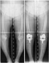
The postoperative radiological evaluation involved the measurement of the tibiofemoral angle, tibial component alignment angle, and tibial component posterior inclination using anteroposterior (AP) and lateral radiographs of the knee joint taken immediately after the operation (Fig. 1). In order to assess the accuracy of the implant, the ratio of the tibiofemoral angle falling to within 6±3 degrees valgus and the ratio of the tibial implant angle being 3 degrees varus to 3 degrees valgus were evaluated.
With regard to clinical results, Hospital for Special Surgery (HSS) score,20 Knee Society Score,21 and range of motion were assessed for 5 months after the operation at the outpatient clinic.
Statistical analysis
Continuous data are expressed as mean and standard deviation, while discrete data are reported as frequencies. Statistical differences in the general characteristics of the patients, analysis of preoperative and postoperative outcomes, clinical evaluation, and radiologic analysis were determined by ANOVA and chi-square tests. In the multiple comparisons of the post-hoc ANOVA, the p value was corrected using the Bonferroni procedure. SAS 9.2 software (SAS Institute, Cary, NC, USA), a statistical package, was used for the analysis, and statistical differences were considered significant when p was below 0.05.
RESULTS
Preoperative evaluation
No differences were found between the groups in terms of age, weight, BMI, and functional scores. However, height, range of motion in the knee joint, pain scores, and HSS scores were significantly smaller in Groups 3 and 4 than in Groups 1 and 2 (p<0.05) (Table 2).
Surgical results
With regard to intraoperative complications, a rupture of the medial collateral ligament occurred in one case each in Groups 1 and 3 during the tibial osteotomy and was repaired with Krackow's method. A 22×4-mm sized piece of cement remained on the medial side of the knee joint in one case in Group 1. Another case in Group 1 was treated with open debridement, irrigation, and replacement of a polyethylene bearing after being infected by Enterobacter intermedius during the 3 months following the surgery.
The surgery lasted 92.5±12.3 minutes on average for Group 4, which was longer than that of the other groups. In addition, the length of the skin incision was longer in Group 4, at 8.5±0.8 cm on average. However, the groups showed no difference in the amount of postoperative blood loss (Table 3 and 4).
A postoperative AP radiograph of the knee joint showed average tibiofemoral angles of 5.2±1.9, 4.7±1.9, 4.9±1.9, and 5.1±2.0 degrees valgus for Groups 1, 2, 3, and 4, respectively, indicating no differences between the groups (p value: 0.419). The alignment angle of the tibial implant averaged 0.2±1.4, 0.1±1.3, 0.1±1.6, and 0.3±1.7 degrees varus for Groups 1, 2, 3, and 4, respectively, showing no differences between the groups (p value: 0.7119). On a radiograph of the lateral side of the knee joint, the posterior slope of the tibial implant was 4.5±1.6, 4.7±1.7, 4.5±1.9, and 5.1±2.0 degrees on average for Groups 1, 2, 3, and 4, respectively, indicating no differences between the groups (p value: 0.1869).
There was no difference in terms of the tibiofemoral alignment, with 83.9%, 82.9%, 85.4%, and 86.7% of each group showing 6±3 degrees valgus angulation (p value: 0.971). However, with respect to the alignment for the tibial implant, 98.1%, 97.6%, 87.5%, and 86.7% of Groups 1, 2, 3, and 4, respectively, had 0±3 degrees varus angulation, demonstrating a lower level of accuracy in Groups 3 and 4 than in Groups 1 and 2 (p value<0.0001) (Table 5).
Clinical results
The range of motion in the knee joint 5 months after the operation measured 128.2±15.2, 127.9±15.0, 127.5±11.7, and 123.0±18.2 for Groups 1, 2, 3, and 4, respectively, indicating no differences between the groups (p value: 0.3754).
The pain scores measured 5 months after the operation showed no differences between the groups, with Groups 1, 2, 3, and 4 at 92.6, 92.8, 92.5, and 89.6, respectively (p value: 0.2599). In addition, the functional scores did not differ between the groups, with Groups 1, 2, 3, and 4 showing 76.1, 77.8, 75.7, and 77.6, respectively (p value: 0.7175). In addition, the HSS scores indicated no differences between the groups, with Groups 1, 2, 3, and 4 showing 90.4, 91.2, 89.7, and 89.4, respectively (p value: 0.3416). In a preoperative and 5-month postoperative comparison, the pain scores improved from 32.1±10.6 to 92.5±7.5, the functional knee scores from 47.2±7.9 to 76.7±17.5, and the HSS scores from 64.9±7.6 to 90.6±7.0 (Table 5).
DISCUSSION
MIS-TKA is a surgical procedure that involves minimum damage to the suprapatellar pouch without patellar eversion and tibiofemoral joint dislocation and minimizes damage and tension in the soft tissue. Thus, it can lessen postoperative pain and reduce recovery and hospitalization periods, thereby increasing patient satisfaction.56789101121 A good surgical method typically embodies basic surgical principles, namely accuracy in terms of proper soft tissue balancing, extension and flexion gap balancing, and recovery of alignment. Conversely, intraoperative complications must not occur, and predicted surgical results must be obtained. Additionally, a comfortable setting is necessary in order for the surgeon to proceed with the operation. However, compared to conventional techniques, MIS-TKA involves a smaller exposed surgical site and partial dependency on a specially-designed alignment guide and modified alignment jig. It requires an experienced surgeon and takes time to obtain the necessary surgical skills. The operation time may be delayed, and it is not a surgeon-friendly operation. The operation is believed to have an intraoperative risk of fracture at the surgical site, damage and rupture of the ligaments or muscles, insufficient removal of cement or bone fragments, skin necrosis due to severe traction, increased chance of infection, improper fixation of implants, and inappropriate implant alignment, all of which have a negative impact on the long-term clinical results, according to some research.1112131415
According to King, et al.,9 MIS-TKA requires more time to master than existing techniques; thus, it carries a higher risk in the context of accuracy and operation time until the surgeon masters it. However, no difference is said to be found once the surgeon becomes skilled in the procedure. Therefore, the successful practice of MIS-TKA requires improved surgical techniques to allow experienced and skilled surgeons to perform the operation with new, modified surgical tools after education via a series of training seminars. Furthermore, it is better to narrow down the surgical view from a wider one, rather than starting with a narrow view initially. It requires very elaborate surgical protocols and problem-solving skills. Moreover, it is critical to accurately locate the modified, improved retractor and pull with the proper level of balanced power with close cooperation of the surgical assistants. In addition, it is important to secure the surgical view through appropriate flexion and extension or internal and external rotation by comparing the narrow surgical view to the mobile window and to pay extra attention in order to minimize intraoperative damage to normal tissue. Instead of using the same surgical view for every patient, it is advisable to extend the incision of soft tissue to prevent problems from arising during the operation due to the surgeon's skill level, patient's characteristics, or difficulties associated with implant insertion. The same applies during intraoperative issues and when problems are expected due to excessive traction of soft tissue. Inappropriate skin traction should be avoided in patients with sensitive skin due to rheumatoid arthritis, diabetes, and steroid dependency due to chronic illness. In addition, extra attention needs to be paid to the removal of any remaining cement or bone fragments in the knee joint. It is advisable that a sufficient incision be made in the soft tissue during the early stage of the operation in cases of inflexibility in the joints or joint stiffness from a previous operation, infectivity, limited range of motion in the joint, revision implant required due to serious bone loss, and severe deformity.
The operation was performed based on the above principles. Minimally invasive surgery was possible in patients who were able to move the knee joint and in whom a mobile window could be created, regardless of the degree of varus deformity. As the medial collateral ligament was located near the tibia, it should have been properly protected during tibial osteotomy. However, rupture of the medial collateral ligament occurred in two cases due to carelessness. In order to prevent this, it is believed that osteotomy should be performed after installing a protective device between the tibia and the medial collateral ligament. Additionally, one should ensure that any remaining foreign substances in the joint are thoroughly removed after surgery. In one case, a large piece of cement remained in the patient due to carelessness, demonstrating the need for meticulous care in the future.
With respect to component alignment, minimally invasive surgery is highly likely to result in malalignment due to the narrow surgical view and difficulty in identifying anatomical markers. Malalignment is more commonly found in patients with severe preoperative varus deformity in the knee joint. Although a small incision was made in the soft tissue, the author was able to achieve recovery of implant alignment, regardless of the degree of varus deformity, by installing an intramedullary alignment guide for the femur and an extramedullary alignment guide for the tibia at the appropriate locations. Furthermore, although each patient had severe varus deformity in the knee joint, they were able to move the joint in the preoperative stage, which allowed for control of soft tissue balancing at the necessary sites through a mobile window.
In terms of surgical accuracy, if the knee is within ±3 degrees of normal alignment on the coronal plane, it is generally considered to have only slight impact on postoperative long-term outcomes.3162223 In the evaluation of all patients, the tibiofemoral angle was 5.0±1.9 degrees valgus, the tibial implant was 0.2±1.4 degrees varus, and the posterior slope of the tibial implant was 4.6±1.7 degrees on average. In 59 previous papers, cases where the knee was more than 3 degrees out of normal alignment on the coronal plane accounted for 10.2% when using navigation and 28.2% when using the conventional technique on average.24 In this study, 83.6% showed 6±3 degrees valgus for the tibiofemoral angle whereas 96.5% showed 0±3 degrees varus for tibial implant alignment, both of which are satisfactory alignment results when compared to the previous reports.
As for alignment accuracy, depending on the severity of the preoperative varus deformity, 83.9%, 82.9%, 85.4%, and 86.7% of Groups 1, 2, 3, and 4, respectively, showed a tibiofemoral alignment of 6±3 degrees valgus, indicating no differences between the groups (p value: 0.971). However, 98.1%, 97.6%, 87.5%, and 86.7% of Groups 1, 2, 3, and 4, respectively, showed a tibial implant alignment of 0±3 degrees varus, with Groups 3 and 4 showing lower accuracy than that of Groups 1 and 2 (p value<0.0001). The lower accuracy in tibial implant alignment was not attributed to the use of minimally invasive surgery in patients with severe varus deformity. Rather, when installing an extramedullary alignment guide, difficulties associated with identifying anatomical markers on a tibial plateau with severe varus deformity might have played a role in not accurately fixing the guide. In addition, severe tibial deformity might have caused difficulties in anchoring the guide at the distal part.
With regards to clinical indicators, more severe cases of preoperative varus deformity in the knee joint were associated with shorter height, smaller range of motion in the knee joint, and lower pain and HSS scores. In the postoperative evaluation, which took place 5 months after the surgery, range of motion in the knee joint, pain scores, functional scores, and HSS scores all improved significantly, regardless of the group. This improvement may be attributed to a shorter recovery period due to minimal soft tissue damage from the minimally invasive surgery and proper recovery of the implant alignment. Higher satisfaction levels were found in patients with more severe preoperative varus deformities, an observation that calls for long-term follow-up studies.
Group 4, a group with many cases of varus deformity in the knee joint, showed a significantly longer operation time and skin incision. Severe cases of deformity are associated with more serious contractures or tibial deformities and difficulties in identifying anatomical markers. Therefore, such cases might have required a longer operation time and a longer skin incision to achieve surgical accuracy.
In conclusion, when minimally invasive surgery was performed in total knee arthroplasty, the accuracy of the tibial implant alignment was slightly reduced in severe cases of preoperative varus deformity. However, it led to relatively satisfactory results, based on radiological outcomes and short-term clinical results, regardless of the preoperative degree of varus deformity.


 XML Download