

 PDF
PDF ePub
ePub Citation
 Print
Print


INTRODUCTION
Despite the wide variety of phakic intraocular lenses (pIOLs) that yield good results, the correction of moderate-to-high astigmatism remains a challenge.1 The foldable iris-fixated pIOL (hereafter, non-toric pIOL; Artiflex, Ophtec BV, Groningen, the Netherlands) can be implanted through a small incision,2 minimizing surgically induced astigmatism, but only corrects spherical error. The toric foldable iris-fixated pIOL (hereafter, toric pIOL; Toric Artiflex, Ophtec BV) is designed to correct cylindrical errors between -5.0 and -1.0 diopters (D) and spherical errors between -13.5 and -1.0 D. Several studies have shown that it effectively and safely corrects myopic astigmatism.234 However, accurate placement of the toric pIOL is critical to obtain satisfactory results concerning residual astigmatism, especially in eyes with high astigmatism. Moreover, toric pIOLs are quite expensive and the manufacturing time between placing the order and receiving the product is somewhat long.
Limbal relaxing incisions (LRIs) are another option for correcting astigmatism surgically. LRIs, also known as peripheral corneal relaxing incisions, flatten the steep meridian and cause coupling of the flat meridian.5 They are easy to perform and relatively inexpensive.67 In addition, LRIs can be combined with non-toric pIOL implantation to correct moderate-to-high astigmatism in myopic eyes. However, LRIs can weaken the cornea and/or decrease visual quality by increasing corneal aberrations and irregular astigmatism.89
If non-toric pIOLs combined with LRIs show comparable refractive and visual outcomes with those of toric pIOLs, non-toric pIOLs could be substituted to overcome the drawbacks of toric pIOLs, including their high cost, the long duration required to receive the pIOL from the manufacturer, and challenging elaborate implantation technique along an accurate axis of astigmatism. To the best of our knowledge, studies comparing the astigmatism-reducing effects of these surgical procedures are rare. Herein, we compared the effectiveness of toric pIOL implantation and non-toric pIOL implantation with LRIs for correcting moderate-to-high astigmatism in myopic eyes.
MATERIALS AND METHODS
Subjects
This retrospective comparative observational study was approved by the Institutional Review Board of Yonsei University College of Medicine (Seoul, Korea). Its protocol adhered to the tenets of the Declaration of Helsinki and followed good clinical practice.
Patients were included if they were older than 20 years and had stable refraction and regular astigmatism between 2.00 and 4.00 D. The exclusion criteria were as follows: history of corneal refractive surgery or ocular disease that may affect visual outcomes (e.g., color vision disturbance, chronic uveitis, glaucoma, and maculopathy); an anterior chamber depth <3.0 mm from the endothelium, corneal endothelial cell density (ECD) <2000 cells/mm2, white-to-white distance <11.0 mm, mesopic pupil diameter >7.0 mm, intraocular pressure (IOP) >21 mm Hg; evidence of acute or chronic corneal infection, corneal inflammation, abnormal iris or pupil function, or cataract; and the development of intraoperative or postoperative complications.
The same surgeon performed toric pIOL implantation (toric group) and non-toric pIOL implantation with concurrent LRIs (LRI group) in the standard fashion at the Eyereum Eye Clinic (Seoul, Korea) between November 2012 and October 2014. The patient's economic preference dictated the choice of surgical method. The groups were further divided into the moderate (2.00 to <3.00 D) and high (3.00–4.00 D) astigmatic subgroups according to preoperative astigmatic severity.
All subjects received complete ophthalmic examinations, including measurements of uncorrected distance visual acuity (UCDVA; Snellen) and IOP (noncontact tonometer NT-530, Nidek Co., Ltd., Aichi, Japan), manifest refraction, slit-lamp biomicroscopy (Haag-Streit AG, Köniz, Switzerland), and dilated fundus examination. Central corneal thickness, keratometric values, and central ECD were measured by ultrasound pachymetry (UP-1000, Nidek Co.), autokeratometry (ARK-530A, Nidek Co.), and specular microscopy (SP-3000P, Topcon Corporation, Tokyo, Japan), respectively. Manifest refraction, UCDVA, and central ECD measurements were repeated 3 and 6 months postoperatively.
Surgical procedures
A small iridotomy was made with consecutive argon green and neodymium-doped yttrium aluminum garnet (Nd:YAG) lasers at least 1 week preoperatively. Immediately before surgery, the axis of astigmatism was marked along the negative axis at the slit-lamp to determine the flat meridian with the patient seated upright.
In the toric group, a 2.8-mm primary limbal incision was made at the marked meridians, and two sub-1.0-mm stab incisions were placed nasally and temporally to the primary incision, as seen fit by the surgeon. A miotic agent was injected intracamerally and 1.0% sodium hyaluronate was used to maintain the anterior chamber and coat the corneal endothelium. Then, the toric pIOL was inserted, fixated with special forceps, and aligned along the orientation marks on the cornea. Finally, the viscoelastic substance was removed by manual aspiration.
In the LRI group, the primary incision was placed on the steep meridian, with the length and location determined by using the Nichamin Age & Pachymetry-Adjusted Intralimbal Arcuate Astigmatic Nomogram.10 The non-toric pIOL was inserted and fixated with special forceps. At the end of the surgery, an additional LRI was placed at the opposite meridian and the viscoelastic material was manually removed.
All surgeries were uneventful and no intraoperative complication was noted. The subjects applied 0.5% moxifloxacin and 0.1% dexamethasone four times daily for the first postoperative week. Thereafter, dexamethasone was replaced with 0.1% fluorometholone and the eye drops were continued four times daily for 1 month.
Statistical analyses
The results are expressed as means±standard deviations, where applicable. The Kolmogorov-Smirnov test was used to confirm data normality. All data were analyzed by using SAS software (version 9.2, SAS Institute, Inc., Cary, NC, USA). Differences between the groups were tested for statistical significance by using independent t tests. Paired t tests were used to test for differences between preoperative and residual astigmatism within each group. Statistical significance was defined as p<0.05.
Astigmatism was analyzed by the power vector method11 and Alpins method.121314 The power vector method aids in visualizing complex changes in refractive error. The manifest refraction data, as written in conventional script notation, were used to calculate power vector coordinates and overall blurring strength (B) by the following formulas: B=(M2+J02+J452)1/2, where M=S+C/2, J0=(-C/2) cos(2α), J45=(-C/2) sin(2α), S=sphere, C=cylinder, and α=axis. The calculated power vector length is a measure of the overall blurring strength of the refractive error.
The Alpins method presents data in standard graphs for reporting astigmatic outcomes of refractive surgery.12131415 The target astigmatism is zero, because emmetropia is the goal. We calculated the target-induced astigmatic vector (TIAV), or the amount and direction of the dioptric force required to achieve emmetropia from the preoperative state, the surgically induced astigmatic vector (SIAV), which is the astigmatic change actually induced by surgery, and the difference vector (DV), which represents the magnitude and axis of the difference in D between the desired and achieved results. The correction index (CI) and success index (SI) were calculated as ratios of the SIAV to the TIAV and the DV to the TIAV, respectively. Free spreadsheets were used to present the data graphically (http://www.standardgraphsforrefractivesurgery.com).15
RESULTS
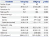
The study included 195 eyes of 146 patients, with 94 eyes (65 subjects) in the toric group and 101 eyes (81 subjects) in the LRI group. There were no significant intergroup differences in the preoperative variables (Table 1).
The groups had comparable visual outcomes. In the toric group, the UCDVA was 20/25 or better in all eyes (100%) and 20/20 or better in 91 eyes (96.8%). In the LRI group, it was 20/25 or better in 99 eyes (98%) and 20/20 or better in 83 eyes (82.2%). The overall blurring strength significantly decreased from 8.69±2.31 D to 0.68±0.60 D in the toric group and from 8.62±1.71 D to 0.72±0.25 D in the LRI group (both p<0.001). The toric group demonstrated a slightly lower blurring strength postoperatively, but the difference was not statistically significant (p=0.469).
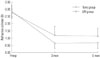
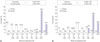
Both groups had significantly reduced astigmatism after the surgery (Fig. 1). The residual cylindrical error was significantly lower (-0.67±0.39 D vs. -1.14±0.56 D; p<0.001) and the mean change in cylindrical error was significantly greater (2.17±0.56 D vs. 1.63±0.72 D; p<0.001) in the toric group than in the LRI group 6 months postoperatively. Furthermore, 47 (50.0%) and 85 (90.4%) of the 94 eyes in the toric group had residual cylindrical errors within 0.50 and 1.00 D, respectively. On the contrary, 19 (18.8%) and 43 (42.6%) of the 101 eyes in the LRI group demonstrated the same respective residual cylindrical error values (Fig. 2). The differences between the groups were significant (both p<0.001).
Subgroup analyses revealed that the mean residual cylindrical errors were -0.64±0.37 and -1.09±0.58 D in toric and LRI group eyes with moderate astigmatism (p<0.001), respectively. For high astigmatism, the mean residual cylindrical errors were -0.72±0.41 and -1.16±0.66 D (p=0.001) in the respective groups. The mean changes in moderate (1.84±0.43 D vs. 1.33±0.61 D; p<0.001) and high (2.55±0.44 D vs. 2.16±0.66 D; p=0.003) astigmatism were significantly greater in the toric group than in the LRI group.
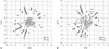
In the analysis of astigmatism by the power vector method,11 most points were concentrated at the center of the graph, especially in the toric group (Fig. 3). The toric group had J0 and J45 vector values between +0.50 and -0.50 D in 86 (91.5%) and 91 (96.8%) of the 94 eyes, respectively. In the LRI group, J0 and J45 vector values in the same range were noted in 76 (75.2%) and 69 (68.3%) of the 101 eyes, respectively. Both differences between the groups were significant (p=0.003 and p<0.001).
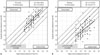
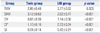
The subjects in the toric group had their astigmatism overcorrected, while those in the LRI group had undercorrected astigmatism (Fig. 4). However, the toric group had corrections much closer to the intended value (p<0.001). The mean CI and SI were significantly different between the groups (both p<0.001) (Table 2).
The postoperative ECDs were 3034.2±236.0 and 3037.5±265.4 cells/mm2 in the toric and LRI groups (p=0.926), respectively. The mean ECD changes were -29.8±120.9 (p=0.019) and -38.1±127.8 (p=0.003) cells/mm2 in the respective groups; the intergroup difference was not significant (p=0.926). Moreover, the percentage change in ECD was -0.92±3.9% in the toric group and -1.19±4.2% in the LRI group (p=0.644).
DISCUSSION
Several articles have reported predictable and effective astigmatic correction with rigid toric iris-fixated pIOLs.1617 However, they have some drawbacks, including a large amount of surgically induced astigmatism, mainly attributable to the length of the primary incision (up to 5.2 mm) required to implant the lens. Although surgically induced astigmatism is accounted for in power calculations, it is not always accurately forecasted. The toric pIOL can be inserted through a 3.2-mm incision, minimizing surgically induced and irregular astigmatism. In one study conducted by Ruckhofer, et al.,1 the mean residual astigmatism was -0.18±0.30 D after toric pIOL implantation. Other studies have also shown relatively low residual cylindrical error with this method (multicenter study, 0.38±0.41 D2; two long-term follow-up studies, -0.39 D and -0.60 D34). In line with these results, we found residual astigmatism of -0.67±0.39 D at 6 months after toric pIOL implantation.
One study showed that toric IOL implantation and LRIs during cataract surgery yielded similar results for astigmatic correction.18 However, Hirnschall, et al.5 reported that toric IOL implantation reduced astigmatism more noticeably and predictably than LRIs after cataract surgery. Further, Mingo-Botín, et al.6 showed that toric IOL implantation resulted in better refractive and visual outcomes in eyes with mild or moderate astigmatism. Consistent with these findings, toric pIOL implantation in our study, led to better astigmatic correction, resulting in lower residual astigmatism and a larger change in cylindrical error, regardless of the preoperative astigmatic severity, albeit the toric and LRI groups had comparable visual acuities and blurring strengths.
The CI is greater than 1.0 if overcorrection occurs, and less than 1.0 if there is undercorrection.12 In the present study, the mean CI was significantly larger in the toric group than in the LRI group (1.10 vs. 0.72). Despite the overcorrection, the refractive outcomes were less deviated from the ideal value (CI=1.0) in the toric group (p<0.001). Furthermore, the mean SI was lower in this group (0.24 vs. 0.42). Considering that the SI is a relative measure of success and preferably zero,12 the reduction in astigmatism was more successful in the toric group, implying that toric pIOL implantation corrects astigmatism more predictably and accurately than non-toric pIOL implantation with LRIs.
Several factors may explain why astigmatism was undercorrected in the LRI group: improper identification of the steep meridian, incorrect calibration of the blade, oblique positioning of the blade instead of apposition perpendicular to the limbus, and the far peripheral placement of incisions.101920 LRIs are associated with other important problems. First, LRIs have a degree of uncertainty because equally relaxing incisions are difficult to create. We examined cases performed by only one surgeon to minimize the influence of variations in surgical style. Second, a relatively long period is needed after LRIs for the corrective effect to stabilize.9 The largest amount of refractive regression occurs between 1 and 3 months after LRIs, and the refractive status stabilizes between 3 and 6 months postoperatively.21 Another study showed that surgically induced astigmatism continued to change up to 10 weeks after LRIs, but remained stable after 10 weeks and up to 3 years postoperatively.22 We used 6-month follow-up data to exclude the possible effects of refractive regression.
The toric pIOL should be implanted along the correct cylindrical axis, and especially in eyes with high astigmatism, because small deviations can result in improper correction. Several studies have indicated that only 1.7–2.4% of eyes implanted with toric pIOLs had greater than 5° of misalignment.24
Endothelial cell loss is a possible complication of both toric and non-toric pIOL implantation. In our study, ECD significantly decreased in both groups, with comparable percent changes from baseline to 6 months. These results are in accordance with previous results regarding iris-fixated pIOLs.123
Our study has some limitations, including its retrospective design and lack of results on visual quality. However, a previous study showed potential improvements in contrast vision and mean contrast sensitivity after toric pIOL implantation.23 In addition, this procedure does not alter or increase higher-order aberrations in myopic eyes.24 On the contrary, LRIs may increase corneal aberrations and decrease functional vision by degrading the optical quality of the cornea.25 Therefore, differences in functional vision may exist between the study groups. A prospective controlled study evaluating both the astigmatism-reducing effects and visual quality of these methods is necessary.
In conclusion, both surgical methods considerably reduced astigmatism and had comparable visual outcomes. However, toric pIOL implantation was more reliable for moderate-to-high astigmatic correction than non-toric pIOL implantation with LRIs in myopic eyes.
 XML Download