Journal List > Yonsei Med J > v.57(6) > 1031875

Choi, Do, Seo, Lee, Oh, Min, and Kang: Prevalence and Risk Factors of Gallbladder Polypoid Lesions in a Healthy Population

Abstract

Purpose

To determine the prevalence of and investigate the risk factors for gallbladder (GB) polypoid lesions in a healthy population.

Materials and Methods

A total of 23827 subjects who underwent abdominal ultrasonography in conjunction with health screening examinations were retrospectively analyzed. The prevalence of risk factors for GB polypoid lesions were evaluated. In addition, risk factors according to the number of polypoid lesions and the presence of stones with polypoid lesions were investigated. To analyze these risk factors, a control group was established with a 1:2 ratio matched for age and sex.

Results

The prevalence of GB polypoid lesions was identified as 9.96%. On multivariate analysis, chronic hepatitis B infection (CHB) and the presence of metabolic syndrome (MS) were risk factors for GB polypoid lesions. CHB and MS were also significant independent risk factors for multiple GB polypoid lesions when compared with solitary GB polypoid lesions. In addition, gastric Helicobacter pylori infection and MS were significant risk factors for GB polypoid lesions with stones when compared with GB polypoid lesions without stones.

Conclusion

The prevalence of GB polypoid lesions in a healthy Korean population was 9.96%. Patients with CHB and MS need to be carefully examined for such lesions.

INTRODUCTION

A gallbladder (GB) polypoid lesion refers to any lesion elevated from the surface of the GB mucosa.1 The presenting symptoms of GB polypoid lesions are nonspecific and vague, and in many cases, asymptomatic. For such reasons, most GB polypoid lesions are detected incidentally on abdominal ultrasonography (US). Although most such lesions are benign, several early GB cancers do present as polypoid lesions.23 Therefore, GB polypoid lesions have important clinical significance.
Recently, with the increasing number of people receiving health screening examinations and the rapid increase in the use of US, the detection of GB polypoid lesions is increasing. The prevalence of GB polypoid lesions varies from 1.3% to 9.5% based on region and race.45678 According to a 2006 Korean report, the prevalence of GB polypoid lesions was 2.2%,8 which was relatively lower than those reported from other regions.
Risk factors for GB polypoid lesions in previous studies included a high body mass index (BMI), male gender, chronic hepatitis B (CHB), chronic hepatitis C, old age, serum cholesterol level, and metabolic syndrome (MS), although the factors varied among the different reports.456789
The aims of this study were to evaluate the prevalence of GB polypoid lesions and to determine risk factors for these lesions in a healthy Korean population. The risk factors for solitary polypoid lesions and the presence of stones in lesions were also analyzed.

MATERIALS AND METHODS

Subjects

Between January 2010 and December 2012, 23827 South Koreans who underwent abdominal US for health screening examinations at Chung-Ang University Hospital in Seoul, Korea, were retrospectively identified. The GB polypoid lesion group was compared with a control group (without lesions), which was established based on a 1:2 ratio matching for age and sex. We performed the matching using random sampling after stratification by age and gender in the control group to compensate for the age and gender differences between the GB polypoid lesion and control groups.

Diagnosis of GB polypoid lesions

Two radiologists with over 10 years of experience each diagnosed the presence of GB polypoid lesions based on the abdominal US (Sequoia 512, Acuson, Mountain View, CA, USA). GB polypoid lesions were identified on abdominal US as fixed, hyperechoic material protruding from the GB wall into the lumen without an acoustic shadow, characterized by the absence of shift with positional change.10 The number and diameter of the largest polypoid lesion were recorded.

Prevalence and risk factor analysis

We evaluated the prevalence of and the risk factors for GB polypoid lesions between the polypoid lesion group and the 1:2-matched control group. We compared overweight status, MS, fasting glucose, CHB, chronic hepatitis C, total cholesterol, triglyceride (TG), high-density lipoprotein (HDL) cholesterol, low-density lipoprotein (LDL) cholesterol, infection with gastric Helicobacter pylori (HP) based on a biopsy of mucosa in the gastric antrum and body or a Campylobacter-like organism test, and presence of colon polyps for those who underwent colonoscopy procedures. In addition, the risk factors for a singular stone and the presence of stones were evaluated in the GB polypoid lesion group. Overweight status was defined as a BMI of over 23 kg/m2.11 According to the National Cholesterol Education Program-Adult Treatment Panel III criteria,9 MS was defined when three or more of the following criteria were satisfied: 1) abdominal obesity: waist circumference of >90 cm in men and >85 cm in women; 2) hypertriglyceridemia: ≥1.7 mmol/ L (150 mg/dL); 3) low HDL cholesterol: <1.03 mmol/L (40 mg/ dL) in men and <1.29 mmol/L (50 mg/dL) in women; 4) high blood pressure: ≥130/85 mm Hg; and 5) high fasting glucose: ≥6.1 mmol/L (110 mg/dL).

Statistics

The sample size was calculated to detect a 1.23-fold increase in the odds of development of a GB polypoid lesion in the presence of one specific risk factor with 5% significance and 80% power. The calculation showed that 2378 cases, each with two matched controls, would be necessary to show such an association.
For continuous variables, the distribution of the data was first evaluated for normality using the Kolmogrov-Smirnov test. As not all of the variables passed the normality test, we additionally checked Q-Q plots. As total and LDL cholesterol did not indicate significant deviation from linearity, we allowed the normal assumption of a parametric test. Normally distributed data are presented as means±standard deviations, and the groups were compared using Student's t-tests. Non-normally distributed data are expressed as medians (P25–P75), and these data were analyzed using Mann-Whitney U tests. Descriptive variables were subjected to χ2 analysis or Fisher's exact test, as appropriate, and results are presented as absolute numbers (%). To identify the significant risk factors for GB polypoid lesions, solitary polyps, and stones with GB polypoid lesions, multiple logistic regression with backward selection was used. The multi-collinearity diagnostic indicated no multi-collinearity issues (condition indices <30; VIF values <10) between the chosen independent variables in this study. Factors that had univariate p values of <0.1 were included for multivariate analysis. A p value of <0.05 was considered to be statistically significant. All statistical analyses were performed using SPSS 18.0 (IBM Corp., Armonk, NY, USA).

RESULTS

Patient characteristics and prevalence of GB polypoid lesions

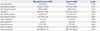
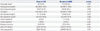
The mean age of the entire cohort was 45.7 years (range, 14–89 years), and 13870 (58.2%) of the patients were men. GB polypoid lesions were identified in 2378 patients among the entire cohort. In the lesion group, the prevalence was 9.96%; 1568 patients (65.9%) were male and 810 (34.1%) were female; and the mean age was 45.9 years (range, 21–76 years) for males and 45.6 years (range, 21–83 years) for females.

Risk factors for GB polypoid lesions

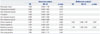
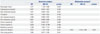
On characteristics and univariate analyses, a low level of serum HDL cholesterol, CHB, and MS were found to be risk factors (Table 1 and 2). On multivariate analysis with logistic regression, CHB [odds ratio (OR) 1.403; 95% confidence interval (CI) 1.095–1.797; p=0.007] was an independent risk factor for GB polypoid lesions, in addition to MS (OR 1.748; 95% CI 1.489– 2.051; p<0.001) (Table 2).

Risk factors for solitary GB polypoid lesions

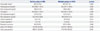
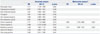
Among 2378 patients, 768 (32.3%) had a solitary polypoid lesion, and 1610 (67.7%) had multiple lesions. In characteristics and univariate analyses, overweight status, serum TG, serum HDL cholesterol, CHB, and MS were factors for the multiplicity of GB polypoid lesions (Table 3 and 4). On multivariate analysis, CHB (OR 1.802; 95% CI 1.135–2.865; p=0.012) and MS (OR 1.414; 95% CI 1.046–1.912; p=0.024) were found to be risk factors for multiple GB polypoid lesions when compared with solitary GB polypoid lesions (Table 4).

Risk factors for the presence of stones with GB polypoid lesions

Among 2378 patients, 119 (5.0%) had GB stones. On univariate analysis, overweight status, MS, and gastric HP infection were found to be risk factors (Table 5 and 6). On multivariate analysis, only gastric HP infection was an independent risk factor for the presence of stones with GB polypoid lesions (OR 2.933; 95% CI 1.185–7.246; p=0.020) (Table 6).

DISCUSSION

Abdominal US is widely used and is the most important tool for the diagnosis of GB polypoid lesions. In recent years, the widespread and frequent use of US has increased the rate of incidental detection of GB polypoid lesions. As most lesions are detected incidentally during US, it is difficult to accurately measure its prevalence. The prevalence of GB polypoid lesions varies among countries and studies. In Denmark, the prevalence was 4.6% in men and 4.3% in women.5 In Japan, the overall prevalence was 5.3% (6.3% in men, 3.5% in women),6 and this was 6.9% in a Chinese study.4 A Korean study in 1999, which evaluated patients who underwent health screening examinations, reported the overall prevalence to be 2.94% (3.63% in men, 2.09% in women).12 Another Korean study in 2012 reported an 8.5% prevalence.13 In our study, the prevalence rate was 9.96%, which was higher than previously reported in Korea. The main reason for this increase may be the wider use of US, which corresponds with the increasing interest among individuals in regular checkups. The incidence of GB polypoid lesions may also be increasing due to the increasing prevalence of MS.14 However, our study evaluated only people who had more interest in their health. This factor have an influence on assessing the prevalence of GB polypoid lesions in the general population.
Cholesterol polyps are the most common type of GB polypoid lesion. Although the etiology is poorly understood, several researchers have suggested that cholesterolosis might be derived from the direct deposition of cholesterol from the blood.15 Others have suggested that alterations in hepatic cholesterol metabolism and altered mucosal esterification of free sterols from bile may contribute to the development of cholesterolosis. Based on these hypotheses, we analyzed several blood lipid components as potential risk factors for GB polypoid lesions. However, blood cholesterol concentration was not an independent risk factor, and this finding was in agreement with previous reports.5781216
There are also reports that hyperglycemia contributes to the formation of biliary stones by hindering the contraction of the GB and inhibiting the secretion of bile from the liver;717 however, hyperglycemia was not a risk factor for GB polypoid lesions in the present study.
MS has become a major public-health concern worldwide, and the prevalence of MS has been increasing.18 As in a previous report,7 we found MS to be a risk factor for GB polypoid lesions in our study. In recent years, the increase in MS due to a high-calorie and high-fat diet has increased the prevalence of GB polypoid lesions as well.
Studies have reported that CHB is a risk factor for GB polypoid lesions,812 and in this study as well, CHB was found to be an independent risk factor for GB polypoid lesions. CHB is known to cause abnormalities in the GB, such as wall thickening and alterations in the lumen size and volume; however, the relationship between CHB and GB polypoid lesions has not been delineated clearly. There are two possible explanations regarding the relationship between CHB and GB polypoid lesions. First, hepatitis B virus that is located in the bile of the GB may have a direct influence on the GB mucosa. Second, the inflammatory changes in liver parenchyma by CHB may affect the GB mucosa.
In general, a solitary polypoid lesion in the GB is more likely to go under malignant transformation than multiple lesions.19 However, the size of the polypoid lesion is a more important factor than the number when estimating the likelihood of malignant transformtion.20 Although MS and CHB may be risk factors for multiple polypoid lesions,2122 as shown in this study, it is hard to conclude that these factors lessen the probability of malignant polypoid lesions.
Gastric HP infection is associated with the development of GB stones2324 and is likely involved in the development of biliary tract cancer as well as GB cancer.25 In this study, the group with both gallstones and polypoid lesions presented with significantly higher rates of HP infection, which implies that HP infection is related to stone occurrence rather than polypoid lesions. However, more studies are needed to establish the potential relationship between HP infection and malignant transformation of GB polypoid lesions, which may be caused by aggravation of the GB mucosal lesions (hyperplasia, metaplasia, or lymphoid infiltration)26 or by the development of gallstones.
In conclusion, the prevalence of GB polypoid lesions in a healthy Korean population was 9.96%. Healthy Koreans with CHB and MS should be screened for GB polypoid lesions. CHB, MS, and gastric HP infection were risk factors for multiple GB polypoid lesions and the presence of stones.
Therefore, cases of CHB and MS need to be carefully examined to screen for GB polypoid lesions. In addition, patients with GB polypoid lesions who have MS or gastric HP positivity should undergo additional workup for the detection of GB stones.

Figures and Tables

Table 1

Characteristics of the Risk Factors for GB Polypoid Lesions (n=7134)

ymj-57-1370-i001
GB polypoid lesion (n=2378) Control (n=4756) p value
Overweight status* 1354 (56.9%) 2642 (55.6%) 0.266
Fasting glucose (mg/dL) 93.0 (87.0–100.0) 94.0 (88.0–100.0) 0.421
Total cholesterol (mg/dL) 196.40±36.56 196.92±35.91 0.571
TG (mg/dL) 96.0 (64.0–146.0) 98.5 (66.0–147.0) 0.162
LDL cholesterol (mg/dL) 122.98±32.44 121.55±31.67 0.074
HDL cholesterol (mg/dL) 49.0 (43.0–57.0) 50.0 (44.0–58.0) 0.001
Chronic hepatitis B 117 (4.9%) 162 (3.4%) 0.002
Chronic hepatitis C 6 (0.3%) 10 (0.2%) 0.723
Metabolic syndrome 307 (12.9%) 373 (7.8%) <0.001
Gastric HP infection 307/646 (47.5%) 529/1140 (46.4%) 0.637
Colon polyp positive 581/1054§ (55.1%) 980/1782§ (55.0%) 0.947

GB, gallbladder; TG, triglyceride; LDL, low-density lipoprotein; HDL, high-density lipoprotein; HP, Helicobacter pylori.

Data show mean±standard deviation, median (P25–P75), or absolute number (%). The level of significance is set below 0.05. Data show median (P25–P75) or absolute number (%). The level of significance is set below 0.05.

*Overweight status: body mass index over 23 kg/m2, Data are presented as median (P25–P75) and analyzed using the Mann-Whiteny U test due to abnormal distribution, Data show positive/esophagogastroduodenoscopy patients, §Data show positive/colonoscopy patients.

Table 2

Univariate and Multivariate Analyses of the Risk Factors for GB Polypoid Lesions (n=7134)

ymj-57-1370-i002
Univariate analysis Multivariate analysis*
OR 95% CI p value OR 95% CI p value
Overweight status 1.058 0.958–1.168 0.266
Fasting glucose (mg/dL) 0.999 0.996–1.002 0.520
Total cholesterol (mg/dL) 1.000 0.999–1.002 0.571
TG (mg/dL) 1.000 0.999–1.000 0.214
LDL cholesterol (mg/dL) 0.999 0.997–1.000 0.074
HDL cholesterol (mg/dL) 0.993 0.989–0.998 0.004
Chronic hepatitis B 1.467 1.151–1.871 0.002 1.403 1.095–1.797 0.007
Chronic hepatitis C 1.201 0.436–3.307 0.723
Metabolic syndrome 1.742 1.485–2.044 <0.001 1.748 1.489–2.051 <0.001
Gastric HP infection 1.048 0.864–1.271 0.637
Colon polyp positive 1.005 0.863–1.172 0.947

GB, gallbladder; OR, odds ratio; CI, confidence interval; TG, triglyceride; LDL, low-density lipoprotein; HDL, high-density lipoprotein; HP, Helicobacter pylori.

*Adjusted for LDL cholesterol, HDL cholesterol, chronic hepatitis B, and metabolic syndrome in logistic regression model, Overweight status: body mass index over 23 kg/m2.

Table 3

Characteristics of the Risk Factors for GB Solitary Polypoid Lesions (n=2378)

ymj-57-1370-i003
Solitary polyp (n=768) Multiple polyps (n=1610) p value
Overweight status* 402 (52.3%) 952 (59.1%) 0.002
Fasting glucose (mg/dL) 93.5 (88.0–101.0) 93.0 (87.0–100.0) 0.055
Total cholesterol (mg/dL) 195.9±37.8 196.6±36.0 0.667
TG (mg/dL) 92.0 (62.0–138.0) 98.0 (65.0–150.0) 0.036
LDL cholesterol (mg/dL) 122.8±32.6 123.2±32.4 0.651
HDL cholesterol (mg/dL) 50.0 (44.0–58.0) 49.0 (43.0–57.0) 0.010
Chronic hepatitis B 24 (3.1%) 93 (5.8%) 0.014
Chronic hepatitis C 1 (0.1%) 5 (0.3%) 0.412
Metabolic syndrome 84 (10.9%) 223 (13.9%) 0.048
Gastric HP infection 74/169 (43.8%) 233/477 (48.8%) 0.258
Colon polyp positive 168/311§ (54.0%) 413/743§ (55.6%) 0.641

GB, gallbladder; TG, triglyceride; LDL, low-density lipoprotein; HDL, high-density lipoprotein; HP, Helicobacter pylori.

Data show mean±standard deviation, median (P25–P75), or absolute number (%). The level of significance is set below 0.05.

*Overweight status: body mass index over 23 kg/m2, Data are presented as median (P25–P75) and analyzed using the Mann-Whiteny U test due to abnormal distribution, Data show positive/esophagogastroduodenoscopy patients, §Data show positive/colonoscopy patients.

Table 4

Univariate and Multivariate Analyses of the Risk Factors for GB Solitary Polypoid Lesions (n=2378)

ymj-57-1370-i004
Univariate analysis Multivariate analysis*
OR 95% CI p value OR 95% CI p value
Overweight status 1.317 1.108–1.566 0.002
Fasting glucose (mg/dL) 0.997 0.993–1.022 0.284
Total cholesterol (mg/dL) 1.001 0.998–1.003 0.667
TG (mg/dL) 1.001 1.000–1.002 0.185
LDL cholesterol (mg/dL) 1.000 0.998–1.003 0.783
HDL cholesterol (mg/dL) 0.991 0.983–0.999 0.026
Chronic hepatitis B 1.771 1.118–2.806 0.015 1.802 1.135–2.865 0.012
Chronic hepatitis C 2.389 0.279–20.487 0.427
Metabolic syndrome 1.309 1.002–1.710 0.048 1.414 1.046–1.912 0.024
Gastric HP infection 1.226 0.861–1.745 0.258
Colon polyp positive 1.065 0.817–1.390 0.641

GB, gallbladder; OR, odds ratio; CI, confidence interval; TG, triglyceride; LDL, low-density lipoprotein; HDL, high-density lipoprotein; HP, Helicobacter pylori.

*Adjusted for overweight status, HDL cholesterol, chronic hepatitis B, and metabolic syndrome in logistic regression model, Overweight status: body mass index over 23 kg/m2.

Table 5

Characteristics of the Risk Factors for the Presence of Stones with GB Polypoid Lesions (n=2378)

ymj-57-1370-i005
Stone (n=119) No stone (n=2259) p value
Overweight status* 84 (70.6%) 1270 (56.2%) 0.002
Fasting glucose (mg/dL) 95.0 (89.0–103.0) 93.0 (87.0–100.0) 0.051
Total cholesterol (mg/dL) 194.87±37.20 196.48±36.54 0.638
TG (mg/dL) 104.0 (69.0–156.0) 96.0 (63.0–146.0) 0.159
LDL cholesterol (mg/dL) 121.13±33.63 123.08±32.38 0.523
HDL cholesterol (mg/dL) 49.0 (43.0–56.0) 49.0 (43.0–57.0) 0.380
Chronic hepatitis B 4 (3.4%) 117 (5.2%) 0.381
Chronic hepatitis C 1 (0.8%) 5 (0.2%) 0.265
Metabolic syndrome 28 (23.5%) 279 (12.4%) <0.001
Gastric HP infection 25/39 (64.1%) 282/607 (46.5%) 0.032
Colon polyp positive 33/62§ (53.2%) 548/992§ (55.2%) 0.757

GB, gallbladder; TG, triglyceride; LDL, low-density lipoprotein; HDL, high-density lipoprotein; HP, Helicobacter pylori.

Data show median (P25–P75) or absolute number (%). The level of significance is set below 0.05.

*Overweight status: body mass index over 23 kg/m2, Data are presented as median (P25–P75) and analyzed using the Mann-Whiteny U test due to abnormal distribution, Data show positive/esophagogastroduodenoscopy patients, §Data show positive/colonoscopy patients.

Table 6

Univariate and Multivariate Analyses of the Risk Factors for the Presence of Stones with GB Polypoid lesions (n=2378)

ymj-57-1370-i006
Univariate analysis Multivariate analysis*
OR 95% CI p value OR 95% CI p value
Overweight status 1.317 1.108–1.566 0.002
Fasting glucose (mg/dL) 0.997 0.993–1.002 0.284
Total cholesterol (mg/dL) 1.001 0.998–1.003 0.667
TG (mg/dL) 1.001 1.000–1.002 0.185
LDL cholesterol (mg/dL) 1.000 0.998–1.003 0.874
HDL cholesterol (mg/dL) 0.991 0.983–0.999 0.026
Chronic hepatitis B 1.567 0.569–4.321 0.384
Chronic hepatitis C 3.820 0.443–32.961 0.265
Metabolic syndrome 1.309 1.002–1.710 0.048
Gastric HP infection 2.932 1.187–7.246 0.031 2.933 1.185–7.246 0.020
Colon polyp positive 1.065 0.817–1.390 0.641

GB, gallbladder; OR, odds ratio; CI, confidence interval; TG, triglyceride; LDL, low-density lipoprotein; HDL, high-density lipoprotein; HP, Helicobacter pylori.

*Adjusted for overweight status, HDL cholesterol, metabolic syndrome, and gastric HP infection in logistic regression model, Overweight status: body mass index over 23 kg/m2.

Notes

The authors have no financial conflicts of interest.

References

1. Christensen AH, Ishak KG. Benign tumors and pseudotumors of the gallbladder. Report of 180 cases. Arch Pathol. 1970; 90:423–432.
2. Aldridge MC, Bismuth H. Gallbladder cancer: the polyp-cancer sequence. Br J Surg. 1990; 77:363–364.
crossref
3. Lee KF, Wong J, Li JC, Lai PB. Polypoid lesions of the gallbladder. Am J Surg. 2004; 188:186–190.
crossref
4. Lin WR, Lin DY, Tai DI, Hsieh SY, Lin CY, Sheen IS, et al. Prevalence of and risk factors for gallbladder polyps detected by ultrasonography among healthy Chinese: analysis of 34 669 cases. J Gastroenterol Hepatol. 2008; 23:965–969.
crossref
5. Jørgensen T, Jensen KH. Polyps in the gallbladder. A prevalence study. Scand J Gastroenterol. 1990; 25:281–286.
6. Segawa K, Arisawa T, Niwa Y, Suzuki T, Tsukamoto Y, Goto H, et al. Prevalence of gallbladder polyps among apparently healthy Japanese: ultrasonographic study. Am J Gastroenterol. 1992; 87:630–633.
7. Chen CY, Lu CL, Chang FY, Lee SD. Risk factors for gallbladder polyps in the Chinese population. Am J Gastroenterol. 1997; 92:2066–2068.
8. Kim SY, Lee HS, Lee YS, Chung KW, Jang BK, Chung WJ, et al. Prevalence and risk factors of gallbladder polyp in adults living in Daegu and Gyeongbuk provinces. Korean J Gastroenterol. 2006; 48:344–350.
9. Lim SH, Kim DH, Park MJ, Kim YS, Kim CH, Yim JY, et al. Is metabolic syndrome one of the risk factors for gallbladder polyps found by ultrasonography during health screening? Gut Liver. 2007; 1:138–144.
crossref
10. Myers RP, Shaffer EA, Beck PL. Gallbladder polyps: epidemiology, natural history and management. Can J Gastroenterol. 2002; 16:187–194.
crossref
11. Western Pacific Regional Office of the World Health Organization, The International Obesity Task Force. The Asia-Pacific perspective: redefining obesity and its treatment. Sydney: Health Communications Australia;2000.
12. Shim SG, Lee KT, Lee JK, Park JH, Ryu JH, Rhee PL, et al. Prevalence and risk factors of gallbladder polyps in health screening subjects. Korean J Med. 1999; 57:1014–1020.
13. Lee YJ, Park KS, Cho KB, Kim ES, Jang BK, Chung WJ, et al. Shifting prevalence of gallbladder polyps in Korea. J Korean Med Sci. 2014; 29:1247–1252.
crossref
14. Hong AR, Lim S. Clinical characteristics of metabolic syndrome in Korea, and its comparison with other Asian countries. J Diabetes Investig. 2015; 6:508–515.
crossref
15. Collett JA, Allan RB, Chisholm RJ, Wilson IR, Burt MJ, Chapman BA. Gallbladder polyps: prospective study. J Ultrasound Med. 1998; 17:207–211.
crossref
16. Braghetto I, Antezana C, Hurtado C, Csendes A. Triglyceride and cholesterol content in bile, blood, and gallbladder wall. Am J Surg. 1988; 156:26–28.
crossref
17. Everson GT. Gallbladder function in gallstone disease. Gastroenterol Clin North Am. 1991; 20:85–110.
crossref
18. Lim S, Park KS, Lee HK, Cho SI. Korean National Health and Nutrition Examination Surveys. Changes in the characteristics of metabolic syndrome in Korea over the period 1998-2001 as determined by Korean National Health and Nutrition Examination Surveys. Diabetes Care. 2005; 28:1810–1812.
crossref
19. Sheth S, Bedford A, Chopra S. Primary gallbladder cancer: recognition of risk factors and the role of prophylactic cholecystectomy. Am J Gastroenterol. 2000; 95:1402–1410.
crossref
20. Koga A, Watanabe K, Fukuyama T, Takiguchi S, Nakayama F. Diagnosis and operative indications for polypoid lesions of the gallbladder. Arch Surg. 1988; 123:26–29.
crossref
21. Esposito K, Chiodini P, Colao A, Lenzi A, Giugliano D. Metabolic syndrome and risk of cancer: a systematic review and meta-analysis. Diabetes Care. 2012; 35:2402–2411.
22. Borena W, Edlinger M, Bjørge T, Häggström C, Lindkvist B, Nagel G, et al. A prospective study on metabolic risk factors and gallbladder cancer in the metabolic syndrome and cancer (Me-Can) collaborative study. PLoS One. 2014; 9:e89368.
crossref
23. Takahashi Y, Yamamichi N, Shimamoto T, Mochizuki S, Fujishiro M, Takeuchi C, et al. Helicobacter pylori infection is positively associated with gallstones: a large-scale cross-sectional study in Japan. J Gastroenterol. 2014; 49:882–889.
crossref
24. Mishra RR, Tewari M, Shukla HS. Association of Helicobacter pylori infection with inflammatory cytokine expression in patients with gallbladder cancer. Indian J Gastroenterol. 2013; 32:232–235.
crossref
25. Murphy G, Michel A, Taylor PR, Albanes D, Weinstein SJ, Virtamo J, et al. Association of seropositivity to Helicobacter species and biliary tract cancer in the ATBC study. Hepatology. 2014; 60:1963–1971.
crossref
26. Hassan EH, Gerges SS, El-Atrebi KA, El-Bassyouni HT. The role of H. pylori infection in gall bladder cancer: clinicopathological study. Tumour Biol. 2015; 36:7093–7098.
crossref
TOOLS
Similar articles