

 PDF
PDF ePub
ePub Citation
 Print
Print


INTRODUCTION
Capsular tension rings (CTRs) of various designs have been widely used for the stable implantation of intraocular lenses (IOLs). CTRs have been shown to provide capsular support for a weak or partially broken zonule during cataract surgery.1 Once positioned in the capsular bag, the CTR exerts centrifugal force against the capsular equator, supporting the area of zonular weakness and recruiting tension from existing zonules for the redistribution of tension over areas of zonulysis.2 Therefore, in cases of zonular dehiscence, lens luxation, a posterior capsular tear during cataract surgery, or previous ocular trauma, use of a CTR is preferred for securing an unstable capsular bag.2
A previous study reported that CTRs provided good predictability and enhanced the optical performance of a multifocal IOL by providing good IOL centration and stability in the capsular bag in eyes without any zonular instability.3 Another study suggested that, in patients with high myopia, refractive outcomes tended to be more accurate after phacoemulsification and simultaneous implantation of an IOL and CTR, compared to surgery without CTR implantation.4 The authors suggested that CTR implantation reduced secondary fibrosis, stabilized the capsule diaphragm during irrigation and aspiration maneuvers, and maintained the position of the IOL.4
The CTR (Ophtec, Groningen, the Netherlands) used in the present study comprised a single-piece polymethyl methacrylate ring that has open rings with eyelets at each end. There are two different models: one (model 275) has a 12-mm non-compressed diameter, and the other (model 276) has a 13-mm non-compressed diameter (compressed diameters of 10 mm and 11 mm, respectively). The former is recommended for use in eyes with an axial length in the normal range, and the latter is recommended for use in myopic eyes and/or eyes with long axial lengths.
The aberration-free single-piece hydrophobic acrylic IOL (model MX60; Bausch & Lomb, Rochester, NY, USA) that was used in the present study is packaged in sterile saline solution (0.9%) for prehydration to equilibrium water content. This ensures that the IOL inserted into the capsular bag is glistening-free by eliminating fluid exchange with the aqueous humor. In this study, we selected the MX60 IOL, considering that it has an aspheric optic and is designed to be free of spherical aberration to minimize ocular aberration, even when it is implanted in a decentered position, thereby making it possible to evaluate the sole effects of the CTR on visual acuity, ocular aberration, and refractive outcomes.
In the present study, we aimed to evaluate clinical outcomes and optical quality after co-implantation of a CTR and a monofocal IOL in comparison to implantation of an IOL only in eyes with cataracts.
MATERIALS AND METHODS
This prospective, comparative, interventional study was approved by the Institutional Review Board of Severance Hospital, and conducted according to the tenets of the Declaration of Helsinki and good clinical practices. All patients gave their informed consent to participate in the study. This study is registered at http://www.clinicaltrials.gov (identification no. NCT02183831).
The inclusion criteria comprised patients scheduled for cataract surgery and aged between 40 and 85 years. The exclusion criteria included previous ocular or intraocular surgery, evidence of trauma, acute or chronic corneal infection, inflammatory conditions of the cornea on slit-lamp examination, and intraoperative or postoperative complications. In addition, patients with a history of any other ocular disease that might affect visual outcomes (color vision disturbance and chronic uveitis) or contrast sensitivity (glaucoma, maculopathy, and high myopia) were excluded.
A total of 52 eyes from 39 patients who met the inclusion and exclusion criteria were allocated randomly into two groups. Randomization sequences were created using EXCEL 2007 (Microsoft, Redmond, WA, USA) with a 1:1 allocation, using random block sizes of 2, 4, and 6, as designated by an independent staff member. After revealing the contents of the envelope in the surgery room, the surgeon was made aware of the allocation and the corresponding surgical technique. Outcome assessors and data analysts were blinded to the allocations. In group 1 (26 eyes of 20 patients), patients underwent phacoemulsification and IOL insertion with a preplaced CTR. In group 2 (26 eyes of 19 patients), patients underwent the same procedures without CTR insertion. A preloaded CTR in a single-use injector (RingJect System, Ophtec), which is convenient for the surgeon, was used in the present study.
All patients were examined for uncorrected distance visual acuity (UCDVA), corrected distance visual acuity (CDVA), and manifest refraction values (spherical error, cylindrical error, and spherical equivalent) before surgery and at 1 and 3 months after surgery. Visual acuity was measured with logMAR UCDVA and CDVA. Autokeratometry (KR-7100; Topcon, Tokyo, Japan) and manifest refraction were performed. Preoperative ocular biometry was performed by means of partial coherence interferometry (IOLMaster; Carl Zeiss Meditec, Dublin, CA, USA) using the Sanders-Retzlaff-Kraff/Theoretical (SRK/T) formula for IOL power calculation.5 The IOL was selected to target emmetropia. To evaluate the accuracy and variance in postoperative refractive outcomes, prediction error was calculated by subtracting the predicted spherical equivalent from the postoperative spherical equivalent obtained by manifest refraction.
Anterior chamber depth (ACD) was measured with a Scheimpflug imaging system (Pentacam; OCULUS Optikgeräte GmbH, Wetzlar, Germany) before surgery and 3 months after surgery. Ocular aberrations were analyzed using a ray-tracing aberrometer (iTrace, Tracey Technologies, Houston, TX, USA) at a pupil size of 4 mm or more under mesopic lighting conditions and without pharmacologic dilation before surgery and at 1 and 3 months after surgery. For comparisons, data were recalculated at a pupil size of 4 mm using software equipped with the iTrace device. In each eye, measurements were repeated three times to obtain a well-focused, properly aligned image of the eye. The parameters analyzed included Zernike coefficients of the third [Z(3,-3) (Trefoil-y), Z(3,-1) (vertical coma), Z(3,1) (horizontal coma), Z(3,3) (Trefoil-x)] and fourth [Z(4,0) (spherical aberration)]. Root mean square values of the total aberration and higher-order aberration, which were analyzed up to the sixth order by expanding the set of Zernike polynomials, were calculated. Additionally, the ocular modulation transfer function (MTF) was measured using a ray-tracing aberrometer. Contrast sensitivity was evaluated at five spatial frequencies (1.5, 3, 6, 12, and 18 cycles per degree) using the Optec 6500 vision testing system (Stereo Optical Co. Inc., Chicago, IL, USA). All measurements were obtained under photopic [target luminance value=85 candelas/m2(cd/m2)] and mesopic conditions(target luminance value=3 cd/m2).
Posterior capsular opacification (PCO) was examined 3 months after surgery. After pupil dilation using a mixture of phenylephrine and tropicamide (Mydrin-P; Santen, Osaka, Japan), PCO values were graded based on a previously reported scale.6
Surgical technique
All surgeries were performed by a single surgeon (T.I.K.) using a temporal clear corneal incision under topical anesthesia. After topical anesthesia with proparacaine hydrochloride ophthalmic solution 0.5% (Alcaine; Alcon Laboratories, Inc., Fort Worth, TX, USA), one-side port incision was created with a 15-degree blade. An ophthalmic viscosurgical device (Healon; Abbott Medical Optics, Inc., Santa Ana, CA, USA) was injected into the anterior chamber before the main incision was created. Three-step clear corneal incision was made using a 2.8-mm Meyco diamond blade (Anton Meyer & Co. Ltd., Biel, Switzerland). A continuous curvilinear capsulorhexis, measuring approximately 5–5.5 mm in diameter, was generated using a 26-gauge bent needle. Hydrodissection, phacoemulsification of the nucleus, and aspiration of the residual cortex were conducted. After the lens capsule was inflated with Healon, a preloaded CTR in a single-use injector was injected, and then, the IOL was implanted (group 1). In eyes with a white-to-white (WTW) up to 11.5 mm, a model 275 CTR was inserted, whereas a model 276 CTR was implanted in eyes with a WTW greater than 11.5 mm. In group 2, the IOL was injected into the capsular bag without insertion of a CTR. Both the main incision and side ports were sealed with stromal hydration using a balanced salt solution (Alcon Laboratories, Inc.) at the end of surgery.
Statistical analysis
The Kolmogorov-Smirnov test was used to confirm the normal distribution of the data. A linear mixed model with a post hoc analysis was used to evaluate possible differences between the groups and time points, all with an unstructured covariance matrix. Even if there were some missing values, it was possible to include all patient data in the linear mixed model. To compare ages, axial lengths, and contrast sensitivity scores between the groups, an independent t test analysis was used. The chi-squared test was used to compare sex, laterality, and PCO occurrence between the groups. The Cochran-Mantel-Haenszel test was performed to determine whether any association was present between the use of a CTR and the occurrence of PCO. The statistical analysis was performed using SAS software (version 9.2; SAS Institute, Inc., Cary, NC, USA). Differences were considered statistically significant when p values were less than 0.05.
RESULTS
Table 1 shows the patients' demographic data. The preoperative logMAR UCDVA and CDVA of both groups were improved at 1 month and 3 months after surgery (UCDVA, p<0.001 for groups 1 and 2; CDVA, p<0.01 for group 1 and p<0.001 for group 2) (Table 2). Actual postoperative refractive outcomes and errors of refractive prediction are shown in Table 2. Predicted spherical equivalents (goal diopters) from the SRK/T formula were -0.46±0.58 D for group 1 and -0.31±0.46 D for group 2 (p=0.337). Regarding spherical error, cylindrical error, and the spherical equivalent, there was no significant difference between the groups at any time point. Refractive prediction errors measured by manifest refraction 1 month after surgery tended to be hyperopic in group 1, compared with those in group 2. At 3 months postoperatively, group 1 demonstrated significantly greater hyperopic changes, compared with group 2. Considering the interaction effect between the two groups and two time points (before and after surgery), there was no significant difference in the measurement of ACD (p=0.326) (Table 3). The ACD measured at 3 months was deeper than the preoperative ACD in both groups (p<0.001) (Table 3). However, there was no significant difference between the groups in ACD measured before surgery or at 3 months after surgery (p=0.283 for before surgery and p=0.846 for 3 months after surgery). Compared with group 2, a larger difference in ACD between the two time points was shown in group 1, although the difference was not significant.
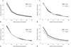
The total, corneal, and internal aberrations did not show any significant difference between the groups at any time point (Table 4). The entire ocular MTF was not significantly different between the groups at 1 or 3 months after surgery (Fig. 1). However, at 3 months, internal MTFs in group 1 were significantly better than those in group 2 at spatial frequencies of 20, 25, and 30 cycles per degree (p=0.034, 0.017, and 0.017, respectively).
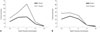
The contrast sensitivity score measured at 3 months postop-eratively under photopic and mesopic lighting conditions showed comparable results between the groups at almost all spatial frequencies (Fig. 2). At 3 months, the PCO grade showed comparable results between the groups (group 1, 0.37±0.60 vs. group 2, 0.38±0.50, p=0.422). The Cochran-Mantel-Haenszel test showed that the use of a CTR was not associated with PCO occurrence (p=0.968).
DISCUSSION
According to the refractive prediction errors that were calculated at 1 month and 3 months after surgery, positive values were observed at all time points and increased as time passed in both groups. That is, the actual postoperative spherical equivalent showed more hyperopic results over time than the predicted spherical equivalent. Especially, co-implantation of a CTR and IOL revealed a greater hyperopic shift, resulting in refractive prediction errors at 3 months after surgery that reached statistical significance.
CTR implantation has been shown to restore vision quality by reducing folds in the posterior lens capsule after IOL implantation.7 Several studies report improvements in the clinical outcomes of cataract surgery after CTR implantation.14 Previous reports regarding eyes with high myopia or multifocal IOL implantation suggested that the use of CTRs yields more precise and consistent refractive outcomes, compared with a control group undergoing cataract surgery without CTR implantation.148 Additionally, the possible effectiveness of CTRs has been suggested even in eyes without zonular instability.9
Postoperative changes in the spherical equivalent are known to relate to IOL decentration and tilt. CTR implantation can avert contracture of the capsular bag, thus preventing IOL decentration.10 A previous study reported that eyes with a CTR had better IOL centration and showed good positioning and stability in the capsular bag, which was demonstrated by a better MTF and minimal aberrations.3 Postoperative changes in the spherical equivalent may also be related to IOL axial shift.11 In our study, structural analysis using a Pentacam showed a statistically significant widening of the ACD after surgery in both groups. Additionally, we observed a greater difference in ACD between the preoperative and postoperative periods in group 1 than in group 2, although the difference did not reach statistical significance. In accordance with the greater difference in ACD between the preoperative and postoperative periods, an axial backward shift of the IOL could be expected in eyes that underwent co-implantation. Thus, considering that axial IOL position is known to be a major factor affecting postoperative refractive changes, we speculated that the backward movement of the IOL in the capsular bag was associated with the widening of the ACD, which probably was caused by subtle movement of the IOL with the aid of the CTR, resulting in the hyperopic refractive change.
The tendency for hyperopic shift could be caused by the IOL design with its posteriorly vaulted optic. The aberration-free single-piece hydrophobic acrylic IOL used in this study has greater surface durability and a greater elasticity modulus. Moreover, the volume of the haptics of the lens is relatively bulky, compared with other IOLs. Additionally, the IOL morphologically features step-vaulted haptics that push in the optic posterior direction for direct contact with the capsular bag.12 That is, the haptics are offset anteriorly with respect to the optic body to enable consistent posterior movement of the optic under haptic compression.12 Although we could not determine the exact configuration of the CTR and IOL in the capsular bag, the unique characteristics of the IOL may contribute to posterior movement of the IOL under conditions of co-implantation with a CTR. Accordingly, we presume that, with a preplaced CTR, posterior movement of an aberration-free IOL in the capsular bag could be consolidated, resulting in the postoperative hyperopic refractive outcome. However, the differences in ACD between the preoperative and postoperative periods did not reach a significant level in either group. Moreover, our study does not provide a precise symmetric rhexis cover for the IOL optic, which is known to affect the effective lens position.13 A longer follow-up duration with more cases would be beneficial to demonstrating the effectiveness of co-implantation of a CTR and IOL.
Eyes that underwent CTR implantation showed no significant superiority in the measurement of postoperative ocular aberration. Moreover, coma of internal aberration, which presents as intra-capsular IOL decentration, was not significantly different between the groups. Regarding the contrast sensitivity score measured 3 months after surgery, both groups demonstrated comparable results at almost all spatial frequencies. However, the group that underwent co-implantation showed a better internal MTF at spatial frequencies of 20, 25, and 30 cycles per degree at 3 months after surgery. The co-implantation of a CTR and IOL is known to show a better MTF by providing greater IOL centration.3 On the other hand, internal aberration is known to be affected by PCO.14 In our study, regarding PCO at 3 months postoperatively, implantation of a CTR showed no significant difference with IOL-only implantation.
In conclusion, implantation of a CTR and aberration-free single-piece hydrophobic acrylic IOL allowed for improvement of the internal MTF; other aberration values were comparable. Co-implantation of the CTR and IOL induced more-hyperopic refractive outcomes than implantation of the IOL alone. The effect of the CTR may not be consistent across different designs and materials of the IOL. Therefore, we suggest reducing the IOL power by 0.5 D lower than the calculated goal diopter for planning the co-implantation of the CTR and aberration-free single-piece hydrophobic acrylic IOL.


 XML Download