

 PDF
PDF ePub
ePub Citation
 Print
Print


INTRODUCTION
Hepatocellular carcinoma (HCC) is the third common leading cancer in Korea.1 And hepatectomy is considered to be the gold standard treatment for the patients who preserved liver function. Recently, many of HCC patients are diagnosed in the state of Child-Pugh grade A due to the advance in diagnosis and the efficient and organized application of nationwide surveillance program in Korea.23 Even the overall survival (OS) is satisfied, nevertheless, the high incidence of tumor recurrence remains a challenge.1 And the OS was mainly influenced by tumor recurrence. Minagawa, et al.4 reported that the OS after second hepatectomy was siginificantly poor in patients whose cancer recurred within 12 months (early recurrence) after first hepatectomy compared to those whose cancer recurred more than 12 months (late recurrence) after initial operation. From that point of view, disease-free survival is an important prognostic factor for survival. The mechanism of high incidence of recurrence for HCC patients and the risk factors leading to early recurrence have not been clearly defined.
An increased evidence of relationship between systemic inflammation and tumor biology has been reported. Neutrophil-to-Lymphocyte Ratio (NLR) was considered as a reliable parameter for monitoring and evaluation of systemic inflammatory response.5 And NLR is easily and repeatedly obtained from peripheral blood. Up to date, NLR has been investigated for its prognostic role in HCC after curative resection,678 transcatheter arterial chemoembolization (TACE),9 radiofrequency ablation (RFA),10 liver transplantation (LT),111213 hepatic arterial infusion chemotherapy,14 and sorafenib monotherapy.15 Since the prognostic value of NLR at Milan criteria level was not inclusive, we assessed the impact of elevated NLR on the outcomes of HCC patients, confirming to Milan criteria after initial hepatectomy during a long follow-up period.
MATERIALS AND METHODS
Patients' inclusion criteria and characteristics
Between March 2001 and December 2011, a total of 621 patients with HCC underwent hepatectomy at the Division of Hepatobiliary Surgery and Liver Transplantation, Department of Surgery, Ajou University School of Medicine, Suwon, Korea. The inclusion and exclusion criteria were as follows: 1) Milan criteria (based on radiological imaging): solitary tumor ≤5 cm in diameter and up to three nodules ≤3 cm in diameter; without any invasion into the major portal/hepatic vein branches and no extrahepatic metastasis. 2) Primary treatment was surgery resection. Patients with preoperative therapy, such as TACE, RFA, second surgical resection or LT for recurrence were excluded. 3) Patents who had clinical evidence of infection or white blood cell count more than >10×109/L, immune system disease, hematology disease, used hematology influenced drugs within 1 month and patients with other cancer were excluded from this study. Finally, two hundred and thirteen patients which consisted of 166 males and 47 females with a median age of 53 years (range 20–79 years) were enrolled in this study. Serum hepatitis B surface antigen (HBsAg) was positive in 162 cases, and anti-Hepatitis C viral antibody in 15 cases.
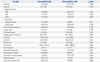
Clinicopathologic characteristics of enrolled patients including gender, age, etiology of cirrhosis, comorbidities [diabetes mellitus (DM), hypertension], surgical procedure (anatomical resection and non-anatomical resection), albumin, total bilirubin, aspartate aminotransferase (AST), alanine aminotransferase (ALT), indocyanine green retention rate at 15 min (ICG R15), alpha-fetoprotein (AFP), tumor size, intrahepatic metastasis, microvascular invasion, microscopic resection margin, capsular invasion and tumor recurrence were collected and detailed in Table 1. In our institute, the procedures of hepatectomy were as follows: anatomical resection: right/left hemihepatectomy or central bisectionectomy (31 cases, 14.6%), left lateral sectionectomy, right anterior/posterior sectionectomy (42 cases, 19.7%) and one segmentectomy (43 cases, 20.2%). Non-anatomical resection: wedge resection (97 cases, 45.5%). All data were retrospectively analyzed from the database of our institute which was prospectively collected.
Follow-up strategy, recurrence and treatment pattern
The median follow up period was 48 months (range 1 to 156 months). And the clinical follow-up strategy followed a strict and stable protocol. All patients in our institute were screened monthly during the first 6 months after surgery for tumor marker such as AFP and laboratory data such as complete blood count (CBC), AST, ALT, albumin, total bilirubin. An enhanced computed tomography scan was performed at 6-month intervals. If recurrence was suspected, additional investigations such as magnetic resonance imaging or positron emission tomography-computed tomography were performed.
The recurrent pattern was defined as early recurrence (recurred within 12 months after initial hepatectomy) and late recurrence (recurred beyond 12 months after initial hepatectomy).
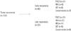
Five types of treatment were adopted to treat the tumor recurrence after initial hepatectomy, such as re-resection, TACE, RFA, salvage LT and conservative management. Finally, fourteen patients with good future liver function were eligible for repeat resection; twenty eight patients received RFA; forty two patients received TACE; ten patients who with multi recurrence tumor and/or stayed at end stage of liver cirrhosis disease received salvage LT; and thirteen patients were received conservative management. The treatment methods for recurrence tumor are listed in Fig. 1.
To determined optimal cutoff of continuous variables
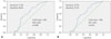
CBC and blood biochemical examination were carried out in all patients within 4 days before operation. The NLR was calculated from the differential ratios by dividing neutrophil to lymphocyte ratio in CBC. In the present study an receiver operating characteristic (ROC) curve indicated that the value of 1.505 was the best cutoff value of NLR for recurrence after hepatectomy. The area under the curve (AUC) was 0.643. The p value was 0.005 (Fig. 2A). The optimal cutoff values of albumin, AST and tumor size for patients overall recurrence-free survival (RFS) after hepatectomy were 4.15 g/L (AUC=0.648, p=0.000), 36.5 U/L (AUC=0.600, p=0.012), and 2.65 cm (AUC= 0.627, p=0.001), respectively. In the 107 recurrence patients, the value of 1.945 determined by ROC curve was the best cutoff value of NLR for early recurrence; the AUC was 0.643, and the p value was 0.005 (Fig. 2B).
The total patients were divided into two groups by optimal cutoff value of NLR (1.505); normal NLR group and elevated NLR group. There were no difference between the two groups in the characteristics, such as gender, age, etiology of cirrhosis, hypertension, non-anatomical resection, albumin, total bilirubin, AST, ALT, ICG R15, AFP, tumor size, microvascular invasion and capsular invasion. As expected, elevated NLR group had a more tumor recurrence patients, as indicated by more patients with intrahepatic metastasis. More patients with DM were found in the elevated NLR group (Table 1).
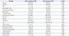
The recurrent patients were divided into two subgroups by recurrence time: early recurrence group (recurred within 12 months) and late recurrence group (recurred beyond 12 months). The OS was significantly different between the early and late recurrence group (p=0.000). The early recurrence group had relatively more patients with elevated NLR, intrahepatic metastasis, microvascular invasion, high grade of cirrhosis (grade 4) and serum HBsAg-positive. On the contrary, the late recurrence group had more patients with high level of AST and significantly longer OS (Table 2).
Statistical analysis
Continuous variables were expressed as mean±standard deviation and compared using Student t test. Categorical data were presented as frequency and analyzed by using the Fisher χ2 test. Multivariate analysis was performed with Cox regression for significant variables on univariate analysis. The Kaplan-Meier method was used to analyses the OS and RFS, and compared using the log-rank test. All statistical analyses were performed with the statistical package for the social sciences (SPSS) 19.0 (SPSS Inc., Chicago, IL, USA). Confidence intervals (CI) were constructed at 95%, and p value <0.05 was considered statistically significant.
RESULTS
Survival outcomes
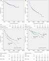
The 1, 3, and 5 year cumulative OS was 95.2%, 86.9%, and 81.3%, respectively (Fig. 3A) whereas the 1, 3, and 5 year cumulative RFS was 79.9%, 60.5%, and 50.1%, respectively (Fig. 3B). The total patients were divided into two groups by optimal cutoff value of NLR (1.505); normal NLR group (NLR<1.505) and elevated NLR group (NLR≥1.505). The RFS time of elevated NLR group was significantly shorter than that of the normal NLR group (the log-rank test, p=0.010) (Fig. 3C). The 1, 3, and 5 year of RFS in normal NLR group was 88.9%, 69.8%, and 57.0%, respectively. In contrast, the 1, 3, and 5 year of RFS in elevated NLR group was 74.0%, 53.5%, and 39.9%, respectively. However, there was no difference in OS between the two groups (the logrank test, p=0.114) (Fig. 3D).
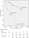
In this cohort study, there were 107 patients suffered from tumor recurrence. The patients who experienced early recurrence had a poor OS (the log-rank test, p=0.000) (Fig. 4). The 1, 3, and 5 year of cumulative OS for the early recurrence group was 78.9%, 46.4%, and 42.5%, respectively. In contrast, the 1, 3, and 5 year of OS for the late recurrence group was 98.5%, 97.0%, and 86.8%, respectively.
Prognostic factors of tumor recurrence in HCC patients within Milan criteria
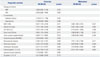
The prognosis factors for RFS and OS of HCC after surgical resection were examined based on the following clinicopathological variables: etiology of cirrhosis (hepatitis B virus, hepatitis C virus), co-morbidities (DM, hypertension), elevated NLR (>1.505), low ablumin (<4.15 g/L), elevated AST (>36.5 U/L), large tumor size (>2.95 cm), multiple tumor number, non-anatomical resection, microvascular invasion, portal vein invasion, intrahepatic metastasis, capsular invasion, high cirrhosis grade (grade 3 and 4),16 and high grade of Edmondson-Steiner (ES; grade 3 and 4).17 The variables of elevated NLR, low ablumin, elevated AST, large tumor size, multiple tumor number, microvascular invasion, portal vein invasion, intrahepatic metastasis and high cirrhosis grade which were significant predictors of RFS analyzed by univariate regression analysis were considered in multivariate regression analysis. And the variables of elevated NLR, microvascular invasion and cirrhosis grade were significant predictors of RFS (Table 3). However, NLR failed to predict OS time analyzed by univariate regression. Univariate regression analysis found that DM, low ablumin, elevated AST, multiple tumor number, microvascular invasion, portal vein invasion and intrahepatic metastasis were significant risk factors for OS. Unfortunately, no risk factor was found by multivariate analysis of OS (Table 4).
Prognostic factors of early recurrence in HCC patients within Milan criteria
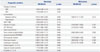
The elevated NLR (1.945), combined with multiple tumor number, mircovascular invasion, portal vein invasion and intrahepatic metastasis were considered as risk factors for early recurrence, when determined by univariate regression analysis. On the other hand, however, microvascular invasion was the only risk factor for early recurrence, when determined by multivatiate regression analysis (Table 5).
DISCUSSION
One and half century ago, the connection of inflammation and malignancy was observed by Rudolf Virchow for the first time18 in leucocytes in neoplastic tissues. An increased comprehension of the relationship between inflammation and cancer was made by experimental and clinical research. Inflammation plays an important role in the development and progression of cancer, including initiation, promotion, malignant conversion, invasion, and metastasis,19 and NLR was considered as a reliable parameter for monitoring and evaluation of systemic inflammatory response.5 Fortunately, NLR could repeatedly and easily be obtained from peripheral blood without costly expenditure. More recently, an increasing number of studies have shown that elevated NLR has adverse prognosis on recurrence and OS for HCC patients after different treatment procedures, such as curative resection,678 TACE,9 RFA10 and LT,111213 hepatic arterial infusion chemotherapy,14 and sorafenib monotherapy.15 In the present study, we further demonstrated that elevated NLR was a prognostic factor of tumor recurrence and early recurrence for HCC patients who meet the Milan criteria after surgical resection, and the optimal cutoff value was 1.505 and 1.945, respectively, calculated by ROC curve (1.505, AUC=0.643, p=0.006; 1.945, AUC=0.641, p=0.015) (Fig. 2).
The underlying mechanism in the association of elevated NLR and adverse outcomes of HCC patients is not clearly defined: recent clinical and experimental researches shed some new light onto their roles. Mano, et al.7 demonstrated that accumulation of tumor-associated macrophages (TAMs) in the HCC lesion is associated with a high NLR after curative resection. TAMs have been reported to be a major component of the tumor inflammatory microenvironment and promote cancer initiation and malignant progression.20 Human TAMs can express various pro-angiogenic factors in tumors, including vascular endothelial growth factor (VEGF)21 and thymidine phosphorylase.22 Peng, et al.23 reported that the count of TAMs was closely related to micro-vessel density in HCC, suggesting that TAMS have a link with HCC angiogenesis and metastasis. The Interleukin (IL)-6 and IL-8 cytokines which are expressed by TAMs in tumors may promote systemic neutrophilia.242526 Mano, et al.7 and Motomura, et al.12 reported a correlation between elevated NLR and upregulation of IL-17 production in peritumoral regions of the liver. The proinflammatory cytokine IL-17 has been demonstrated to foster tumor immune escape27 and promote tumor growth in HCC.28 IL-17 could be indirectly upregulated by promoted expansion of IL-17-producing CD8+ T cell by TAMs in HCC patients.29 In summary, TAMs plays an important role for tumor progression, and NLR could indirectly reflect in peripheral blood of TAMS through the cytokines such as IL-6, IL-8, and IL-17.
In this cohort study, the median follow-up period was 48 months (range 1 to 156 months), and the cumulative 1, 3, and 5 year OS was 95.2%, 86.9%, and 81.4%, respectively (Fig. 3). However, the RFS remains unsatisfactory. Total 107 patients experienced tumor recurrence. The cumulative 1, 3, and 5 year RFS was 86.1%, 68.7%, and 44.2%, respectively (Fig. 3). When patients were divided into two groups by optimal cutoff value of NLR (1.505), and the elevated NLR group was found to have more tumor recurrence patients (p=0.000) and more patients with intrahepatic metastasis (p=0.014). As expected the variable of elevated NLR (1.505) together with microvascular invasion, low albumin, and high grade of cirrhosis were found to be significant predictors for RFS analyzed by multivariate regression analysis (Table 3). Our present finding is consistent with previous reports of Sumie, et al.30 and Poon, et al.31 that microvascular invasion and cirrhosis were independent risk factors for tumor recurrence.
Minagawa, et al.4 reported that early recurrence patients whose cancer recurred within 12 months after initial hepatectomy have a worse OS than late recurrence, it is believed that intrahepatic metastasis represents early recurrence and is associated with vascular invasion and subsequent intrahepatic venous spread.32 Similar outcome was also found in our present study. In our experience of 40 early recurrence patient treatments, 90% of (36/40) cases developed to multiple recurrence and the outcome was unsatisfactory even when treated by salvage LT. The OS of early recurrence patients was significantly worse than the late recurrence (p=0.000). The NLR was found to be an independent risk factor [hazard ratio (HR), 1.678, p=0.004] for early recurrence in univariate regression analysis with 1.945 cutoff value of NLR (Table 5). As mentioned above, elevated NLR showed a strong relationship with tumor progression: underlying mechanism might be that angiogenesis and metastasis of the tumor cell in the tumor lesion are promoted by TAMs.
In our present study, more patients with DM were found in elevated NLR group. DM is considered to be a risk factor for OS by univariate analysis but it failed by multivariate analysis. The relationship between systematic inflammatory and DM has been proved by earlier studies33 that systematic inflammation may induce insulin resistance and decrease insulin secretion. However, a question of whether DM is an adverse factor for HCC remains controversial. Two resent studies3435 identified that DM is an independent factor for poor survival in HCC patients who underwent curative therapy. On the contrary, however, Poon, et al.36 reported that DM did not seem to influence either the risk of recurrence or long term OS after resection of HCC.
In the present study, NLR failed to predict for prognosis for OS of the HCC patients confirming to Milan criteria after surgical resection. The potential reasons maybe that the inclusion criterion of this study was Milan criteria and the patients preserved liver function well. The primary good liver function would facility to choose an optimal treatment for tumor recurrence. Fig. 1 shows the types of treatment for tumor recurrence patients. Fourteen patients received repeated resection and the remaining patients who were not suitable for resection received TACE, RFA, systemic chemotherapy, oral chemotherapy, supportive treatment and/or salvage LT. The 1, 3, 5-year OSs of repeated resection were 100%, 92.3%, and 92.3%, respectively. However, the 1, 3, 5-year OS of the remaining patients were 91.2%, 78.1%, and 67.5%, respectively (p=0.022). Therefore the repeated resection seems to significantly prolong the OS of HCC recurrent patients and 8 of 14 patients belonged to the elevated NLR group.
In conclusion, the inflammatory biomarker of preoperative elevated NLR indirectly reflect vascular infiltration factors in the tumor microenvironment, and preoperative elevated NLR is useful in predicting tumor recurrence and early recurrence after surgery resection in patients with HCC confirming to Milan criteria.
 XML Download