

 PDF
PDF ePub
ePub Citation
 Print
Print


INTRODUCTION
Sepsis is a devastating clinical condition characterized by systemic inflammation occurring during a severe infection. Severe sepsis and septic shock are leading causes of morbidity and mortality in the intensive care unit (ICU).12 The reason that some patients die while others survive similar insults is partially understood, although some of this patient outcome variability may be caused by genetic variation. Several reports have confirmed that susceptibility and outcomes from infectious disease are inheritable.345 Indeed, numerous studies have demonstrated that innate immune responses to pathogens exhibit interindividual variability strongly influenced by genetic factors, which may affect disease susceptibility and severity.6789101112 In the pathophysiology of sepsis, the innate immune system is activated prior to the acquired immune system: cells of the innate immune system, such as monocytes, macrophages, and neutrophils, represent the front line of the host response to infection, invasion, and injury.13
High mobility group box 1 (HMGB1) is a highly conserved, ubiquitously expressed protein, originally discovered as a nonhistonal nuclear DNA binding protein.141516 It is located on chromosome 13 and present in the nuclei and cytoplasm of nearly all cell types.17 In response to infection and injury, HMGB1 is secreted by activated innate immune cells and/or passively released by necrotic or damaged cell.18 Some studies have demonstrated that HMGB1 is a late mediator of sepsis in amplifying the inflammatory response and that serum/plasma HMGB1 concentrations are elevated in patients with sepsis.1920 Accumulating evidence supports a central pathogenic role for HMGB1 in the pathogenesis of sepsis and multiple organ dysfunction syndromes.21222324
The past decade has witnessed important advances in the understanding of genetic polymorphisms in sepsis; numerous studies have identified that these sepsis-related genetic polymorphisms are associated with severity and/or outcomes.1112 Although, several studies reported the clinical relevance of HMGB1 genetic variation,252627 there are limited data on the relationship between single nucleotide polymorphisms (SNPs) of HMGB1 and clinical outcomes in patients with sepsis. Moreover, the characteristics of these polymorphisms differ according to ethnicity, although few data have been reported in the Korean population. Therefore, we hypothesized that SNPs of HMGB1 could influence clinical outcomes in Korean patients with sepsis.
In this study, we genotyped a SNP of known genetic variants within HMGB1 in patients diagnosed with sepsis (including severe sepsis and septic shock), and analyzed its relationship with various clinical parameters, including disease severity and prognosis. We also investigated the relationship between this HMGB1 polymorphism and serum concentrations of HMGB1 and various cytokines.
MATERIALS AND METHODS
Study subjects
Inclusion criteria were adult patients diagnosed with sepsis, including severe sepsis and septic shock. There was no exclusion criterion. In total, 212 patients were enrolled from March 1, 2011 to October 31, 2012. All patients were >20 years of age [median 67.5 (range 29-95) years, M:F=149:63] and had been admitted to the ICU of a Asan Medical Center (Seoul, Korea). Sepsis, severe sepsis, and septic shock were defined using American College of Chest Physicians/Society of Critical Care Medicine.2829 All patients were managed according to therapeutic recommendations based on early goal-directed therapy and lung-protective ventilator strategy.2930 Survivors were defined as patients who had survived for 28 days after ICU admission. The study objectives and procedures were fully disclosed, and a case report form for this study was completed. All data were collected from the medical records and laboratory and radiographic findings in all patients. This study was approved by the Institutional Review Board (IRB) of the Asan Medical Center (2012-0878). Informed consent was confirmed by the IRB, and written informed consent was obtained from all study participants or their surrogates.
Data collection
The following data were gathered from the medical records of patients: age, gender, the primary cause of sepsis on initial admission, underlying comorbidities, duration of mechanical ventilation, and lengths of stay in the ICU and hospital. All patients were categorized as sepsis, severe sepsis, and septic shock on ICU admission. Acute Physiology and Chronic Health Evaluation (APACHE) II and Sequential Organ Failure Assessment (SOFA) scores were calculated on the sampling day for this study.3132 We also identified the causative pathogen for sepsis in patients with positive blood culture, and classified them accordingly. In addition, we recorded laboratory data (complete blood count, lactate, C-reactive protein, procalcitonin) on sampling day, and surveyed for the presence of neutropenia (absolute neutrophil count <1500/mm3).
SNPs genotyping
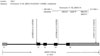
Blood samples were drawn within 24 hrs after ICU admission. Genomic DNA was isolated from 5 mL of ethylenediaminetetraacetic acid (EDTA)-anticoagulated venous blood by the standard method using proteinase K and phenol/chloroform extraction. SNP data for the HMGB1 gene [chromosome 13, position 29930000-29939000 (9 kb total)] was obtained from the HapMap data (version 2, release 21) for 45 unrelated Han Chinese individuals from Beijing, China (CHB) and 44 unrelated Japanese individuals from Tokyo, Japan (JPT) samples. From the database, a total of three SNPs with a minor allele frequency >0.05 (rs1045411, rs3742305, rs2249825) were identified in HMGB1 (excluding 5'- and 3'-flanking regions) and selected for genotyping; all are common SNPs with a minor allele frequency >0.05. Among the three SNPs, rs1045411 located in the 3'-untranslated region (Fig. 1) was chosen for further analyses, because this SNP seemed to show the most significant difference in allele frequencies between patients and normal healthy persons (χ2 test, p<0.05) with the lowest p value and highest odds ratio, compared to the other two SNPs (data not shown). Genotypes for rs1045411 in all patients were determined by Sanger sequencing. The target region of HMGB1 was amplified using forward primer 5'-TGGAAGTGGGAGGCAAT TTA-3' (HMGB1_1045411_F) and reverse primer 5'-TGCTGT GCAAA GGTTGAGAG-3' (HMGB1_1045411_R). Amplification conditions were one cycle of 95℃ for 7 min, 30 cycle of 95℃ for 30 s, 56℃ for 30 s, 72℃ for 1 min, plus one cycle of 72℃ for 5 min. Amplification conditions were one cycle of 95℃ for 7 min, 30 cycles of 95℃ for 30 s, 56℃ for 30 s, and 72℃ for 1 min, plus one final cycle of 72℃ for 5 min. Amplified products of 303 bp were confirmed by agarose gel electrophoresis and purified by treating with Exo-SAP (10:1 U ratio mixture of exonuclease I and shrimp alkaline phosphatase, USB corp., Cleveland, OH, USA) at 37℃ for 15 min, 80℃ for 15 min, and a 4℃ hold. Sequencing was carried out using an ABI 3730XL sequencer by Cosmogenetech, Seoul, Korea. Briefly, sequencing amplifications were performed using the BigDye terminator (ver. 3.1) cycle sequencing kit (Applied Biosystems, Foster City, CA, USA). PCR products of 30-90 ng were used as templates and the cycling reaction consisted of one cycle of 95℃ for 90 s, 25 cycles of 95℃ for 30 s, 50℃ for 5 s, and 60℃ for 4 min, followed by a 4℃ hold with HMGB1_ 1045411_F and HMGB1_1045411_R. After purifying the cycle sequencing reaction products with magnetic beads (MagneSil GREEN, Promega, Madison, WI, USA), the sequencer instrument was run according to the manufacturer's protocol.
Serum HMGB1 and cytokines measurement
Blood samples for cytokine measurement were immediately centrifuged, and serum was frozen -80℃ until the assay could be performed. We measured serum HMGB1 levels and several inflammatory [interleukin (IL)-1β, IL-6, IL-17, and tumor necrosis factor (TNF)-α] and anti-inflammatory (IL-10) cytokines from 190 patients, because we did not have sufficient quantities of samples; we could not measure serum levels of HMGB1 or other cytokines in 22 patients. The serum HMGB1 level was measured using a sandwich enzyme-linked immunosorbent assay (HMGB1 ELISA; IBL International, Hamburg, Germany). Intra-assay and inter-assay coefficients of variation (CV) were 5.5-13.7% and 7.6-13.7%, respectively. Also, functional sensitivity was 0.2 ng/mL (lowest HMGB1 concentration with ≤20%). The serum levels of cytokines were determined by Luminex® Performance Assay multiplex kits (R&D Systems, Minneapolis, MN, USA). Analyses were performed in accordance with manufacturer's protocol.
Statistical analysis
The genotype frequencies of three SNPs were tested for Hardy-Weinberg equilibrium using Haploview (v4.2) (http://www.broadinstitute.org/haploview). No significant deviation from Hardy-Weinberg equilibrium was observed. Continuous variables are expressed as median with range. Student's t-test or the Mann-Whitney U-test, depending on the normality of distribution, were used to compare continuous variables between two groups, and the Kruskal-Wallis test was used for comparisons among three groups. Also, the χ2 and Fisher's exact tests (for small numbers) were used to compare categorical variables. To evaluate whether these SNPs could influence clinical outcomes, univariate and multivariate Cox regression analyses were performed including all clinical data from medical records, with adjustment for age, gender. Survival curves were obtained with using the Kaplan-Meier method with the log-rank test. All statistical analyses were performed using the Statistical Package for the Social Sciences (ver. 19.0; IBM, Armonk, NY, USA). A two-tailed p value<0.05 was considered to indicate significant difference.
RESULTS
Baseline characteristics of the study subjects
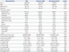
The clinical characteristics and comparisons of survivors and non-survivors are presented in Table 1. The median durations of stay in the ICU and hospital were six (range 1-104) and 20 (1-830) days, respectively. The ICU and hospital mortality rates were 29.2% and 36.3%, respectively. In total, 150 patients (70.8%) were diagnosed with septic shock; 143 (67.5%) received ventilator care. Also, 86 (40.6%) and 6 (2.8%) patients had renal failure and neutropenia on admission, respectively. The most common primary site of infection was respiratory tract (57.5%), and 72 patients (34.0%) had positive blood cultures (Table 1). The underlying diseases present in all the patients are shown in Table 2. Oncologic and chronic heart diseases were the most common.
Analyses according to rs1045411 genotypes
When we performed rs1045411 genotyping, the proportion of patients groups by genotype was GG (71.2%), GA (26.4%), and AA (2.4%), respectively. There was no significant difference in other clinical characteristics and outcomes among these three groups (data not shown). When patients were categorized into those with GG (n=151) versus those with GA+AA (n=61), the proportions of A alleles among patients with septic shock, severe sepsis, and sepsis were 31.3%, 23.7%, and 20.8%, respectively. However, there were no significant differences in this allele according to sepsis severity (data not shown).
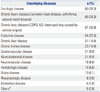
As underlying comorbidities or primary site of infection could influence clinical outcomes, we also evaluated whether the categories of SNPs (GG and GA+AA) had different implications according to specific comorbid conditions or primary site of infection. In patients with chronic lung diseases or diabetes, patients with the variant A allele had higher positive blood culture rates than those with GG genotype. This allele was also associated with a higher Gram-positive blood culture rate in male patients and Gram-negative culture rate in female and non-survivors. Patients with chronic renal disease had a higher incidence of shock. In cytokine analysis of patients with chronic lung diseases, patients with the variant A allele had significantly higher levels of IL-1β, IL-6, IL-10, IL-17, and TNF-α than those with GG genotype. In analysis of those with diabetes, patients with the variant A allele had higher IL-17 levels. In patients with sepsis caused by respiratory tract infection, patients with this allele had higher levels of IL-10 and IL-17 than those with GG genotype (Table 3).
Influence of rs1045411 genotypes on patient survival
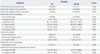
A univariate Cox proportional hazards model was performed to evaluate the influence of the variant A allele of rs1045411 on survival in all patients, as well as some subgroups that showed significant findings. However, the A allele was not significantly associated with survival (Table 4), and thus, we could not clarify a significant Kaplan-Meier survival curve for this allele (data not shown).
DISCUSSION
Although HMGB1 has been investigated extensively as an extracellular protein with cytokine-like activity that plays a pivotal role as a late mediator of sepsis,192022232433 few studies have suggested a role for HMGB1 genetics in critically ill patients.2526 To our knowledge, this is the first reported study to investigate the clinical relevance of rs1045411, one of the known HMGB1 SNPs, in patients diagnosed with sepsis, severe sepsis, and septic shock in Korean populations. Because of the lack of reported data regarding any association between this SNP and various cytokines, our study also assessed this relationship. In view of our results, the rs1045411 genotype likely plays a distinct role in the pathophysiology of sepsis in the Korean population.
In this study, there was no significant difference in the clinical characteristics examined according to rs1045411 genotype in all patients; however, the variant A allele of the rs1045411 polymorphism was associated with higher positive blood culture rates and elevated cytokine levels, particularly in patients with chronic lung diseases or diabetes as comorbidities (Table 4). These findings suggest that patients with the variant A allele might experience more severe inflammatory responses under specific conditions. Patients without these comorbidities, however, showed no significant difference in blood culture rates or cytokine levels between the GG genotype and the GA+AA genotype. Although we could not determine the mechanism underlying the association of this polymorphism with various causes of chronic lung diseases or diabetic control, our rs1045411 results might be more useful for predicting sepsis severity in patients with these comorbidities. It is also important to evaluate the impact of this polymorphism on prognosis through further analysis of a larger number of sepsis patients with these conditions.
In studying human HMGB1, Kornblit, et al.25 identified six SNPs (rs1412125, rs41369348, rs2249825, rs1060348, rs3472305, rs41376448), and reported significant associations of rs1060348 and rs41369348 with early and late mortality in patients who were diagnosed with systemic inflammatory response syndrome in Denmark.26 Zeng, et al.27 studied the clinical relevance of HMGB1 in patients with major trauma in South China. Among the three SNPs (rs1412125, rs2249825, rs1045411) selected using the HapMap database, they reported that rs2249825 and the haplotype TCG might be used as a relevant risk factor for the development of sepsis and multiorgan dysfunction syndrome in patients with major trauma. Compared with these reports, our study enrolled patients with sepsis (including severe sepsis and septic shock) and evaluated the clinical utility of genotype using subgroup analysis and cytokine level measurements. In addition, severity would have differed among the three studies because additional mechanical ventilation, higher SOFA and APACHE II scores, and longer ICU stays were included in our report.
This study had several limitations. First, we expected that there would be significant differences in clinical outcomes (e.g., ICU and hospital length of stay, duration mechanical ventilation, organ dysfunctions, and causative organisms) according to rs1045411 polymorphism; however, we found no such difference, possibly due to the small sample size. Second, this study was performed in only one hospital and the sample size was small; thus, it may not be representative of the general population of Korea. However, all patients were managed according to the "Surviving Sepsis Campaign Guideline for Management of Severe Sepsis and Septic Shock,"29 and standardized procedures in this hospital were performed as a rule to reduce any confounding factors related to the outcome of sepsis. Third, we could not measure serum HMGB1 levels using Western blot analysis, which shows higher values than those using ELISA. Fourth, the number of total enrolled patients was not based on our preliminary sample size analysis.
In conclusion, we investigated the clinical relevance of rs10 45411 genotypes, one of the known HMGB1 SNPs, in patients diagnosed with sepsis, severe sepsis, or septic shock. In this study, patients with the variant A allele had higher positive blood culture rates and higher cytokine levels than did those with the GG genotype under some specific conditions. Although this polymorphism had no significant impact on survival, patients with the variant A allele might experience an aggravated inflammatory response to these conditions. Also, there may be interactions between our studied haplotype and other genes involved in immune responses. Large-scale studies are required to determine the clinical significance of this polymorphism.

 XML Download