

 PDF
PDF ePub
ePub Citation
 Print
Print


INTRODUCTION
Peritoneal dialysis (PD) is a well-established and effective therapy for patients with end stage renal disease (ESRD). However, PD is underused in clinical field, despite having several advantages compared with hemodialysis (HD), which include the preservation of residual renal function, hemodynamic stability, better survival in the early dialysis period, and better quality of life.1234 The availability of timely PD catheter insertion, low rate of complication related with procedure and the low rate of catheter failure with effort of nephrologists are important requisites that will increase the utility of PD and reduce patient morbidity.156 The blind percutaneous PD catheter insertion by nephrologists shows similar outcome to catheter insertion performed by surgeons, and it is advantageous in that it can timely initiate dialysis therapy and does not require an operating room.56789101112
The trocar and cannula method was first practiced and widely used by nephrologists for PD catheter insertion. While it is simple to perform and easy to learn, serious complications including bowel perforation and major bleeding can occur, because sharp and thick trocar is used to blindly access to the abdominal cavity.11013 Consequently, for several years, we have performed a modified PD catheter insertion technique to reduce the number of complications associated with this procedure.
In this study, we conducted a retrospective analysis of patients who had undergone PD catheter insertion using a modified percutaneous catheter insertion technique and compared the outcomes with those who had their PD catheters inserted using the conventional trocar and cannula method. The purposes of this study are; first, to describe our modified percutaneous PD catheter insertion method, and second, to show its clinical advantages compared with conventional trocar and cannula method, based on experience from a single center.
MATERIALS AND METHODS
Patients
We retrospectively analyzed patients who underwent percutaneous PD catheter insertion by nephrologists at a single center, Yonsei University College of Medicine, Severance Hospital, Seoul, Republic of Korea, from January 2006 to September 2012. A total of 107 patients had their PD catheter inserted by modified method (group M) and 81 patients by conventional trocar and cannula method (group C). Patients with previous abdominal surgery or previous abdominal infection, which can lead to adhesion, were excluded from PD catheter insertion by nephrologists. The patients who had undergone emergency procedures in intensive care unit were also excluded from both groups; thus, 25 patients were excluded in group M and 15 patients were excluded in group C. Catheter insertion failure did not occurr in any of the patients. The study was approved by the Institutional Review Board of Severance Hospital.
Covariates
The patients were evaluated for their baseline characteristics, including demographics, medical history, drug history, and laboratory examination. The complication rate, procedure time, Numeric Pain Intensity Scale (NPIS) score (which is to evaluate pain in subjective number from 0 to 10) immediately after procedure, time needed for ambulation, and hospitalization period were compared between group M and group C. Then, the complications were subcategorized into intra-operational bowel injury, PD fluid leakage, catheter tip migration, catheter obstruction, intra-abdominal hemorrhage, complicated hemorrhage, and wound infection to compare the complication in more detail. Complications that occurred during day 0 to day 7 were defined as early complication, while those during day 8 to 3 months were defined as late complications. Intra-abdominal hemorrhage was defined as red blood cell counts >10000 cells/µL in PD fluid. And, complicated hemorrhage was defined as hemorrhage that led to additional procedures that delayed PD initiation.
PD catheter insertion method
A straight, double cuffed Tenckhoff catheter was used. Immediately after PD catheter insertion, continuous ambulatory peritoneal dialysis (CAPD) was performed in all patients without break-in procedure.14 PD catheter insertion method was performed as follows;
Conventional trocar method for PD catheter insertion
A 2-cm vertical incision was made over the lateral rectus muscle just above the level of the umbilicus, on the side opposite to the dominant hand and either above or below the belt line. The subcutaneous tissue was freed from the anterior rectus sheath. A 14-gauge angiocatheter was inserted in a perpendicular direction into the abdomen, and the needle was removed, leaving the plastic tube. Two liters of 1.5% glucose solution for dialysis were infused into the peritoneal cavity through the plastic tube. The Tenckhoff catheter introducer trocar was inserted obliquely through the rectus in the direction of the left pelvic gutter. The left pelvis is preferred because the bowel will tend to push the catheter into place as it undergoes normal peristalsis. This track created through the rectus muscle was sequentially dilated. The PD catheter with a stylet was then placed through the introducer sheath and was directed toward the left pelvis. The catheter is introduced into the peritoneal cavity until the deep antimicrobial cuff is located within the rectus muscle (just superficial to the posterior sheath). Then, stylet is removed from the PD catheter. The catheter was secured by pursestring sutures around the deep cuff. Inferolaterally to the first skin incision, either above or below the patient's belt line, a second, small, horizontal skin incision was made. This incision is made caudal and parallel to the floor to allow gravity drainage of any subcutaneous fluid. The nephrologist then tunneled the PD catheter from the first to the second skin incision, ensuring that the second superficial antimicrobial cuff was placed deep within the subcutaneous tissue between the two skin incisions. The PD catheter insertion site was closed with nylon suture.
Modified method for PD catheter insertion
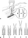
Preparation to free anterior rectus sheath and infusion of dialysis solution were identical as conventional trocar method. The patient's abdomen was carefully palpated to avoid major vessels and confirm vagility of the bowel. After 0.4 cm vertical incision of anterior rectus sheath, a mosquito hemostat was used to spread through rectus muscle, and puncture posterior rectus sheath and peritoneum (Fig. 1). After confirming enough opening with inner nail of trocar, a Tenckhoff catheter with a stylet was introduced into the deep pelvis, aiming at the angle of 10 degrees off the perpendicular toward the patient's coccyx. Then, stylet was removed to confirm the function of Tehnckhoff catheter. If the Tenckhoff catheter didn't ensure the expected flow, the catheter was reinserted with stylet until proper function was acquired. While the patient tensed the abdominal wall, the deep cuff was pushed into the rectus muscle immediately below the anterior rectus sheath. The catheter was secured by pursestring sutures around the deep cuff. After performing second incision, the procedure was identical to the conventional method.
CAPD maintenance
PD was performed with 500 mL of 1.5% glucose dialysis solution and heparin (1000 U/L), every 6 hours for the first 3 days in supine position. All patients were on absolute bed rest during the first 3 days. After 3 days, the PD dialysis dosage was adjusted according to patients' conditions. Patient training was performed during 7-14 days after CAPD catheter insertion by CAPD nurse and the patients were acquainted with the apparatus afterwards.
Statistical analysis
All variables were analyzed using SPSS for Windows version 18.0 (SPSS Inc., Chicago, IL, USA). The categorical data were presented as percentage of the number of patients, and the continuous data were presented as mean±standard deviation (SD). Statistical differences between the groups were assessed using chi-square test for categorical data and Student's t-test for continuous data. A p value<0.05 was regarded statistically significant.
RESULTS
The baseline characteristics of two groups are shown in Table 1. There were no difference between the two groups regarding mean age, gender, body weight, body mass index, number of hypertension patients, use of anti-coagulation agent and estimated glomerular filtration rate. However, the proportion of patients with diabetes mellitus was significantly higher in group M (56.10%) than in group C (31.82%) (p=0.003), and the use of anti-platelet agents was significantly higher in group M (40.24%) than in group C (21.21%) (p=0.012).
The complications related to PD catheter insertion procedure were investigated (Table 2). The overall complication rates were not significantly different between two groups. However, when the complications were subdivided to early and late complications, where an early complication was defined as an event occurring from day 0 to day 7 after the catheter insertion procedure, and a late complication was defined as an event occurring from day 8 to 3 months after the PD catheter insertion procedure, the early complication rate in group M (1.2%) was significantly lower than in group C (19.7%) (p<0.001). One hemorrhagic event, in which the red blood cell count was >10000 cells/µL in PD fluid during routine analysis, occurred in group M (1.2%) during early complication period. However, in group C, PD fluid leakages (3.0%), catheter obstructions (4.6%), and hemorrhages (12.1%) occurred during early complication period. The rate of hemorrhage after PD catheter insertion was significantly lower in group M (1.2%) than in group C (12.1%) (p=0.010). Complicated hemorrhagic events which lead to additional procedure or delay in PD initiation, occurred more frequently in group C (4.6%) than group M (0%), but it was not statistically significant. There was no significant difference in late complication between group M (28.0%) and group C (30.3%).
PD catheter revision events associated with the complication of PD catheter insertion were assessed during early and late complication periods (Table 3). There was no significant difference in total PD catheter revision rate between the two groups. However, the PD catheter revision rate in early complication period was significantly lower in group M (0%) compared with group C (6.1%) (p=0.024). All PD catheter revision events in group M occurred during late complication period and caused by catheter obstruction. In group C, four PD catheter revision events occurred during early complication period and one PD catheter revision event occurred during late complication period. In one of four early PD catheter revision events in group C, one patient suffered from hypovolemic shock caused by bleeding, underwent emergent surgery, and was treated in an intensive care unit.
As for patient's convenience and time spent in hospital between two groups, the procedure time (63±16 min vs. 96±19 min, p<0.001), immediate post procedural pain (NPIS; 2.43±1.80 vs. 3.14±2.07, p=0.030), and post procedure days until ambulation (3.95±1.13 days vs. 6.17±1.34 days, p<0.01) were significantly lower in group M than in group C (Table 4). There was no significant difference in total hospitalization period between group M and group C (14.71±7.05 days vs. 13.86±3.7 days, respectively).
DISCUSSION
There are various methods to implant PD catheters, which include open dissection or laparoscopic method by surgeon, the peritoneoscopic method, percutaneous trocar and cannula method or Seldinger method by nephrologists. Each method has its advantages and disadvantages and there is no proven standard method for PD catheter insertion. The advantages of percutaneous PD catheter insertion over surgical insertion include lower catheter-related mechanical and infectious complication rates, higher long-term catheter patency and the absence of needs for surgery and general anesthesia.1115 However, the percutaneous PD catheter insertion technique has not been widely accepted due to the early mechanical complications and the potential risk for bowel perforation associated with its blind puncture method.113 Hence, there have been several attempts to maximize the advantages of percutaneous PD catheter insertion, while minimizing its disadvantages.14
In this study, we assessed the advantages of modified percutaneous PD catheter insertion technique. The patients who had undergone modified percutaneous PD insertion technique suffered fewer complications than those in group C who had undergone conventional trocar. While only one patient in group M experienced complication, four patients in group C suffered complications, which included a massive bleeding that resulted in shock, emergency operation and admission to intensive care unit. As early complications within 1 week after procedure are assumed as complications directly related to the procedure itself, modified percutaneous PD insertion technique appears to be a safer and less traumatic way to insert PD catheter. Rate of revisions due to complications were also in favor of modified percutaneous PD insertion technique. The patients in group M did not require revision within 7 days, while the patients in group C required 4 revisions within 7 days, which was statistically significant. The obstructions in early complication period were probably caused by blood clots from hemorrhage by the trocar. Since trocar injures the muscle fiber with its blunt tip when puncturing rectus muscle, trocar and cannula method can easily result in bleeding. However, modified percutaneous PD insertion technique allows us to insert PD catheter with minimal possibility of bleeding and bowel perforation without any add-on apparatus.
The modified percutaneous PD insertion technique was also superior in regards with patient's convenience. The procedure time for group M patients was significantly shorter than that for group C patients. Furthermore, NPIS pain score, which was measured immediately after procedure, was significantly lower for patients in group M than patients in group C. Post-procedural days until ambulation in group M patients were significantly lower than that in group C patients. The hospitalization period was similar in both groups, however, the reason for the similarity was not because of patients' outcome, but because of the time needed for patients to be educated and acquainted with the PD procedure itself or the time required to organize support groups for the patients.
Three limitations may have influenced this study. First, the study included a total of 148 procedures from a single center. Eighty-two procedures were performed with the modified percutaneous PD insertion technique and 66 procedures were performed with the conventional trocar and cannula method, which was rather a small number of procedure. Secondly, since this was a retrospective study, there might have been many variables that might not have been accounted for. For instance, the proportion of patients with diabetes mellitus and usage of anti-platelet agent were higher in group M. However, incidence of intra-abdominal hemorrhage, complicated hemorrhage, wound infection and other complication showed that group M was superior to group C. Hence, if there had been a prospective study, the advantage of modified method would have been more prominent. Finally, there may have been a bias that arose during the course of the procedures which was related to the skills of nephrologists who performed the procedure, since the modified method was performed by a single nephrologist, while conventional trocar and cannula method was performed by multiple nephrologists. Nevertheless, all nephrologists had over 100 cases of experiences in PD catheter insertion, which was sufficient to qualify them as experienced nephrologists.
In conclusion, our modified method for percutaneous PD catheter insertion may be more suitable for both patients and physician, and has lower early complication rate than with the conventional trocar and cannula method.


 XML Download