

 PDF
PDF ePub
ePub Citation
 Print
Print


INTRODUCTION
Congenital cystic adenomatoid malformation (CCAM) of the lung, which was first described as a distinct disease entity by Ch'In and Tang,1 is a rare anomaly of the lower respiratory tract characterized by cystic adenomatous overgrowth of the terminal bronchioles and airspaces.2 Approximately 80% of CCAM cases are usually identified during the first 2 years of life, and typical manifestations include progressive respiratory distress or recurrent respiratory tract infection.
Adult presentation, however, is uncommon, and only a few adult cases have been reported sporadically. CCAM, in adults, usually involves the unilateral lobes of the lung and can be complicated with recurrent respiratory tract infections and abscesses.3,4,5,6,7 CCAM has also been noted to be associated with the development of malignancies such as adenocarcinoma8,9 and pleuropulmonary blastoma.10 Therefore, surgical resection of the affected part of the lung is the treatment of choice, even in asymptomatic patients, because it may prevent recurrent respiratory tract infections and malignant transformation.11
To date, however, no accurate data exist on the clinical characteristics, or courses and outcomes, of patients with CCAM complicated by pneumonia. Thus, in the current study, we investigated the clinical characteristics, overall responses to preoperative antibiotic treatment, and the outcomes of patients treated via subsequent surgical resection in patients with CCAM complicated by pneumonia.
MATERIALS AND METHODS
Study patients and data collection
We retrospectively reviewed the medical records of consecutive adult patients with newly diagnosed, histologically proven CCAM, confirmed by surgical resection, between March 2005 and July 2013 at the Armed Forces Capital Hospital, an 874-bed referral military hospital in Gyeonggi province, South Korea. Data were retrospectively collected regarding epidemiology, underlying conditions, initial presenting symptoms, chest radiography, contrast-enhanced chest computed tomography (CT), surgical treatment (including type of surgical approach and extent of surgical resection), and duration from the radiological diagnosis to surgery. Of all such patients, those with CCAM complicated by pneumonia were evaluated.
Pneumonia was defined as the presence of a new infiltrate on chest radiography, together with at least one of the following: fever (temperature≥38.0℃); hypothermia (temperature<35.0℃); a new cough with or without sputum production; pleuritic chest pain; dyspnea, or altered breath sounds on auscultation.12 Data for acute pneumonia symptoms or signs, initial inflammatory markers [such as the white blood cell (WBC) count, C-reactive protein (CRP) level, and erythrocyte sedimentation rate (ESR)], microbiological tests, abnormal findings of initial chest CT scans, and the initial empirical antibiotics and the duration of antibiotic treatment were evaluated. Follow-up data regarding the changes in clinical, laboratory, and radiological findings during antibiotic treatment before surgical resection were evaluated. Complications associated with surgery and survivals were also evaluated. Follow-up data were last obtained on August 1, 2013.
The present study was approved by the Institutional Review Board of the Armed Forces Capital Hospital, which permitted review and publishing of information from the patients' records. The requirement for informed consent of the individual patients was waived given the retrospective nature of the study.
Patient management and treatment outcomes
Of all CCAM patients with or without pneumonia, those with CCAM complicated by pneumonia initially received antibiotic treatment after microbiological testing of peripheral blood and sputum. During antibiotic treatment, non-surgical interventions, such as catheter drainage of an abscess pocket to relieve symptoms or flexible bronchoscopy, with bronchoalveolar lavage to evaluate accurate causative agents, were performed. Follow-up chest radiographs and CT scans were usually obtained approximately every 3-5 days initially, and then at 2-3 weeks, to evaluate the radiological response after commencement of antibiotic treatment. Patients with CCAM complicated by pneumonia subsequently underwent elective or emergency surgery with diagnostic and curative therapeutic intent, considering the response to antibiotic treatment.
The response to antibiotic treatment was evaluated based on clinical, laboratory, and radiological improvements. Clinical improvement was defined as disappearance of the symptoms or signs noted at initial evaluation, and laboratory improvement was defined as a decrease in the WBC count, CRP level, or ESR during antibiotic treatment. Radiological improvement was defined as partial or complete disappearance of radiographic findings associated with pneumonia (parenchymal infiltration around the cystic lesion, air-fluid level, and pleural effusion), although multiple cystic lesions related to CCAM could persist.
Patients who initially presented with blood-tinged sputum alone, without acute pneumonia symptoms, received conservative treatment without antibiotic treatment, and subsequently underwent elective surgery with diagnostic and curative therapeutic intent. All non surgical interventions and determination of the timing of surgery in patients with CCAM with or without pneumonia depended on the decision of the attending physician.
RESULTS
Baseline characteristics of patients with CCAM
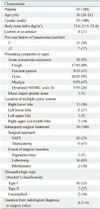
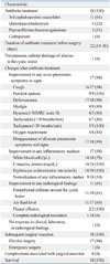
Nineteen adult patients with CCAM were identified during the study period. The clinical characteristics of the patients are shown in Table 1. The median age of these patients was 20 years (IQR, 20-21 years) and all were males. Four (21%) patients were current or ex smokers. Seven (37%) patients had a history of recurrent pneumonia occurring at least twice. Of these 19 patients, 18 initially presented with acute pneumonia symptoms or signs including cough, purulent sputum, fever, myalgia, and/or dyspnea, and received preoperative antibiotic treatment. The remaining patient presented with only blood-tinged sputum, without evidence of pulmonary infection on chest CT scan, and underwent conservative treatment without prescription of antibiotics. Eventually, all 19 patients underwent surgical resection. The most common location of multiple cystic lesions was the right lower lobe (n=11, 58%), followed by the left lower lobe (n=5, 27%), right upper and middle lobe (n=2, 10%), and left upper lobe (n=1, 5%). Approximately half (n=10, 53%) of the patients underwent video-assisted thoracoscopic surgery (VATS), and most patients (n=16, 85%) underwent lobectomy. The median duration from radiological diagnosis to surgery was 4 weeks (IQR, 2-6 weeks). Upon histopathological evaluation, 10 of the 19 (53%) patients were found to have CCAM type I, and 7 (37%) CCAM type II (Stocker's classification). We were unable to identify the CCAM type in two patients (10%).
Characteristics of the patients with CCAM complicated by pneumonia
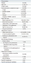
The characteristics of the 18 patients with CCAM complicated by pneumonia are shown in Table 2. All 18 patients presented with pulmonary or systemic symptoms, such as cough (n=17, 94%), purulent sputum (n=9, 50%), fever (n=18, 100%), myalgia (n=9, 50%), and/or dyspnea (n=5, 28%). Seven (39%) patients had tachycardia (>100 beats/min) and five (28%) tachypnea (>30 breaths/min) at initial presentation. The median oxygen saturation value on room air was 94% (IQR, 88-96%).
The WBC count and CRP and ESR levels as inflammatory markers were elevated in all patients, with median values of 11510/µL (IQR, 8990-15145/µL), 11.3 mg/dL (IQR, 3.3-19.4 mg/dL), and 45 mm/h (IQR, 29-67 mm/h), respectively. As a causative agent, Streptococcus pneumoniae was positive in microbiological culture tests in seven (39%) patients, comprising four bronchoalveolar lavage fluid samples and three sputum samples.
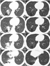
On chest CT images, all patients had parenchymal infiltration around cystic lesions, 17 (94%) had an air-fluid level, and 2 (11%) had pleural effusion. Fig. 1 shows typical CT findings in patients with CCAM complicated by pneumonia. The chest CT image shows parenchymal infiltration around multiple cystic lesions, and an air-fluid interface in the right lower lobe. After 27 days of broad-spectrum antibiotic treatment, CT imaging revealed near-disappearance of the parenchymal infiltrate around the multiple cystic lesions, and the air-fluid interface in the right lower lobe.
Responses to treatment and outcomes in patients with CCAM complicated by pneumonia
The clinical, laboratory, and radiological responses to treatment and outcomes in patients with CCAM complicated by pneumonia are summarized in Table 3. At admission, all 18 patients received broad-spectrum antibiotic treatment; third-generation cephalosporins±macrolides (n=11, 61%), quinolone±clindamycin (n=4, 22%), piperacillin/tazobactam±quinolone (n=2, 11%), and carbapenem (n=1, 6%). The median duration of antibiotic treatment before surgical resection was 22 days (IQR, 14-30 days). One patient received percutaneous catheter drainage during antibiotic treatment to relieve symptoms.
Upon antibiotic treatment for a median time of 22 days (IQR, 14-30 days) prior to surgical resection, all acute pneumonia symptoms and abnormal signs disappeared in 17 (94%) patients, at a median time of 10 days (IQR, 8-15 days); the median times to disappearance of cough, purulent sputum, fever, myalgia, dyspnea, tachycardia, tachypnea, and oxygen requirement were 8, 7, 5, 4, 4, 4, 3, and 5 days, respectively. Improvement or normalization of all inflammatory markers occurred in 17 (94%) and 9 (50%) patients at median times of 8 days (IQR, 5-19 days) and 17 days (IQR, 13-24 days), respectively. Radiological improvement was evident in 11 (61%) patients at a median time of 18 days (IQR, 14-27 days); partial radiological improvements occurred in 10 (56%) patients and complete radiological resolution in only 1 (6%).
The 17 (94%) patients who exhibited clinicolaboratory improvement after preoperative antibiotic treatment underwent elective surgery to treat CCAM. However, the clinical, laboratory, and radiological findings of one patient (6%) did not improve despite 13 days of antibiotic treatment; this patient subsequently underwent emergency surgery with diagnostic and curative therapeutic intent. With 17 (94%) elective and 1 (6%) emergency surgeries in patients with CCAM complicated by pneumonia, all patients survived without complications associated with surgery.
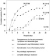
As shown in Fig. 2, we observed serial changes in clinical, laboratory, and radiological findings in response to preoperative antibiotic treatment in patients with CCAM complicated by pneumonia. The median time of resolution of all symptoms and abnormal signs in half of the patients was approximately 5 days. The times to improvement and normalization of any inflammatory marker in half of the patients were 8 and 24 days, respectively. The time to any radiological improvement in half of the patients was 20 days.
DISCUSSION
In the current study, we investigated the clinical characteristics and courses of patients with CCAM complicated by pneumonia, and showed that surgery may be performed safely without ensuing complications after clinicolaboratory improvement was attained by appropriate antibiotic treatment, regardless of a lack of complete radiological resolution. Of the 18 study patients with CCAM complicated by pneumonia, preoperative antibiotic treatment caused all acute pneumonia symptoms and abnormal signs to disappear; at least one inflammatory marker improved in 17 patients (94%). Partial radiological improvement occurred in 10 (56%) patients, but complete radiological resolution in only 1 (6%), However, the remaining patient (6%) exhibited no improvement in terms of clinical, laboratory, or radiological findings, despite preoperative antibiotic treatment. Nevertheless, 17 (94%) underwent elective and 1 (6%) emergency surgeries, and all patients survived without complications.
To date, no accurate data on the clinical course or adequate duration of antibiotic treatment prior to subsequent surgical resection in adult patients with CCAM complicated by pneumonia are available, and a few reports have discussed the treatment response and timing of subsequent surgical interventions.4,5,13,14,15 For example, Herrero, et al.5 reported two cases of CCAM in adults, including one patient presenting with recurrent pneumonia. This 46-year-old female with recurrent pneumonia was initially treated with broad-spectrum antibiotics and clinically improved, but her lung consolidation did not resolve completely. After 3 months, when a follow-up chest CT scan showed almost complete resolution of the consolidation, the patient subsequently underwent lobectomy. Huang, et al.14 reported a 51-year-old male with recurrent lower respiratory tract infection symptoms and intermittent febrile episodes for more than 10 years who underwent planned surgical resection using a VATS approach because the symptoms did not respond to antibiotic treatment. However, these previous studies have some limitations; they were small case series or not focused on analysis of the response to, or duration of, medical treatment and the timing of surgical intervention.
Thus, the data in Fig. 2 contain useful information on the times required for improvements in clinical, laboratory, and radiological findings in response to antibiotic treatment in patients with CCAM complicated by pneumonia. The median times to disappearance of all acute pneumonia symptoms and abnormal signs, decrease in the level of any inflammatory marker, and radiological improvement in half of the patients were 5, 8, and 20 days, respectively. Patients with complicated pneumonia (such as those with an abscess) usually show clinical improvement with decreased fever within 3 to 4 days after antibiotic treatment16,17 and Hirshberg, et al.18 showed that factors predictive of the mortality of 77 patients with a lung abscess indicated the importance of surgical intervention; however, the adequate duration of antibiotic treatment and appropriate timing of subsequent surgical resection remain controversial. Therefore, our data may have clinical significance and facilitate to estimate the response to medical treatment and determinate adequate duration of antibiotic treatment and appropriate timing of subsequent surgical intervention in patients with CCAM complicated by pneumonia.
During our study period, 19 patients with CCAM with or without pneumonia were identified; interestingly, more than half (n=10, 53%) of these patients underwent right lower lobectomy, 5 (27%) underwent left lower lobectomy, 2 (10%) underwent bilobectomy (right upper and middle lobe), 1 (5%) underwent left upper lobectomy, and 1 (5%) underwent right lower segmentectomy. Surgical resection was done by VATS in 10 (53%) patients and thoracotomy in 9 (47%) patients. It is generally known that CCAM has no predilection for a particular location or side and usually involves a single lobe.19,20 However, most of our patients had involvement of the lower lobes (n=16, 85%) and right lower lobes (n=10, 53%), and 2 (1%) patients had involvement of the upper and middle lobes. Studies similar to ours regarding lower lobe or right side predominance have been reported;21,22 however, they involved short case series. Thus, further studies concerning the epidemiological tendencies are required.
The current study had a number of limitations. First, there were epidemiological biases for age and gender in the patient group because this study was conducted at a military hospital, and all enrolled patients were young males, which may limit the applicability of our data to general populations, although most of the reported adult CCAM patients were not elderly individuals. Second, no long-term follow up of the study patients was performed. Because young Korean males are obliged to serve in the military for 2 years, the median postoperative follow-up period was less than 2 years at our hospital, which may have been insufficient to evaluate treatment outcomes. Thus, to perform a more accurate analysis, further studies of the response to medical and surgical treatment in adult patients with CCAM complicated by pneumonia are needed.
In conclusion, we described in the current study, the clinical characteristics and courses of patients with CCAM complicated by pneumonia, and showed that surgery may be performed safely without ensuing complications after clinicolaboratory improvements were achieved by appropriate antibiotic treatment, even in the absence of complete radiological resolution.
 XML Download