Abstract
Purpose
The expression of p53 in patients with rectal cancer who underwent preoperative chemoradiationand and its potential prognostic significance were evaluated.
Materials and Methods
p53 expression was examined using immunohistochemistry in pathologic specimens from 210 rectal cancer patients with preoperative chemoradiotherapy and radical surgery. All patients were classified into two groups according to the p53 expression: low p53 (<50% nuclear staining) and high p53 (≥50%) groups.
Results
p53 expression was significantly associated with tumor location from the anal verge (p=0.036). In univariate analysis, p53 expression was not associated with disease-free survival (p=0.118) or local recurrence-free survival (p=0.089). Multivariate analysis showed that tumor distance from the anal verge (p=0.006), ypN category (p=0.011), and perineural invasion (p=0.048) were independent predictors of disease-free survival; tumor distance from the anal verge was the only independent predictor of local recurrence-free survival. When the p53 groups were subdivided according to ypTNM category, disease-free survival differed significantly in patients with ypN+ disease (p=0.027) only.
Total mesorectal excision after preoperative chemoradiation remains the cornerstone of treatment for patients with potentially resectable, locally advanced rectal cancer, as it allows for increased control of local disease, greater sphincter preservation rates, and improved disease-free survival.1,2,3 Pathologic variables related to tumor responses to chemoradiation, including ypT category, ypN category, and tumor response grade, remain important prognostic indicators for long-term oncologic outcomes.4,5,6 However, these measures are insufficient, as patients at the same disease stage may exhibit different clinical outcomes and different responses to adjuvant therapy. Therefore, identification of additional prognostic markers in radiated rectal cancer specimens at the molecular level is needed to supplement the standard tumor staging system.
The p53 tumor suppressor gene is the most frequently mutated tumor-associated gene in malignant human tumors, including colorectal cancer.7 The prognostic significance of p53 expression in rectal cancer remains unclear,8,9,10,11,12,13 although a few studies have reported associations of p53 expression with prognosis in patients with rectal cancer after preoperative chemoradiation.9,11 Thus, this study attempted to outline the prognostic role of immunohistochemical evaluation of expression of p53 in pathologic specimens from patients with rectal cancer after preoperative chemoradiotherapy and radical surgery.
A total of 568 patients with rectal cancer underwent surgical resection after preoperative chemoradiation at Samsung Medical Center in Korea between January 2007 and March 2011. Eligibility criteria were as follows: 1) histologically confirmed adenocarcinoma, 2) tumors located within 10 cm of the anal verge, 3) locally-advanced (cT3-4 or N-positive) tumors, 4) curative-intent treatment for rectal cancer, and 5) no evidence of distant metastatic disease. Individuals who met the following criteria were excluded: 1) history of any other malignancy associated with hereditary colon cancer syndrome, 2) history of previous chemotherapy or radiotherapy, or 3) a complete pathologic response after preoperative chemoradiation due to an inability to perform immunohistochemical staining. Ultimately, 210 patients were included in the final analysis. This study was approved by our Institutional Review Board.
All patients received preoperative chemoradiation. Details of the preoperative chemoradiation protocol followed by our institution have been published previously.14,15 Briefly, preoperative radiotherapy was delivered to the whole pelvis region at a dose of 40.4-50.4 Gy. Preoperative chemotherapy was concurrently administered with radiotherapy based on a 5-fluorouracil or capecitabine regimen. All patients underwent radical surgery 6 to 8 weeks after preoperative chemoradiation. Of the 210 patients, 195 (92.9%) received postoperative adjuvant chemotherapy. The tumors were staged according to the 7th American Joint Committee on Cancer TNM classification. Assessment of the tumor response to chemoradiation was evaluated according to the tumor response grade (TRG), as described by Mandard, et al.16: TRG0 (no regression), TRG1 (minimal regression), TRG2 (moderate regression), TRG3 (near total regression), and TRG4 (complete regression). TRG3-4 scores were defined as a good TRG response in this study.
p53 expression was evaluated by immunohistochemical staining, as previously described.13 Immunohistochemistry was performed on formalin-fixed, paraffin-embedded tissue. From each paraffin-embedded block, a 2-mm punch was taken for tissue microarrays, as described by Hendriks, et al.17 Each section of normal and tumor tissues (4 µm thick) was assessed by tissue microarray and mounted on a glass slide. The tissue microarray slides were deparaffinized, and the tissues were then rehydrated with xylene and ethanol. The slides were washed with water and phosphate-buffered saline (PBS). Endogenous peroxidase activity was blocked by incubating sections in 3% H2O2 in PBS for 30 min, and the slides were then washed with water, after which heat-induced epitope retrieval was performed. The slides derived from normal and tumor tissues (4-µm thick) were stained with mouse monoclonal antibodies specific for p53 (clone DO-7, 1:100; Dako, Glostrup, Denmark). p53 expression was described as low if <50% nuclear staining was observed and high if ≥50% nuclear staining was observed.18 Normal colonic epithelial tissue adjacent to tumor tissue and lymphocytes served as internal positive controls.
Statistical evaluation was carried out using the statistical package SPSS for Windows (version 14.0; SPSS Inc., Chicago, IL, USA). Association between p53 immunostaining and clinicopathological features were explored with Student's t-test and the chi-square test, as appropriate. Disease-free survival curves were calculated using the Kaplan-Meier method, and differences between curves were evaluated using the log-rank test. To identify the significant independent prognostic factors associated with disease-free survival, variables with p-values less than 0.2 on univariate analysis were entered into a multivariate analysis of stepwise logistic regression. All values of p<0.05 were considered statistically significant.
The analysis included 151 men (71.9% of the sample), and the median participant age was 56 years (range 31-79). Of the 210 patients, 181 (86.2%) underwent sphincter-saving operations. The median number of retrieved lymph nodes from each patient was 11 (1-39). Using the 7th UICC TNM staging system, 16, 64, 125, and 5 patients had ypT1, ypT2, ypT3, and ypT4 cancers, respectively; 138, 55, and 17 patients had ypN0, ypN1, and ypN2 tumors, respectively.
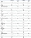
The relationships between tumor p53 expression and the clinicopathological features of colorectal adenocarcinoma are summarized in Table 1. The expression of p53 in tumors was significantly associated with tumor location from the anal verge (p=0.036). No significant association was detected between p53 expression and age, gender, histology, tumor diameter, ypT category, ypN category, tumor regression grade, lymphatic invasion, vascular invasion, perineural invasion, or tumor budding.
During study follow-up (median of 48 months), the factors associated with lower disease-free survival in univariate analysis were tumor distance from the anal verge (p=0.022), ypT category (p=0.022), ypN category (p<0.001), lymphatic invasion (p=0.002), perineural invasion (p=0.005), and tumor regression grade (p=0.006) (Table 2). The factors associated with lower local recurrence-free survival in univariate analysis were tumor distance from the anal verge (p=0.011), lymphatic invasion (p=0.049), and perineural invasion (p=0.046) (Table 2). No significant association was observed between both disease-free survival and local recurrence-free survival and p53 expression (Table 2). A multivariate analysis showed that tumor distance from the anal verge (p=0.006), ypN category
(p=0.011), and perineural invasion (p=0.048) were independent predictors of disease-free survival; tumor distance from the anal verge was the only independent predictor of local recurrence-free survival (Table 3).
When the low and high p53 groups were further subdivided according to their ypTNM category (Fig. 1), disease-free survival significantly differed in relation to p53 expression for ypTanyN+ disease (p=0.027), but not for ypT1-2N0 and ypT3-4N0 diseases (p=0.122 and p=0.676, respectively).
The product of the p53 tumor suppressor gene is a nuclear phosphoprotein that indirectly regulates cell growth and inhibits cells with mutagenic damage from entering S-phase by arresting the cell cycle in G1.7 The clinical significance of p53 in the progression and metastasis of colorectal cancer is controversial.8,13,19,20,21,22,23 A number of studies have reported worse clinical outcomes in patients with p53 overexpression or mutations; however, this association has not been found to be universally true, as several investigations, including a large series of 541 patients with colorectal cancers with a long follow-up period by Soong, et al.,23 have suggested that the presence of p53 accumulation is associated with improved survival, independently of tumor stage or grade, in distal colorectal cancers. Moreover, Watanabe, et al.8 failed to demonstrate a significant association between p53 overexpression and clinical outcomes in patients with locally advanced colon cancer. In a previous publication, we reported that p53 expression, as well as the mismatch repair system, had no prognostic impact in patients with non-radiated colorectal cancer.13 In this study, we found that p53 expression in radiated specimens was not an independent predictor of survival, although it had a significant prognostic impact on survival in patients with ypTanyN+ rectal cancer.
The prognostic value of p53 expression for the outcomes of treatment for rectal cancer is still unknown.9,10,11,12,13,24 Moreover, few publications have examined the prognostic impact of p53 expression in patients with radiated rectal cancer. Schwandner, et al.10 suggested that immunohistochemical assessment of both p53 and Bcl-2 status was a significant predictor of prognosis after curative surgery for rectal cancer and that p53 was a stronger predictor of prognosis than Bcl-2. In contrast, Morgan, et al.24 reported that the immunohistochemical p53 status of rectal cancer was not associated with clinicopathological variables, nor predictive of oncologic outcomes. Saw, et al.12 found no correlation between p53 expression and survival in 60 patients with rectal cancer treated with preoperative chemotherapy or radiotherapy. They also concluded that predicting the prognosis of patients with locally advanced low rectal cancer who have received preoperative therapies remains a challenge. We concur with this, as, in our study of a relatively large consecutive cohort of patients with radiated rectal cancer, p53 expression was not shown to be an independent predictor of survival.
The prognostic role of p53 expression according to the tumor location remains unclear. In colon cancer, proximal colon tumors, when compared with distal tumors, are more often found in older patients and benefit from adjuvant chemotherapy.25,26 In this study, although p53 expression was not independent predictor for survival, distance from the anal verge had a significant prognostic impact in rectal cancer patients with preoperative chemoradiation. We found that patients with proximal rectal cancer showed better survival and greater p53 expression than patients with distal rectal cancer. This finding is consistent with the study by Hilska, et al.,27 in which p53 overexpression showed better prognosis in rectal cancer than in colon cancer.
The differences in the results of previous studies may have been related to subjectivity in scoring, the absence of a uniform cut-off value for the definition of positive tumors, and differences in antibodies used, immunohistochemical methods, patient materials, and durations of follow-up. For nuclear markers such as p53, arbitrary values ranging from 5% to 75% have been used. Sometimes the cut-off point is based on a mean or a median staining index,28 and sometimes the cut-off is set somewhere between the indisputable negatives and all other samples.29 Occasionally, a cut-off is chosen on the basis of earlier studies.
We acknowledge that this study was not a proper trial with a large population sample size, and thus, there is a possibility for Type II error. Although our findings are insufficient to draw definite conclusions, they may still be of value. Further investigations with larger sample sizes are necessary to assess the prognostic role of p53 expression in patients with rectal cancer and to validate their possible value as novel therapeutic targets. In conclusion, p53 expression in resected specimens had a significant prognostic impact on survival in patients with stage III rectal cancer after preoperative chemoradiation, though it was not an independent predictor of survival. Confirmation of these results in larger sample sizes is needed, however.
Figures and Tables
Fig. 1
Disease-free survival according to the p53 expression and ypTNM category. (A) ypT1-2N0, (B) ypT3-4N0, and (C) ypTanyN+.

Table 1
Correlation between a p53 Expression and Clinicopathological Parameters

Table 2
Univariate Analysis of the Prognostic Factors for 5-Year Disease-Free Survival (DFS) and Local Recurrence-Free Survival (LRFS)

Table 3
Multivariate Analysis of the Prognostic Factors for 5-Year Disease-Free Survival (DFS) and Local Recurrence-Free Survival (LRFS)

ACKNOWLEDGEMENTS
This research was supported by Basic Science Research Program through the National Research Foundation of Korea (NRF) funded by the Ministry of Education, Science and Technology (grant number 2012R1A1A1004888).
References
1. Roh MS, Colangelo LH, O'Connell MJ, Yothers G, Deutsch M, Allegra CJ, et al. Preoperative multimodality therapy improves disease-free survival in patients with carcinoma of the rectum: NSABP R-03. J Clin Oncol. 2009; 27:5124–5130.

2. Sebag-Montefiore D, Stephens RJ, Steele R, Monson J, Grieve R, Khanna S, et al. Preoperative radiotherapy versus selective postoperative chemoradiotherapy in patients with rectal cancer (MRC CR07 and NCIC-CTG C016): a multicentre, randomised trial. Lancet. 2009; 373:811–820.

3. Sauer R, Becker H, Hohenberger W, Rödel C, Wittekind C, Fietkau R, et al. Preoperative versus postoperative chemoradiotherapy for rectal cancer. N Engl J Med. 2004; 351:1731–1740.

4. García-Aguilar J, Hernandez de Anda E, Sirivongs P, Lee SH, Madoff RD, Rothenberger DA. A pathologic complete response to preoperative chemoradiation is associated with lower local recurrence and improved survival in rectal cancer patients treated by mesorectal excision. Dis Colon Rectum. 2003; 46:298–304.

5. Huh JW, Park YA, Jung EJ, Lee KY, Sohn SK. Accuracy of endorectal ultrasonography and computed tomography for restaging rectal cancer after preoperative chemoradiation. J Am Coll Surg. 2008; 207:7–12.

6. Habr-Gama A, Perez RO, Nadalin W, Nahas SC, Ribeiro U Jr, Silva E Sousa AH Jr, et al. Long-term results of preoperative chemoradiation for distal rectal cancer correlation between final stage and survival. J Gastrointest Surg. 2005; 9:90–99.

7. Nigro JM, Baker SJ, Preisinger AC, Jessup JM, Hostetter R, Cleary K, et al. Mutations in the p53 gene occur in diverse human tumour types. Nature. 1989; 342:705–708.

8. Watanabe T, Wu TT, Catalano PJ, Ueki T, Satriano R, Haller DG, et al. Molecular predictors of survival after adjuvant chemotherapy for colon cancer. N Engl J Med. 2001; 344:1196–1206.

9. Spitz FR, Giacco GG, Hess K, Larry L, Rich TA, Janjan N, et al. p53 immunohistochemical staining predicts residual disease after chemoradiation in patients with high-risk rectal cancer. Clin Cancer Res. 1997; 3:1685–1690.
10. Schwandner O, Schiedeck TH, Bruch HP, Duchrow M, Windhoevel U, Broll R. p53 and Bcl-2 as significant predictors of recurrence and survival in rectal cancer. Eur J Cancer. 2000; 36:348–356.

11. Jakob C, Liersch T, Meyer W, Becker H, Baretton GB, Aust DE. Predictive value of Ki67 and p53 in locally advanced rectal cancer: correlation with thymidylate synthase and histopathological tumor regression after neoadjuvant 5-FU-based chemoradiotherapy. World J Gastroenterol. 2008; 14:1060–1066.

12. Saw RP, Morgan M, Koorey D, Painter D, Findlay M, Stevens G, et al. p53, deleted in colorectal cancer gene, and thymidylate synthase as predictors of histopathologic response and survival in low, locally advanced rectal cancer treated with preoperative adjuvant therapy. Dis Colon Rectum. 2003; 46:192–202.

13. Kim HR, Kim HC, Yun HR, Kim SH, Park CK, Cho YB, et al. An alternative pathway in colorectal carcinogenesis based on the mismatch repair system and p53 expression in Korean patients with sporadic colorectal cancer. Ann Surg Oncol. 2013; 20:4031–4040.

14. Park CH, Kim HC, Cho YB, Yun SH, Lee WY, Park YS, et al. Predicting tumor response after preoperative chemoradiation using clinical parameters in rectal cancer. World J Gastroenterol. 2011; 17:5310–5316.

15. Lee H, Park HC, Park W, Choi DH, Kim YI, Park YS, et al. Negative impact of pretreatment anemia on local control after neoadjuvant chemoradiotherapy and surgery for rectal cancer. Radiat Oncol J. 2012; 30:117–123.

16. Mandard AM, Dalibard F, Mandard JC, Marnay J, Henry-Amar M, Petiot JF, et al. Pathologic assessment of tumor regression after preoperative chemoradiotherapy of esophageal carcinoma. Clinicopathologic correlations. Cancer. 1994; 73:2680–2686.

17. Hendriks Y, Franken P, Dierssen JW, De Leeuw W, Wijnen J, Dreef E, et al. Conventional and tissue microarray immunohistochemical expression analysis of mismatch repair in hereditary colorectal tumors. Am J Pathol. 2003; 162:469–477.

18. Rashid A, Zahurak M, Goodman SN, Hamilton SR. Genetic epidemiology of mutated K-ras proto-oncogene, altered suppressor genes, and microsatellite instability in colorectal adenomas. Gut. 1999; 44:826–833.

19. Huh JW, Lee JH, Kim HR. Expression of p16, p53, and Ki-67 in colorectal adenocarcinoma: a study of 356 surgically resected cases. Hepatogastroenterology. 2010; 57:734–740.
20. Bouzourene H, Gervaz P, Cerottini JP, Benhattar J, Chaubert P, Saraga E, et al. p53 and Ki-ras as prognostic factors for Dukes’ stage B colorectal cancer. Eur J Cancer. 2000; 36:1008–1015.

21. Zeng ZS, Sarkis AS, Zhang ZF, Klimstra DS, Charytonowicz E, Guillem JG, et al. p53 nuclear overexpression: an independent predictor of survival in lymph node--positive colorectal cancer patients. J Clin Oncol. 1994; 12:2043–2050.

22. Ahnen DJ, Feigl P, Quan G, Fenoglio-Preiser C, Lovato LC, Bunn PA Jr, et al. Ki-ras mutation and p53 overexpression predict the clinical behavior of colorectal cancer: a Southwest Oncology Group study. Cancer Res. 1998; 58:1149–1158.
23. Soong R, Grieu F, Robbins P, Dix B, Chen D, Parsons R, et al. p53 alterations are associated with improved prognosis in distal colonic carcinomas. Clin Cancer Res. 1997; 3:1405–1411.
24. Morgan M, Koorey D, Painter D, Findlay M, Newland R, Chapuis P, et al. p53 and DCC immunohistochemistry in curative rectal cancer surgery. Int J Colorectal Dis. 2003; 18:188–195.

25. Birkenkamp-Demtroder K, Olesen SH, Sørensen FB, Laurberg S, Laiho P, Aaltonen LA, et al. Differential gene expression in colon cancer of the caecum versus the sigmoid and rectosigmoid. Gut. 2005; 54:374–384.

26. Kapiteijn E, Liefers GJ, Los LC, Kranenbarg EK, Hermans J, Tollenaar RA, et al. Mechanisms of oncogenesis in colon versus rectal cancer. J Pathol. 2001; 195:171–178.

27. Hilska M, Collan YU, O Laine VJ, Kössi J, Hirsimäki P, Laato M, et al. The significance of tumor markers for proliferation and apoptosis in predicting survival in colorectal cancer. Dis Colon Rectum. 2005; 48:2197–2208.


 PDF
PDF ePub
ePub Citation
 Print
Print


 XML Download