

 PDF
PDF ePub
ePub Citation
 Print
Print


INTRODUCTION
Interstitial lung diseases (ILDs) are a heterogeneous group of diffuse parenchymal lung disorders that are frequently caused by infection, environmental or occupational toxins, certain medications, radiation therapy of the chest, or connective tissue diseases (CTDs).1 Unlike external causative factors such as a pertinent exposure or drug history, a CTD may occasionally confuse the diagnostic process because some patients will present with ILD years prior to receiving a diagnosis of CTD or may have presentations limited to pulmonary manifestations of an autoimmune disease.2,3 In these cases, a delayed diagnosis or difficult-to-diagnose state of underlying CTD sometimes leads to a categorization of ILD as idiopathic or sometimes unclassifiable. However, most importantly, CTD-ILD is associated with a more favorable prognosis than idiopathic ILD of equivalent severity4 and the prognosis, degree of reversibility, and an optimal therapy also differ for each type of CTD.5 Therefore, it is crucial to rigorously evaluate the underlying CTD in all patients presenting with ILD.
Idiopathic inflammatory myopathies (IIMs) are a group of systemic diseases involving the skeletal muscle and internal organs classified within the group of CTDs. Other than skin involvement, the lung is the most common extramuscular target in IIM6 and the prevalence of ILD in patients with IIMs has been reported to be from 20 to 65%.7,8,9 The only prospective study conducted so far revealed a prevalence of 78%.10 Anti-synthetase syndrome, characterized by myositis, ILD, fever, Raynaud's phenomenon, arthritis, and mechanic's hand combined with positive anti-synthetase antibody,11 has especially been shown to be associated with higher rates of ILD than those without anti-synthetase antibody.12 Although it is known that the myopathic manifestations of IIMs often precede lung involvement, 18% of patients ultimately diagnosed with IIM-associated ILD did not have muscle-related symptoms at the time of radiographical or physiological confirmation of lung involvement in one series13 and might have been diagnosed as idiopathic ILD.
It will not be sufficient to detect CTD in patients presenting with ILD by simply screening for nonspecific autoantibodies, such as antinuclear antibody or rheumatoid factor, because it has been reported that the prevalence of nonspecific antibodies was not different between the group that developed CTD and the group that did not,14 and also antinuclear antibody-negative patients could still present with autoantibodies to cytoplasmic antigens.15 In particular, the autoimmune response in myositis is directed to several heterogeneous antigens. Many of which are poorly detectable by conventional assays because they are expressed in low amounts in cell/tissue extracts or are highly sensitive to degradation/denaturation.16 Additionally, myositis autoantibodies are predominantly localized in the cytoplasm, thus leading to poor representation in cell extracts.17
In this study, we evaluated the frequency of myositis autoantibodies using an immunoblot assay to detect multiple autoantibodies in a sensitive manner, and investigated the clinical significance of the presence of antibodies in patients diagnosed with idiopathic ILD.
MATERIALS AND METHODS
Patients
A total 32 patients diagnosed with idiopathic ILD were included in this study. We collected consecutive serum samples from patients who were diagnosed with idiopathic ILD and for whom fluorescent antinuclear antibody test (FANA) or anti-Jo-1 antibody tests were requested at Samsung Medical Center from April 2013 to October 2013. In this study, idiopathic ILD was defined as acute and chronic lung disorders collectively referred to as ILD or diffuse parenchymal lung diseases of unknown etiology in which a patient did not fulfill the classification criteria for any specific CTD or vasculitis, and in whom lung diseases were not potentially caused by drugs or occupational-environmental exposure.18 Screening for CTD and a review of patient history with respect to pulmonary complaints were performed by experienced rheumatologists and pulmonologists. The diagnosis was made using the comprehensive clinical evaluation and the findings of high resolution computed tomography.
We obtained the clinical information and laboratory data, including patient demographic features of age and gender, clinical features of pulmonary and extrapulmonary manifestations at the time of diagnosis, smoking history and treatment regimen, white blood cells (WBC), erythrocyte sedimentation rate (ESR), C-reactive protein (CRP), creatine kinase (CK), lactate dehydrogenase (LD), rheumatoid factor (RF), FANA, anti-neutrophil cytoplasmic antibody (ANCA), pulmonary function test (PFT) for forced vital capacity (FVC), diffusing capacity for carbon monoxide (DLCO), total lung capacity (TLC), and blood gas analysis for partial pressure of oxygen in arterial blood (PaO2).
The study was approved by the Institutional Review Board of our institution.
Measurement of myositis-specific autoantibodies and myositis-associated autoantibodies
We examined the recruited patients for a panel of 11 myositis-associated autoantibody antibodies (MAAs) or myositis-specific autoantibodies (MSAs). MAAs, which are frequently encountered in rheumatic disorders associated with myositis, include anti-Ro52, anti-U1RNP, anti-PM/Scl-100 and -75, and anti-Ku. MSAs, which are specific for IIMs, include anti-synthetase autoantibodies, anti-Mi-2, and anti-signal recognition particle (anti-SRP).19 Anti-synthetase autoantibodies encompass anti-Jo-1 (histidyl-), anti-PL-7 (threonyl-), anti-PL-12 (alanyl-), anti-EJ (glycol-), and anti-OJ (isoleucyl-tRNA synthetase).17 For the assessment, a line blot immunoassay kit (EUROLINE Myositis Profile, Euroimmun AG, Luebeck, Germany), which detects human immunoglobulin G autoantibodies in serum, was used according to the manufacturer's instructions. Test nitrocellulose strips were printed with lines of purified, biochemically characterized antigens. Each antigen was coated onto a separate membrane fragment at the optimized efficiency of antibody detection with a specificity from 95% to 100%. The EUROLineScan program, providing automated evaluation, was used to recognize the position of the strips, and then identify the bands and measure their intensity. The results were defined as positive when the signal intensity was more than 11. The intensity of autoantibodies are graded into weakly (+), moderately (++), and strong (+++) according to the signal intensity of 11-25, 26-50, and >50, respectively, values assigned by the EUROLineScan program which was provided by the manufacturer.
Statistical analysis
For categorical variables, clinical information including demographic and clinical features, smoking history and treatment regimen, and laboratory data results including RF, FANA, and ANCA positivity were expressed as frequencies with percentages. Comparisons were performed between the groups with and without myositis autoantibodies using a chi-square test. For continuous variables, the other laboratory data were expressed as a median with 25-75 percentiles. Comparisons were performed between the groups with and without myositis autoantibodies using a Mann-Whitney test. Results were considered significant when the two-tailed p-value was <0.05 in both continuous and categorical variables. MedCalc software version 13.0.0.0 (MedCalc Software, Ostend, Belgium) was used for the statistical analysis.
RESULTS
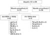
One or more autoantibodies encompassing nine specificities, except for anti-Mi-2 and anti-PM-Scl 100, were present in 12 of the 32 patients (12/32, 38%). MSAs with or without MAAs were present in eight patients (8/32, 25%) and MAAs alone were present in four patients (4/32, 13%). Anti-synthetase autoantibodies including Jo-1, EJ, OJ, PL-7, and PL-12 were present in seven patients (7/32, 22%). Among them, anti-Jo-1 and anti-EJ were each encountered in two patients, and anti-PL-7, anti-PL-12, and anti-OJ were each encountered in one patient. Another myositis-specific autoantibody, anti-SRP, was detected in one patient. Although anti-Ro-52 was most frequently detected, being found in 12 patients with myositis autoantibodies, it was found to mostly occur with other autoantibodies, except in one case (Fig. 1).
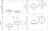
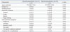
Clinical characteristics and laboratory findings of patients at the time of diagnosis were compared according to the presence of myositis autoantibodies and the results are summarized in Table 1 and 2, Fig. 2. In terms of clinical characteristics, we found that patients with myositis autoantibodies showed a mechanic's hand more frequently than those without autoantibodies (p=0.027). Other differences were not significant. In laboratory findings, WBC, ESR, CRP, CK, PaO2, and the frequencies of RF, FANA, and ANCA positivity revealed no statistically significant differences between the two groups. However, PFT results for all three parameters, FVC, DLCO, and TLC, were significantly lower in the group with myositis autoantibodies (p=0.022, 0.006, and 0.008, respectively) and also LD was significantly higher in the group with myositis autoantibodies (p=0.046).
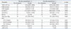
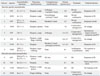
All individual cases with myositis autoantibodies are summarized in Table 3 with respect to clinical characteristics and progressions.
DISCUSSION
To our knowledge, this is the first report that evaluates the presence of various myositis autoantibodies in Korean idiopathic ILD patients. Our study showed that, among idiopathic ILD patients, 38% were found to have myositis autoantibodies. Compared to other studies investigating myositis autoantibodies in groups of idiopathic ILD patients, this proportion is quite high. However, the prevalence of myositis autoantibodies in idiopathic ILD has been reported to vary widely, mainly due to the enrollment of heterogeneous patient populations that were diagnosed and categorized differently and due to differences in test methodology and between target panels of myositis antibodies.12,20,21
In the study by Nakashima, et al.,20 anti-ARS antibodies were detected in 10.7% of idiopathic interstitial pneumonia using an enzyme-linked immunosorbent assay (ELISA) system with a mixture of six recombinant ARS antigens, and in the study by Watanabe, et al.,12 6.6% were positive for six anti-ARS antibodies in idiopathic interstitial pneumonia using an RNA immunoprecipitation procedure (IPP). Another study by Fischer, et al.21 showed that 24% of patients presenting with idiopathic interstitial pneumonia and features of anti-sythetase syndrome were positive for anti-PL-7 or PL-12 antibodies. In this study, we included a significant number of patients who had positive serological results on the FANA and ANCA tests. Many patients had clinical characteristics of CTD including mechanic's hand and arthralgia in addition to the presence of autoantibodies related to specific CTD categories but these findings were insufficient to make a diagnosis of a specific CTD category such as undifferentiated connective tissue disease (UCTD) or IIM. This explanation is supported by the short disease duration, as shown in Table 3.
The line blot immunoassay, the diagnostic method used in this study, also seemed to detect a higher proportion of myositis autoantibodies among idiopathic ILD patients compared to other studies that used the RNA IPP or ELISA.12,20 According to the previous reports, evaluating myositis autoantibodies by commercial line blot assay, especially anti-Ro52 anti-Ku, was known to have higher sensitivity compared to that of a highly specific IIP method or in-house immunoblot, although it did not have enough sensitivity for the non-anti-Jo-1 anti-ARS antibodies.17,22 In comparison with the ELISA results (Orgentec, Mainz, Germany), performed as routine clinical test in our laboratory and only available for the anti-Jo-1 among autoantibodies evaluated in this study, the patient sample which had weakly positive anti-Jo-1 autoantibody on the line blot immunoassay (case no. 2 in Table 3) had a negative result on the ELISA test, while only a strong positive anti-Jo-1 autoantibody (case no. 1 in Table 3) was detected by the ELISA test (data not shown). A previous study reported that the prevalence of myositis autoantibodies was greater than 50% among IIM patients when investigated using the same commercial line blot assay kit as the one used in the current study23 and the results seem to indicate the high sensitivity of the commercial line blot assay.
The group with myositis autoantibodies showed the mechanic's hand and abnormal pulmonary function test results, with low FVC, DLCO, and TLC, and higher LD values in the blood samples than the group without myositis autoantibodies. Although CTD-ILD is known to be associated with a more favorable prognosis than idiopathic interstitial pneumonia of equivalent severity,4,5 there are few publications that found significantly better PFT results in both the CTD-ILD or UCTD-ILD groups at diagnosis or during the clinical course.12,24 In this study, a significant number of patients who had positive serological results on the FANA and ANCA tests were included. These patients may potentially be classified as UCTD. This might have influenced the difference in PFT between groups, although we compared the groups with or without myositis autoantibodies.
In our study, the anti-synthetase antibodies were the most frequent of the myositis autoantibodies, supporting many case reports describing anti-synthetase antibody-positive ILD without myositis.25 Many previous reports on patients with anti-synthetase syndrome have revealed an association between anti-synthetase antibody specificity and distinctive phenotypic features. In our cases, both patients with anti-Jo-1 had joint impairment, which was not seen in both patients with anti-PL7/PL12. The anti-Jo-1 patients also had higher CK levels (data not shown) than those of the patients with anti-PL7/PL12, showing that the presence of the anti-Jo1 antibody results in more severe myositis and joint impairment than with anti-PL7/PL12.26 Also, our case with anti-OJ showed a spontaneous improvement in symptoms and PFT results, in agreement with a previous study reporting that anti-OJ-positive patients had good clinical characteristics, such as a lack of Raynaud's phenomenon and a good response to corticosteroid treatment.27 No patient presented with more than one anti-ARS antibody simultaneously. Patients with anti-SRP can present with an acute onset of severe myopathy with significant muscle enzyme elevation, which can be refractory to standard treatments and may appear to have a lower frequency of interstitial pneumonia.28 Similarly, in our cases, one patient with anti-SRP showed refractory and worsening pulmonary symptoms in spite of treatment with azathioprine or pirfenidone, an oral derivative of pyridine having anti-fibrotic properties. The presence of anti-Ro52 antibody in patients with anti-synthetase syndrome was reported to cause more severe ILD29 as it is the most common additional autoantibody detected in anti-synthetase syndrome.30 Our case with both anti-EJ and anti-Ro-52 received rigorous corticosteroid and immunosuppressant treatment.
The detection of autoantibodies can be made by several methods, including standard indirect immunofluorescence, immunoprecipitation, immunodiffusion or counter immunoelectrophoresis, or by automatized immunoassays, such as an ELISA or immunoblotting.16 The commercial line blot immunoassay has recently been reported to be a valid screening test for autoantibodies in CTDs and also as a suitable alternative to the more complex and time-consuming confirmatory test.16 Furthermore, the test's multiparametric platform makes the simultaneous detection of multiple analytes in one sample possible. Although there is a view that anti-Ro52 antibody has a poor diagnostic value for autoimmune myositis and is nonspecific, it can be useful as an indicator of the development of a more severe ILD in patients with anti-synthetase syndrome.
There are a few limitations in this study. First, the small number of patients included in this study may not be enough to represent idiopathic ILD patients generally. Second, we could not confirm our results generated by the line blot immunoassay with other sensitive and specific assays which employ different techniques or principles, except for anti-Jo-1. Considering that there is a limited availability of clinical test systems, it may be an issue of continuous interest to investigate the presence of myositis antibodies in idiopathic ILD with or without CTD and their clinical significance. This study is the first report in Korea that evaluates myositis autoantibodies among idiopathic ILD patients, so further studies in a larger number of Korean patients may be warranted.
In conclusion, we suggest that the evaluation of myositis autoantibodies needs to be performed in patients who are diagnosed with idiopathic ILD in the presence of the clinical characteristics including mechanic's hand, arthralgia, and autoantibodies, which are insufficient to make a diagnosis of a specific CTD category. Considering the highly variable serologic heterogeneity of IIM, the line blot immunoassay which enables the simultaneous detection of multiple antibodies would be an useful diagnostic method because it is both efficient and objective.
 XML Download