

 PDF
PDF ePub
ePub Citation
 Print
Print


INTRODUCTION
Atrial fibrillation (AF) is the most common form of cardiac arrhythmia in clinical practice and is associated with significant morbidity.1 Many studies have investigated mechanisms underlying AF development, and growing evidence supports that genetic variations play a role in its pathogenesis.2 For instance, mutations in mitochondrial DNA (mtDNA) have been shown to be associated with the same factors that are considered critical in development of AF: aging process and oxidative stress. So far, a greater number of mtDNA mutations have been found in individuals with greater age,3 heart failure,4 and ischemic heart disease.5 Among the known mutations in mtDNA, 4977bp deletion mutation (mtDNA4977-mut) is one of the most frequently detected genetic alterations,6,7 and it has been identified in various human tissues, including skeletal muscle,8 brain,9 and heart.10,11 Fittingly, the mtDNA4977-mut has been increasingly associated with AF. While a number of studies have been conducted to establish a relationship between mtDNA4977-mut and AF, characterizations of AF patients positive for this mutation are still inadequate and applicability of peripheral mtDNA4977-mut as a biomarker in AF remains untested. Therefore, in this study, we set out to analyze the frequency of somatic mtDNA4977-mut in Korean patients with non-valvular AF and in age-matched controls to outline an association between AF and mtDNA4977-mut in peripheral blood, to correlate the pathophysiological characteristics of AF patients with mtDNA4977-mut, and finally, to validate the utility of the mutation as a biomarker for atrial remodeling.
MATERIALS AND METHODS
Patient selection
The study protocol was approved by the Institutional Review Board of Severance Cardiovascular Hospital, Yonsei University Health System, and adhered to the Declaration of Helsinki. All patients provided written informed consent. The study enrolled 212 consecutive patients with AF included in the Yonsei AF Ablation Cohort (83.5% male, 51.1±12.5 years old) and 212 age-matched controls (50.0% male, 51.1±13.2 years old). All AF patients underwent radiofrequency catheter ablation (RFCA). Of the AF patients, 153 had paroxysmal AF (PAF) and 59 had persistent AF (PeAF). We examined all 212 AF patients with 3D spiral computed tomography (CT; 64 Channel, Light Speed Volume CT, Philips, Brilliance 63, Eindhoven, the Netherlands) to visually define the anatomy of left atrium (LA), and used trans-thoracic echocardiography (Sonos 5500, Philips Medical System, Andover, MA, USA or Vivid 7, GE Vingmed Ultrasound, Horten, Norway) to evaluate patients for additional structural heart disease, LA remodeling, and ventricular function. The control group consisted of 212 healthy subjects without structural heart disease who were age-matched with the patient group. We confirmed the absence of any potential structural heart disease in the control group with echocardiography and treadmill exercise ECG. Whole blood samples, collected using EDTA as an anticoagulant, were taken for DNA extraction and genetic analyses.
Electrophysiological mapping and radiofrequency catheter ablation
We conducted electrophysiologic mapping in the AF group. For guidance during the RFCA procedure, a 3D electroanatomical map was produced. By double trans-septal puncture, multi-view pulmonary venograms were obtained. Systemic anti-coagulation was achieved with intravenous heparin by maintaining an activated clotting time of 350-400 seconds. The 3D spiral CT images were merged with NavX-generated 3D geometry of the LA and pulmonary veins (PV) (NavX, St. Jude Medical Inc., Minnetonka, MN, USA) to acquire an electroanatomical map. Also, intracardiac electrograms were collected with the Prucka Cardio Lab™ electrophysiology system (General Electric Medical Systems Inc., Milwaukee, WI, USA). We generated an LA voltage map by obtaining contact bipolar electrograms from 350-500 points on the LA endocardium during atrial pacing at 500 ms and calculated mean LA voltage as previously described.12,13 To calculate local conduction velocity, conduction distance was measured on the anterior-posterior and posterior-anterior views of the isochronal map and then divided by the time difference as described before.13 In all patients, the RFCA was conducted with an open irrigation 3.5 mm-tip deflectable catheter (Celsius, Johnson & Johnson Inc., Diamond Bar, CA, USA; Coolflex, St. Jude Medical Inc., Minnetonka, MN, USA; 30-35 W; 47℃). All patients initially underwent circumferential PV isolation and cavo-tricuspid isthmus block. For those patients with PeAF, we added a roof line, posterior inferior line, and anterior line14 as a standard lesion set. At the operator's discretion, additional ablations of the superior vena cava, a non-PV foci or complex fractionated electrogram was conducted.
Detection of mtDNA4977-mutation in whole blood
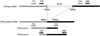
Total DNA was extracted from the whole blood using the commercially available QIAamp DNA Mini kit (Qiagen, Valencia, CA, USA). Two primer sets were designed using Primer3 (http://frodo.wi.mit.edu/primer3/), and each forward primer was labeled with the fluorescent dye 6-FAM (Macrogen Inc., Seoul, Korea) for PCR amplification of mtDNA4977-mut (Fig. 1). The primer sequences were as follows: mtDNA4977bp-Forward 1:5'-FAM-CAGTGAAA TGCCCCAACTAAA-3', mtDNA4977bp-Reverse 1: 5'-TCGATGATGTGGTCTTTGGA-3', and mtDNA4977bp-Forward 2:5'-FAM-ATGGCCCACCATAATTACCC-3', mtDNA4977bp-Reverse 2:5'-GATAGGGCTCAGGCG TTTGT-3'. PCR amplification was performed with a final volume of 10 µL that contained 1.0 µL Gold ST*R buffer (Promega, Madison, WI, USA), 1.0 U AmpliTaq Gold DNA polymerase (Applied Biosystems, Foster City, CA, USA), 0.6 µM of each of the primers, and 10 ng of total DNA as the template. Thermal cycling was conducted on a PCR machine (Bio-Rad Laboratories, Hercules, CA, USA) under the following conditions: 95℃ for 11 min, followed by 33 cycles at 94℃ for 20 sec, 60℃ for 30 sec, and 72℃ for 30 sec, and a final extension at 72℃ for 7 min. After PCR had finished, 1.0 µL aliquots of each of the PCR products and 0.2 µL of GeneScan 500 LIZ size standard (Applied Biosystems, Foster City, CA, USA) were added to 20 µL de-ionized formamide. The mixture was denaturated and separated by capillary electrophoresis on a 3130xI Genetic Analyzer (Applied Biosystems, Foster City, CA, USA), and the size and area of the specific fragments were displayed as peaks on an electropherogram that was generated using the GeneScan Analysis Software 3.1.2 (Applied Biosystems, Foster City, CA, USA).
Biochemical analysis
Peripheral blood samples were taken before RFCA and the plasma levels of the following protein markers were measured using enzyme-linked immunosorbent assay kits: tissue inhibitor of metalloproteinases-1 (TIMP-1; R&D Systems, Minneapolis, MN, USA), transforming growth factor-β (R&D Systems, Minneapolis, MN, USA), and pro-atrial natriuretic peptide (pro-ANP; Biomedica, Antony, France).
Statistical analysis
Multiple parameters including clinical features, echocardiographic parameters, electro-anatomical remodeling of the LA, and the plasma levels of protein biomarkers were compared between patients with AF and their age-matched controls. These parameters were also compared within the AF patient group between those with and without the mtDNA4977-mut. Comparisons between groups were analyzed using the t-test for continuous variables or the chi-squared test for nominal variables. All continuous variables were expressed as mean±SD, whereas all categorical variables were expressed as absolute and relative frequencies (%). In order to examine the association between the parameters and mtDNA4977-mut in AF, both univariate and multivariate logistic regression analyses were performed. All statistical analyses were conducted using SPSS version 15.0 (SPSS Inc., Chicago, IL, USA), and all p-values <0.05 were considered statistically significant.
RESULTS
Frequency of peripheral mtDNA4977-mut in patients with AF vs. control
The somatic mutation associated with oxidative stress mtDNA4977-mut was detectable in the peripheral blood of both AF patients and their age-matched controls. Overall, 21.9% (93/424) of patients included in the study tested positive for mtDNA4977-mut. When AF patients were compared to the control group, the prevalence of peripheral mtDNA4977-mut was not significantly different overall (24.5% for AF vs. 19.3% for control, p=0.197). As further outlined in Table 1, body mass index (p=0.003), frequency of hypertension (p=0.007), and LA dimension (p<0.001) were all greater and left ventricular ejection fraction (p=0.001) was smaller in AF patients than their age-matched controls.
Peripheral mtDNA4977-mut in AF
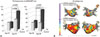
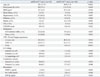
While the AF and control groups overall did not show significant differences in the frequency of mtDNA4977-mut in peripheral blood, the two groups did significantly differ when they were further broken down according to age (Fig. 2A). Patients were first divided according to the median age of 51 years. Within the AF patient group, the prevalence of mtDNA4977-mut was significantly higher in those 51 years and older (35.5% vs. 12.7%, p<0.001) than in those younger than 51 years old. The mutation was still more frequent in older AF patients when they were compared to their age-matched controls (35.5% AF vs. 22.7% control, p=0.038). These findings were significant when we divided the AF patient population between those younger than 65 or those older than 65 years old, where the cut-off point was suggested by a commonly utilized risk score for stroke, CHA2DS2-VASc score (Fig. 2A). Within the AF group, AF patients with mtDNA4977-mut were on average older (p<0.001), exhibited a higher frequency of diabetes (p=0.008), and were more likely to take angiotensin converting enzyme inhibitor/angiotensin II receptor blocker (p=0.007) or statin (p=0.001) than those without mtDNA4977-mut (Table 2).
mtDNA497-mut and electro-anatomical remodeling of the left atrium
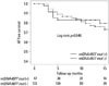
Table 2 shows comparisons of electroanatomical characteristics between AF patients with and without mtDNA4977-mut. Patients with mtDNA4977-mut had a greater LA size (p=0.014) and higher mitral inflow peak velocity (E)/diastolic mitral annular velocity (Em) ratio (p<0.001) than those without the mutation. Endocardial voltage (p=0.035) as well as conduction velocity (p=0.048) on the posterior LA were lower in patients possessing the somatic mutation (Fig. 2B). Upon multivariate logistic regression analysis, E/Em ratio [odds ratio (OR) 1.113, 95% confidence interval (CI) 1.011-1.225, p=0.029] was found to be independently associated with mtDNA4977-mut (Table 3). In protein biomarker assay, plasma levels of TIMP-1 (p=0.004) and pro-ANP (p=0.036) were higher in AF patients with mtDNA4977-mut than those without the mutation (Table 2), and TIMP-1 was independently associated with mtDNA4977-mut in patients with AF (OR 1.896, 95% CI 1.094-3.284, p=0.023) (Table 3). However, clinical recurrence rates after AF catheter ablation were not significantly different between patients with and without mtDNA4977-mut (Fig. 3).
DISCUSSION
In the current study, we reported that a somatic mutation, mtDNA4977-mut, detected in peripheral blood is associated with AF, the presence of which varied depending on age. Additionally, peripheral mtDNA4977-mut was associated with advanced electro-anatomical remodeling of the LA, elevated left ventricular filling pressure estimated by E/Em, and high plasma levels of TIMP-1. To the best of our knowledge, our study is first to demonstrate an association between peripheral blood mtDNA4977-mut, AF, and remodeling, suggesting the potential applicability of the mtDNA mutation as a biomarker of cardiac arrhythmia.
AF is a degenerative disease and is related to mtDNA4977bp deletion mutation
AF is now commonly recognized as a disease of aging; advanced age increases one's predisposition to this arrhythmia. According to recent studies, the mechanism underlying aging is primarily a progressive decline in mitochondrial function.15 Leakage of superoxide from the mitochondrial electron transport chain induces oxidative damage and accumulation of damage over time results in mtDNA deletion.16,17 mtDNA4977-mut is one of the most common deletion mutations identified in mitochondria. This mutation is frequently found in aging human tissues, especially those vulnerable to increased oxidative stress, like the heart.18,19 In the mutation, deletion of a sequence that encodes subunits of ATPase and NADH dehydrogenase disrupts aerobic metabolism and ultimately generates increased amounts of radical oxidative stress (ROS).11,20 Similarly, many studies attempting to reveal the pathophysiology of AF have pointed to mitochondrial dysfunction and ROS as important mediators thereof: for example, NADPH oxidase,21,22 NOS,23,24 and MPO,25,26 previously discovered as major sources of ROS in the heart,27 have now been shown to be critical in arrhythmogenesis. Furthermore, a growing body of evidence supports the idea that mitochondrial dysfunction can directly alter cardiomyocyte excitability and cell-to-cell coupling.28,29,30 As aging and AF exhibit surprising similarities, subsequent studies have attempted to investigate whether there indeed exists an association between aging, mitochondrial dysfunction, and AF. Lai, et al.10 examined right atrial appendage tissues and found that both aging and AF were independently associated with accumulation of mtDNA4977-mut. Lin, et al.,11 also observed increased oxidative damage, including the mtDNA deletion mutation, in atrial muscles from fibrillating hearts in comparison to tissue in sinus rhythm. However, whether ROS and mitochondrial dysfunction as result of the ageing process alone are sufficient to produce an arrhythmogenic atrial substrate remain in question.15 In our study, we observed that among AF patients, those with the mtDNA mutation were older on average. In comparison to their age-matched controls, elderly AF patients still maintained a higher prevalence of mtDNA4977-mut.
Electroanatomical remodeling and diastolic dysfunction in mtDNA4977-mut
Over the course of AF, the presence of mtDNA4977-mut was associated with more accelerated electroanatomical remodeling. Our current study revealed that parameters reflecting electroanatomical remodeling, such as atrial voltage and conduction velocity, are significantly different between patients with and without the mtDNA4977-mut. In line with our results, Tsuboi, et al.31 postulated that a rapid atrial rate or AF induced hypoxia in the atrium, increasing the generation of oxygen radicals. Further deterioration of mitochondrial function ensued as damage to mtDNA accumulated. The level of ATP in atrial muscle subsequently fell, resulting in impaired calcium handling, increased calcium in the cytoplasm, and reduced L-type calcium current.31 Ultimately, atria were electrically remodeled, beginning a vicious cycle in which AF begets AF.32 In addition to electrical remodeling, mtDNA4977-mut appears to be also associated with structural remodeling of the LA in AF. E/Em ratio and LA size, according to our observation, were significantly elevated in the mtDNA4977-mut positive AF patient group, and E/Em ratio was independently associated with the mutation in AF. From our analysis, we speculated that higher LV filling pressure indicated by higher E/Em evoked more advanced structural remodeling of LA in compensation.33,34,35 We previously reported that impaired LV diastolic function significantly contributed to electroanatomical remodeling of LA in patients with PAF.36 In the current study, this interaction between the two chambers was especially prominent in AF patients with the mtDNA mutation. When we compared AF types, mtDNA4977-mut was present in 24.8% of PAF patients and in 24.1% of PeAF patients (p=0.867). This suggests that mtDNA4977-mut is more likely to be associated with ageing, metabolic factors, ventricular diastolic dysfunction, or left atrial remodeling, rather than AF burden itself.
Clinical implications
Over time, surrogate markers have gained increasing clinical importance, as detection allows for not only early diagnosis of a disease but also prognosis. This is especially true in chronic degenerative diseases like AF. Several protein biomarkers have been shown to reflect various aspects of AF, such as electro-anatomical remodeling37 or chronicity.38 However, multiple confounding factors, such as transient inflammation and associated systemic disease, affect plasma levels of these protein biomarkers and ultimately prevent effective and accurate use in the clinic. In contrast to protein biomarkers, genetic markers are reproducible and stable enough to characterize the state of disease in patients with greater certainty. As our study showed, mtDNA4977-mut may serve as a stable indicator of patients with AF or at risk of rapid remodeling of the LA due to AF. Furthermore, the results of the current study suggest the potential utility of peripheral blood for the detection of mtDNA4977-mut in cardiac disease or arrhythmias. Previous studies on the association between mtDNA mutation and AF have been conducted by acquiring atrial tissue for analysis,10,11 which is clinically impractical. As these results have been reproduced in our study with peripheral blood, we demonstrated that mtDNA mutation can be readily assessed as a valuable biomarker of AF; further studies are need to confirm our results in different populations.


 XML Download