

 PDF
PDF ePub
ePub Citation
 Print
Print


INTRODUCTION
Laparoscopic cholecystectomy (LC) is the gold standard approach for treating benign gallbladder diseases.1 With advancements in laparoscopic techniques and experiences, the indication of LC has expanded to acute severe cholecystitis, and even early gallbladder cancer.2,3 In addition, reduced-port LC has been actively applied for cosmetic purposes.4,5 Recently, single-port LC has been introduced with the goal of providing patients with minimally invasive cosmetic surgery, stimulated by several pioneering gastrointestinal endoscopists who tried to perform surgical procedures using endoscopic routes; this technique is known as natural orifice transluminal endoscopic surgery.6 However, several technical difficulties such as inter-instrumental cloudiness from laparoscopic movements within the limited space (single port) require specialized laparoscopic instruments for improving surgeons' ergonomics and the effectiveness of dissection. Therefore, while it is true that single-port LC is regarded as feasible and safe, it is not very popular.
A robotic surgical system has been introduced to overcome the limitations of conventional laparoscopic surgery, and its clinical application is currently available for single-port surgery.7,8 The most outstanding features of the robotic single-site surgical system are that it has curved flexible laparoscopic instruments for switching between surgeons' right and left orientations to improve ergonomics, yet it does not allow wrist-like motion at the tip of the instrument. There are also ready-made trocar insertion sites in specially designed silicone-ports that make it easy to establish robotic settings. The accessory trocar can be controlled by assisting surgeons and is designed to provide active retraction of the gallbladder to open Calot's triangle. In addition, the single-site robotic surgical system is incorporates a new technique for biliary tree visualization, consisting of a preoperative intravenous injection of indocyanine green (ICG) and the use of a near-infrared (NIR) light during surgery for real-time fluorescent cholangiography.9
To the best of our knowledge, our current experiences of robotic single-site cholecystectomy (RSSC) are thought to be the first to be reported in Asia. In this article, we report our initial experiences of RSSC and compare them with our conventional single-port technique (single-fulcrum laparoscopic cholecystectomy10,11) to validate the technical feasibility of RSSC.
MATERIALS AND METHODS
From October 2013 to November 2013, five consecutive cases of RSSC were performed by a single surgeon in a single institution. We carefully selected patients for RSSC based on limited indication regarding patients' concern for cosmetic outcome after surgery, the presence of an asymptomatic gallbladder (GB) stone, and the presence of a GB polyp or GB adenomyomatosis without inflammation.
Robotic single-site surgical system
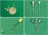
The da Vinci Single-Site™ Instrumentation and the da Vinci® Si™ System (Intuitive Surgical®, Sunnyvale, CA, USA) was adopted for RSSC. A single-site port was used for RSSC that included five lumens consisting of an 8.5-mm endoscope, a 5-mm or 10-mm accessory port, a curved cannula, and an insufflation adaptor. Additionally, a curved 5-mm instrument cannula designed for optimizing triangulation toward the operative field and several 5-mm semi-rigid instruments were employed for the operative procedure (Fig. 1).
Surgical procedure
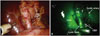
The robot single-site surgical system was applied to RSSC with a specialized single port for robotic surgery and a curved cannula with flexible instruments. A vertical 2-cm transumbilical skin incision was made, and the fascia layer was opened in same direction. After the single port consisting of a pliable silicone architecture was inserted into the fascial opening using Kelly forceps and an Army retractor, a pneumoperitoneum was created by carbon dioxide gas inflation. A camera was inserted to explore the peritoneal cavity and localize the fundus of the gallbladder after inserting the 8.5-mm camera port. The patient table was rotated to align with the orientation of the umbilicus to the fundus of the gallbladder (main axis). The patient-side cart of the robotic surgical system was moved to the patient table along the main axis. The robotic arm was docked to the camera port, curved cannula, and accessory trocar, according to predetermined sequences.7 Flexible robotic instruments were inserted through the curved cannula, and the robotic system provided a switching motion between right-hand and left-hand orientations to improve the surgeon's ergonomics. The assistant surgeon performed gallbladder traction toward the lateral and upward direction to expose Calot's triangle. After dissecting around the GB neck and cystic duct, the cystic duct and cystic artery were ligated securely by intracorporeal tie ligation and a Hem-o-lok clip and then divided with robotic scissors. Compared to laparoscopic single-port surgery, the robotic single-port surgical system provided a stable environment to perform intracorporeal tie ligation, despite the absence of EndoWrist movement. Additionally, the robotic single-port surgical system has a special feature, called Intraoperative Firefly™ Fluorescence Imaging. This feature allows the surgeon to visualize the biliary system during the operation by intravenous injection of ICG before the operation and a NIR light for real-time fluorescent cholangiography (Fig. 2, Supplementary Video 1). After dividing the cystic duct and the cystic artery, the gallbladder was dissected meticulously from the liver bed to prevent perforation of the gallbladder and spillage of bile into the peritoneal cavity. An Endo-pouch was inserted into the operative field via an accessory trocar, which was handled by the assistant surgeon. The specimen was retrieved using the Endo-pouch and delivered through the transumbilical incision. The fascia and skin were closed layer by layer.
Comparison between SFLC and RSSC
To compare the improvement of proficiency and learning period for early experiences of the new surgical technique, the first 20 cases that received a single-fulcrum laparoscopic cholecystectomy (SFLC) were obtained in our prospective data set.10,11 We compared factors including clinicopathologic factors and operative outcomes between RSSC and SFLC. The total operation time was defined as the length of time from the beginning of the skin incision to the final closure of the wound. The actual dissection time included the period from the dissection of Calot's triangle to the retrieval of the specimen.
Statistics
Continuous variables are expressed as the mean±the standard deviation and categorical variables are shown as frequencies and percentages. The Mann-Whitney U test for continuous variables and the chi-square test were employed to explore statistically significant associations between parameters. Statistical significance was determined if the p-value was less than 0.05.
RESULTS
General characteristics of patients with robotic single-site cholecystectomy
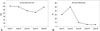
RSSC was performed consecutively in 5 patients by a single surgeon between October 2013 and November 2013. Four patients were females and one was male; the median age of the patients was 35 years old (range: 29-53) (Table 1). Despite strict selection criteria for patient enrollments, we observed moderate inflammatory changes around the Calot area and dilated cystic duct requiring intracorporeal tie ligation before endoscopic clipping in most cases. Additionally, the surgical system induced an upward medial retraction of the GB by the assisting surgeon, which caused a narrowing of Calot's triangle, leading to prolonged dissection time, in contrast to SFLC (Table 2). Neither bile duct injury nor bile spillage occurred during any of the operations. Robotic setting (docking) time, actual dissection time, and intraoperative blood loss seemed to decrease gradually as case numbers increased (Fig. 3). There were no open conversion cases, and most of the patients were discharged the day after the operation.
Comparison between initial experiences of RSSC and SFLC
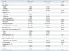
We compared the perioperative outcomes of RSSC (n=5) with the first 20 outcomes of SFLC (n=20) in order to evaluate the technical validation of the robotic single-site surgical system when applied to laparoscopic cholecystectomy (Table 2). Based on previous analysis of the learning curve for SFLC,11 we obtained 20 initial cases as the training phase in the database for SFLC. In comparisons between RSSC and SFLC groups, there were no statistically significant differences in most patient-related factors, such as age, sex, Body Mass Index, diagnosis, and American Society of Anesthesiologist score (p>0.05). The operative outcomes regarding intraoperative blood loss, bile spillage during operation, postoperative pain scale values, postoperative complications, and length of hospital stay did not show significant differences between the two groups (p>0.05). However, there were significant differences in operative time (RSSC: 132.6±25.2 min; SFLC: 39.75±15.6 min; p=0.001) and actual dissection time (RSSC: 53.4±8.4 min; SFLC: 32.2±13.4 min; p=0.003) between the two groups.
DISCUSSION
It is interesting to note that our unique technique of SFLC may be a prototype for current RSSC in that the operative view is similar except for the GB retraction method and ergonomic issues. In SFLC, two working instruments cross each other at the fascia layer (single fulcrum). With traction of the gallbladder neck performed by the right hand, the surgeon needs to use non-dominant left-hand movement to avoid inter-instrumental cloudiness.11 However, in the robotic system, this right-left orientation problem in our SFLC was completely solved: the surgeon's right-hand motion in the console controlled the left-sided robotic arms, yet the effector movement was noted in right side of the patient due to the curved configuration, maximizing surgeons' ergonomics when performing single-site laparoscopic surgery. In addition, intraoperative stable 3-D images, no tremor, and real-time fluorescent cholangiography provided optimal conditions for safe laparoscopic single-site cholecystectomy. Even in cases of dilated cystic duct, intracorporeal tying of the cystic duct was feasible (in fact, 4 patients out of 5 in the present series required intracorporeal tying due to dilated cystic duct) (Table 1), indicating the possible of expansion of this minimally invasive cosmetic surgery.12
Due to technical difficulties in laparoscopic single-site surgery, surgeons' experience levels are very important in performing laparoscopic single-site cholecystectomy. It is estimated that at least about 5-20 cases are necessary for overcoming the learning curve,13,14,15 and most learning curves in laparoscopic single-site cholecystectomy are thought to be overcome during surgery. On the contrary, the learning curve of robotic single-site cholecystectomy is mostly related to the robotic setting period. Once the operative setting is optimally defined, the unique characteristics of the robotic surgical system make it easy to perform single-site cholecystectomy.16,17 In addition, we just applied Firefly Fluorescence Image in last 2 cases. It was felt that this system can enhance the safety of laparoscopic cholecystectomy to avoid unnecessary bile duct injury. We need to accumulate further experiences about it.
However, it should be noted that there are several disadvantages of the robotic surgical system when performing single-site cholecystectomy. Surgeons are free from instrumental cloudiness; however, effector instruments cannot be angulated. Therefore, the degree of freedom in the intracorporeal field is thought to be the same as a conventional laparoscopic system, or even worse, as the extracorporeal direction of the effector instrument's movement cannot be changed. In our laparoscopic single-site cholecystectomy technique, SFLC, a right-angled instrument was very useful for overcoming this limitation. It allows surgeons to change the direction of dissection when identifying the cystic duct and cystic artery. However, there is no reliable robotic instrument to overcome this technical pitfall to date. The energy source design is also limited to a mono-polar cautery hook. Thus, the electric cautery cannot be used freely, and the robotic arm should be changed in order to switch the site of the energy source. Additionally, the accessory trocar is too close to the port for the robotic camera and the left side of the camera port, resulting in ineffective traction of the gallbladder due to limitations in right-left movement. The optimal direction of gallbladder traction should be towards the upward lateral side in order to open Calot's triangle. However, with the current robotic surgical system, we observed that the robotic camera crosses under the laparoscopic instrument for the retraction of the gallbladder, resulting in an upward medial traction of the gallbladder and narrowing Calot's triangle. This operative view made surgical dissection of the Calot area very difficult, leading to prolonged operation time in our series.
In addition, difficult operations were expected in cases of gallbladder rupture or bleeding during procedure. In such cases, the optimal operative view could not be maintained, as the accessory port should be used for suction and manipulation for preventing bile spillage or for suctioning bleeding to ensure the operation field. GB traction could not remain steady in those cases. Therefore, strict patient selection and careful dissection are thought to be important in taking advantage of RSSC, which is why our present series includes more asymptomatic GB polyps than the SFLC series in our retrospective data set (p<0.004) (Table 2). Of course, the high cost of the robotic surgical system would be also another major obstacle to expanding RSSC to routine clinical practice.12 In the near future, it is highly expected that new surgical instruments, such as a right-angle dissector, as well as angulated motion of effector instruments, and the available energy source system in the Maryland dissector needs to be improved in order to be more effective and safe when used to perform single-site cholecystectomy.
In conclusion, the robotic single-site surgical system generally provides a more comfortable environment and improved ergonomics for surgeons performing laparoscopic cholecystectomy. Special features of the robotic system, such as intraoperative fluorescence imaging and ergonomic normal hand orientation, can contribute to safe and comfortable operation in single-site minimally invasive cosmetic surgery. However, several potential disadvantages should be considered when performing RSSC. Nevertheless, with advancement of technical innovation and strict case selection, RSSC could be an alternative that may become a potential means of safe and effective minimally invasive cosmetic surgery. More experiences must be carefully performed in order to exactly address the role of RSSC in the advanced laparoscopic era.

 XML Download