

 PDF
PDF ePub
ePub Citation
 Print
Print


INTRODUCTION
Cervical spondylotic myelopathy (CSM) is a disorder resulting from cervical spinal cord compression. Laminoplasty is a widely used procedure for the surgical treatment of cervical spinal cord compression in order to achieve safe decompression and postoperative stability. Since 2005, we have been using open-door en bloc laminoplasty (Itoh and Tsuji) with hydroxyapatite (HA) spacers. We previously reported good clinical results using this laminoplasty with preservation of the C2 and C7 spinous processes.1
This open-door en bloc laminoplasty procedure requires the creation of a hole that allows a thread to be placed through a lateral mass for HA spacer fixation. The lateral mass is occasionally damaged when creating the hole during cases of reoperation, foraminotomy, or osteoporosis. Here we report results using an HA spacer fixation system.
MATERIALS AND METHODS
First, a preliminary study was conducted to examine whether ceramic spacers can be affixed safely using anchors in cervical open door laminoplasty. The second study was prospectively performed to evaluate the feasibility of this new technique based on the results of the preliminary study.
Study 1
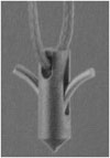
Sixteen consecutive patients who underwent laminoplasty for cervical myelopathy between November 2007 and July 2008 at our hospital were included in this preliminary study. Their age ranged from 33 to 85 years old. For HA spacer fixation, we fixed a Mini Quick Anchor Plus system to a lateral mass with either 2-0 or 0 Ethibond (Johnson & Johnson, Piscataway, NJ, USA) (Fig. 1). One spacer was used in 14 patients, and two spacers were used in two patients. One ceramic spacer was usually used at C5 if the extent of laminoplasty was between C4 and C6 and associated with laminotomy at C3 and C7 to preserve the spinous processes and the attached muscles. Two ceramic spacers were used if laminoplasty was extended to C3 and/or C7. Plain radiography and CT were used to confirm the site of anchor insertion.
Study 2
Inclusion criteria for this prospective study were as follows: definite myelopathy on physical examination and load compression observed for at least three disk levels between C3-4 and C6-7. Forty-five consecutive patients with CSM and 43 consecutive patients with OPLL were prospectively followed (6-49, average: 26 months). They consisted of 62 men and 26 women, aged 40-85 years (average age 67 years). The anchors of the Mini Quick Anchor Plus system were used in 20 patients in the early study period, and 5.5-mm Twinfix anchors (Smith and Nephew) were used in patients in the later study period.
Clinical outcomes were estimated using the Japanese Orthopedic Association scoring system (JOA score). The rate of recovery, which indicated the degree of normalization after surgery, was calculated using Hirabayashi's formula: (postoperative score-preoperative score)×100/[17 (full score)-preoperative score]. The symptoms were evaluated at more than 6 months after surgery to avoid early complaints that might be directly related to surgical intervention. When the complaints persisted for more than 1 month, the symptoms were judged to be "present". Radiological findings were analyzed for postural anomalies of the cervical spine, which was defined in the lateral view in the neutral position as lordosis and kyphosis using the angle between the lines passing through the posterior margin of the C2 vertebral body and C6 or C7. The lordotic angle was reported as a positive value, and the kyphotic angle as a negative value.
RESULTS
Study 1
Postoperative CT showed that the anchor was displaced outside the bone in 2 patients, however, this displacement did not result in any clinical problems. Plain radiographs showed no dislodgment of the anchor during the follow-up period. A spacer dislodged due to a suture rupture in one early patient, who received a 2-0 Ethibond anchor. Once 0 Ethibond anchors were used, no suture ruptures were observed.
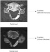
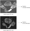
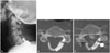
Insertion sites were examined for anchors displaced outside the bone. For 16 patients, an anchor was inserted into the pedicle of the upper vertebral arch in eight patients and in the upper intervertebral foramen in the other eight. The first eight patients had anchors inserted within the bone, while two of the latter eight patients had anchors that were displaced outside of the bone (Fig. 2). Thus, the angles of anchor insertion were examined in eight patients who had anchors inserted into the upper intervertebral foramen. The anchors were within the bone in four patients with an insertion angle of greater than 30° from the sagittal plane. Displacement in the intervertebral foramen was seen in two of four patients with an insertion angle of 30° or less (Figs. 3 and 4).
Study 2
In the CSM group, the average operation time was 111 minutes and the average blood loss was 119 mL; the corresponding values for the OPLL group were 112 minutes and 149 mL, respectively. In the CSM group, the average JOA score improved from 9.5 points preoperatively to 13.3 at follow-up (recovery 51%). In the OPLL group, the average JOA score improved from 10.1 (5-14) points preoperatively to 14.4 (11-16) at follow-up (recovery 62%). In all patients, postoperative CT scans showed that anchors were securely inserted in the bone. None of the patients complained of axial neck pain with a VAS score >30/100. In the CSM group, the mean lordosis angle decreased from 13.4° (range -4.5° to -35.8°) preoperatively to 11.9° (range -5.6° to -33.1°) at follow-up. In the OPLL group, the mean lordosis angle decreased from 14.4° (range 0.4° to -33.5°) preoperatively to 11.9° (range -7.2° to -43.5°) at follow-up. There were no serious complications.
Case presentation
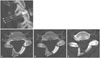
A 41-year-old woman presented with OPLL. The patient had previously undergone posterior decompression for thoracic vertebral lesions. During the study period, cervical laminoplasty was performed using two spacers. Placement of a spacer in the lower cervical vertebra was facile, as an anchor was used that did interfer with the rod (Fig. 5).
DISCUSSION
Representative procedures of open-door cervical laminoplasty are those proposed by Hirabayashi and by Itoh and Tsuji.23 The Hirabayashi procedure involves suture fixation of the opened laminae. After its expansion, there is a concern that sutures in the muscle can push back on the vertebral canal. The advantage of the Itoh-Tsuji procedure is the maintenance of stable support, as the spinous process is used for bone grafting; therefore bone harvesting from other sites is unnecessary. However, the fixation procedure is considered to be complex. The Itoh-Tsuji procedure using spacers is used at our hospital. It is very important to firmly place the spacers in the opened side and to affix them securely to prevent dislodgement into the spinal canal and restenosis. In patients with conditions such as osteoporosis, failure can occur when creating a hole in the lateral mass through which the suture thread passes for spacer fixation. Refixation is considered to be difficult once such failure occurs.
There are many techniques for maintaining and securing the expanded spinal canal, such as a fascial or joint-capsule anchoring suture, spacer interposition, allograft, autograft, or miniplate fixation.45678 Yang, et al.910 achieved fixation using a suture anchor on the hinge side in an open-door laminoplasty. They reported the relative technical ease of the procedure and only a small number of complications. Similarly, Khan, et al.11 reported the utility and ease of using the suture-anchor system.12 Kurokawa, et al.13 reported the validity of using suture anchors in laminoplasty. Hu, et al.14 conducted a study to compare open-door laminoplasty between patients with anchors on the hinge side and patients with titanium miniplates. They reported that titanium miniplate fixation is more effective than suture anchor fixation in preventing laminar closure, accompanied by a higher JOA recovery rate at 1-year follow-up and a lower incidence of axial symptoms.15 In our current study, the first report of the use of suture anchor fixation at the open side of laminoplasty, the recovery rate of the JOA score were 51% in the CSM group and 62% in the OPLL group. The losses of cervical lordosis were 1.5° and 2.5°, respectively. None of the patients complained of axial neck pain with a VAS score >30/100. These clinical results were equivalent or superior to the previous studies. By fixing the suture anchor at the open side, the clinical results were equivalent to the Tsuji-Itoh method and superior to the methods fixing the suture anchor at the hinge side.16
In conclusions, the use of the suture anchor system made it unnecessary to create a hole in the lateral mass and enabled reliable and faster fixation of the HA spacers in open-door laminoplasty. The technique did not appear to require a significant learning curve. However, in the first study, anchor misplacement occurred in several cases. The results of this study indicated that an anchor should be inserted in the pedicle area of the vertebral arch (cranial side of the lateral mass), with an insertion angle in the cross section of approximately 30° laterally and in the sagittal section of approximately 30° superiorly.
 XML Download