

 PDF
PDF ePub
ePub Citation
 Print
Print


INTRODUCTION
Elastin is involved in the maintenance of vascular compliance. A change in the balance between elastin synthesis and degradation may lead to the development of a pathological vascular condition.1 Elastin degradation has been reported to be a determinant of arterial stiffness and has prognostic value for clinical outcomes in high-risk populations.2 In many studies, elastin degradation peptides have been proposed as participating in the progression of atherosclerosis through the activation of various biological processes.3
For decades, anti-elastin antibodies have been assayed by immunological techniques. Levels of the antibody and the changes that occur in normal and pathological conditions have also been investigated in prior studies.456 Therein, blood levels of elastin peptides and those of the antibodies against elastin peptides did not correlate.78 Moreover, elastin degradation by diverse enzymes can release various epitopes, and any antibody-disease association may depend on the origin of the epitopes.9 To date, the clinical relevance of antibody levels is not thoroughly understood and interpretation of antibody levels warrants caution. Although a few studies have investigated anti-elastin antibody levels in atherosclerotic cardiovascular disease, across these studies, the relationship between antibody levels and atherosclerosis has been inconsistent.5610
The aim of this study was to evaluate the association between anti-elastin antibody levels and coronary artery disease (CAD). We also assessed relationships between antibody levels and cardiovascular risk factors using a newly developed enzyme-linked immunosorbent assay (ELISA). We found that lower levels of anti-elastin are related to CAD and that this relation is linked to arterial stiffness.
MATERIALS AND METHODS
Study patients
This study included 171 patients with CAD and 174 control subjects without CAD. Participants were drawn from the database of the Cardiovascular Genome Center at Yonsei University Health System, Seoul, Korea. CAD patients were recruited when undergoing coronary angiography for chest discomfort or chest pain. Subjects aged 30-70 years with stenosis >50% in at least one epicardial coronary artery were included. The exclusion criteria included uncontrolled high blood pressure (BP) (systolic BP >180 mm Hg or diastolic BP 110 mm Hg); uncontrolled diabetes mellitus (fasting blood glucose 180 mg/dL); a history of structural heart disease with or without heart failure; thyroid [thyroid stimulating hormone <lower limit of normal or upper limit of normal (ULN)], liver (serum aminotransferase >2×ULN) or kidney disease (serum creatinine >1.5 mg/dL); acute or chronic inflammatory disease; and malignant neoplasm. Control subjects were matched for sex and age (±5 years), and selected from the database of a community health check-up center in Mapo-gu, Seoul, Korea. Written informed consent was obtained from all participants, and the study protocol was approved by the Institutional Review Board of Severance Hospital, Seoul, Korea.
Collection of clinical and angiographic data
At the time of enrollment, subjects were interviewed regarding their individual medical histories and underwent complete physical examinations. Hypertension was defined as a BP >140/90 mm Hg on two or more occasions or undergoing antihypertensive treatment. Diabetes mellitus was defined as a fasting blood glucose ≥126 mg/dL, postprandial blood glucose ≥200 mg/dL, or current treatment with hypoglycemic medications. Hyperlipidemia was defined as low-density lipoprotein-cholesterol ≥160 mg/dL. CAD characteristics were evaluated by a cardiologist who was blinded to the study's purpose. The cardiologist first evaluated the most severe clinical presentation of CAD in the patient's history, and then the number of coronary arteries with at least one stenosis of >50%. After a 12-h fasting period, venous blood samples were collected, centrifuged, and stored at -80℃.
Measurement of augmentation index and pulse wave velocity
The augmentation index (AI) was measured in the sitting position after a 5-min rest using a radial artery tonometry device (SphygmoCor, AtCor Medical, Sydney, Australia), as described previously.11 The central BP was calibrated to the brachial BP measured using the OMRON HEM 7080IT (Omron Healthcare, Kyoto, Japan). Briefly, using a high-fidelity micromanometer (Millar Instruments, Houston, TX, USA), peripheral pressure waveforms from the radial artery were recorded at the wrist of the dominant arm. Central systolic BP, diastolic BP, pulse pressure, augmentation pressure, and AI were acquired from the pulse waveform analysis. Augmentation pressure is the difference between the second and first systolic peak pressures, and the AI is defined as the ratio of augmentation pressure to aortic pulse pressure.
The heart-to-femoral pulse wave velocity (hfPWV) was determined by a VP-2000 pulse wave unit (Nippon Co. Ltd., Komaki, Japan), as described previously.12 Briefly, after an overnight fast and a 5-min rest, the PWV was measured in a supine position. Carotid and femoral artery pressure waveforms were recorded from multi-element tonometry sensors at the left common carotid and left femoral arteries. The electrocardiogram was monitored from electrodes placed on both wrists. Heart sounds were detected by a microphone placed on the left sternal edge in the third intercostal space. The hfPWV, a marker of central aortic stiffness, was calculated using the equation Lhf/(Thc+Tcf). Lhf refers to the distance from the heart to the femoral artery, Thc is the interval between S2 and the notch of the carotid pulse wave, and Tcf is the time interval between the carotid and femoral artery pulse wave.
Enzyme-linked immunosorbent assay (ELISA) for anti-elastin antibody titer
The anti-human elastin antibody quantification assay was performed using a modified ELISA protocol.13 Briefly, human aortic elastin peptides were purchased from EPC (Owensville, MO, USA) and dissolved and used to coat ELISA plates (Greiner, Kremsmunster, Austria). After incubation and washing with phosphate-buffered saline (PBS) containing Tween 20 (BioRad, Bedford, MA, USA) (PBS/Tween), plates were blocked by I-block (Tropix, Bedford, MA, USA) and incubated. After washing with PBS/Tween, human serum samples were diluted and incubated. After washing, biotinylated chicken anti-human IgG antibody (Immunology Consultants Laboratory, Newberg, OR, USA) was added and the samples were incubated. Plates were washed again, then alkaline phosphatase-conjugated streptavidin (BD bioscience, San Diego, CA, USA) was added, and the samples were incubated. After a final wash, alkaline phosphatase substrate (Sigma, St. Louis, MO, USA) was added and the plates were allowed to develop until the standard curve was readily apparent. The colorimetric reaction was terminated by adding sodium hydroxide, and the optical density of the individual wells was determined. We selected one sample from a subject with emphysema that showed a very high optical density as a standard for relative quantification in all assays.
Statistical analysis
Differences between cases and controls were evaluated using chi-square tests for categorical variables and Student's t-tests for continuous variables. As anti-elastin levels had a log-normal distribution, they were log-transformed before analysis. The relationship between anti-elastin titers and clinical or vascular characteristics was analyzed by Student's t-test or an analysis of variance test. To identify independent associations between anti-elastin levels and vascular characteristics, univariate and multivariate linear regression analyses were performed. Variables that showed p<0.15 in the univariate analysis were entered into the multivariate analysis. SPSS software, version 18 (SPSS Inc., Chicago, IL, USA) was used for all statistical analyses.
RESULTS
Characteristics of the study subjects
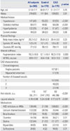
The characteristics of the 345 study subjects (mean age: 51.0±7.7 years, men: 63%) are presented in Table 1. Hypertension, diabetes mellitus, and hyperlipidemia were observed with greater frequency in the CAD group than in the control group. CAD patients also had a higher diastolic BP, AI, and hfPWV. The blood anti-elastin antibodies levels were significantly lower in the CAD group than in the control group (p<0.001). In the CAD group, 39% of subjects experienced myocardial infarction and 41% of coronary angiographies revealed multi-vessel involvement.
Determinants of anti-elastin
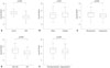
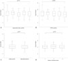
The log anti-elastin levels were significantly lower in CAD patients, men, and individuals with hypertension, diabetes mellitus, hyperlipidemia, or high hfPWV (Figs. 1 and 2). In CAD patients, anti-elastin levels were not dependent on myocardial infarction history or the number of diseased vessels (Fig. 2). In univariate analysis, male sex, hypertension, diabetes mellitus, hyperlipidemia, and hfPWV were inversely correlated with log anti-elastin levels. In multivariate analysis, male sex, diabetes mellitus, hyperlipidemia, and AI were identified as determinants of log anti-elastin levels (Figs. 1 and 2, Table 2). In the analysis of potential AI determinants, the relationship between index and log anti-elastin levels was again found to be significant and independent of other clinical risk factors (Supplementary Table 1, only online).
DISCUSSION
The present study assessed the relationship between anti-elastin antibody levels and CAD and its characteristics. This study revealed that anti-elastin levels are lower in CAD patients than in matched controls. Anti-elastin levels were associated with sex, hypertension, diabetes mellitus, hyperlipidemia, and arterial stiffness, but not with myocardial infarction history or angiographic severity of CAD. AI, a parameter of arterial stiffness, was identified as a determinant of antibody levels independent of other confounding risk factors. Our results suggest that lower anti-elastin levels are related to CAD and that this relation is linked to arterial stiffness rather than advancement of atherosclerosis. We quantified this antibody using a novel assay against aortic elastin peptides, and our result may be used as basic data for further validation studies.
One major finding of our study was lower anti-elastin antibody levels in CAD patients than in controls. This result is consistent with that reported by Baydanoff, et al.,6 which indicated that anti-human aortic elastin antibody levels were lower in patients with atherosclerosis. Although its mechanism was not fully elucidated at that time, the authors suggested an adaptive restriction of the immune response to avoid aggravation of the atherosclerosis as a possible cause. However, the antibody levels and elastin-derived peptide levels did not correlate. In the same patients with atherosclerosis, their elastin degradation peptide levels were higher than those in other subjects.8 In another study,7 the antibody and peptide levels did not correlate either. Clarification of the relationship between antibody levels and elastin-derived peptide concentrations may help better understand their physiology. Meanwhile, a few studies have shown results that differ from ours. In one study, anti-elastin peptide antibody levels were higher in patients with ischemic heart disease and obliterative arteriosclerosis of the legs.5 In another, a significant increase in serum alpha-anti-elastin antibody levels was demonstrated in patients with carotid plaque destabilization.10 It was also reported that cathepsins K and S at vascular matrix remodeling can degrade elastin, and this supports that elastolytic products can be elevated in progressed atherosclerosis.14 Furthermore, studies have shown that atherosclerosis-associated cytokines augment cysteinyl cathepsin activity, which can promote elastin degradation, in various vascular diseases.15 Taken together, caution should be exercised when interpreting antielastin antibody levels in clinical situations.
As mentioned above, the mechanisms and implications of varying anti-elastin antibody levels have not been fully understood. However, several possible roles have been proposed. Anti-elastin antibody binds elastin peptides and may block their chemotactic effect on inflammatory cells.16 Thus, lower antibody levels may be insufficient to block this effect of elastin peptides. Secondly, hyperactivity of the immune response to elastin may cause more vascular damage. Therefore, a decrease in antibody levels may result from the impaired immune reaction against elastin to reduce the aggravation of atherosclerosis.6 Nevertheless, uncovering the significance of the lower antibody levels in our study requires further analysis. For instance, in the current study, antibody levels were independently associated with sex, diabetes mellitus, hyperlipidemia, and an arterial stiffness index. Although antibody level was significantly associated with CAD, other cardiovascular risk factors also showed correlations with antibody levels. Thus, it is assumed that the relationship between antibody levels and CAD can be influenced in part by the vascular conditions developed by other risk factors. We found arterial stiffness to be an independent determinant of anti-elastin. Risk factors can coexist in a single patient and influence one another. Therefore, caution should be taken in interpreting the level of an antibody that is interrelated to other factors. Another point to consider is whether the antibody levels reflect a general vascular pathology or CAD specifically. Although the antibody levels were correlated with the presence of CAD in our study, they were not associated with disease advancement. The antibody concentration was linked to the arterial stiffness index, and this concentration may indicate functional change of non-coronary vasculature as well as the risk of CAD.
The relationship between elastin degradation and arterial stiffness has been investigated in many studies. Ruseva, et al.17 showed that the anti-elastin antibody level is lower when elastin degradation is experimentally slowed in rats. Maclay, et al.4 measured cutaneous elastin degradation and reported that this was positively associated with PWV. Our results are not in agreement with these reports, and therefore, clarification of the association between antibody level and stiffness is needed. Notably, whether the blood antibody level correlates with arterial stiffness, whether changes in antibody levels have clinically significant implications, and which clinical factors may influence antibody levels remain to be clarified. In this regard, our analysis of the association between antibody levels and clinical risk factors might serve as helpful basic data.
A prior study demonstrated that female mice have higher anti-elastin antibody levels than males, which is consistent with our data.18 The authors suggested that reproductive organ remodeling in female mice may be the cause of this finding, although this does not sound plausible in our subjects. Previous studies on the association between diabetes mellitus and antibody levels did not show consistent results. The fibulin-1 level, an elastin-associated protein, was lower in patients with type 2 diabetes than in controls.19 Conversely, in another study conducted in diabetic children, anti-elastin IgG level was positively correlated with diabetes duration, hemoglobin A1c level, and the level of antibodies to advanced glycation end-products. Although low-density lipoproteins interact with arterial elastin during the development of atherosclerosis,20 the data on the blood levels of elastin peptide or the antibody against it in hypercholesterolemia are limited. Interestingly, Fülöp, et al.5 showed that anti-elastin antibody levels decreased in subjects with IV hyperlipoproteinemia with hypertriglyceridemia, whereas we found that antibody levels were lower in patients with a history of hypercholesterolemia. On the other hand, it was shown in the study of Baydanoff, et al.8 that anti-elastin antibody levels decrease after the age of 60 years. In their study, no significant change in antibody levels was noticed until that age. In our study, antibody levels did not vary with subjects' age. However, because the age range in our subjects was relatively narrow, it is difficult to say whether age is significantly associated with antibody levels.
Our study was designed with well-matched cases and control subjects, and systemically assessed the effects of CAD or individual cardiovascular risk factors on anti-elastin antibody levels. In particular, the analysis of associations with detailed vascular characteristics is the strength of this study. Several findings were demonstrated for the first time in the current study, although they require further validation. Some limitations of our study can be pointed out. The contribution of AI to anti-elastin levels was significant but weak (beta=-0.006) and its clinical impact could be modest. In our study, we could only measure anti-elastin antibody levels. A simultaneous examination of elastin peptide level may have given us more insight into the pathological role and clinical interpretation of antibody levels. In addition, patients with CAD had slightly but significantly higher diastolic BP. Although it did not show significant relationship with arterial stiffness in our data, it could be one of confounders in the analyses. Lastly, we excluded subjects with uncontrolled hypertension or diabetes mellitus and these factors with medications might have partially affected our results.
To summarize, anti-elastin antibody levels were lower in CAD patients and associated with male sex and a history of hypertension, diabetes mellitus, or hyperlipidemia. Antibody levels negatively correlated with arterial stiffness, but not with myocardial infarction history or the severity of CAD. In conclusion, a lower anti-elastin antibody titer is related to CAD and this relation is linked to arterial stiffness rather than to the advancement of atherosclerosis. Our results may provide the basic data for further validation studies of this novel assay.

 XML Download