

 PDF
PDF ePub
ePub Citation
 Print
Print


INTRODUCTION
To successfully rejuvenate an aging face, contributing factors to changes associated with aging must be thoroughly assessed. The Asian face is markedly different from Caucasian face in many aspects, including skin tone, texture, elasticity, skin thickness, subcutaneous fat contents, and skeletal framework.123 Therefore, the aging process in Asian faces could be remarkably different from that in Caucasian faces.456
Three-dimensional (3D) CT studies revealed that the midfacial skeleton shows angular changes with aging in Caucasians.789 These changes may play a role in the loss of soft-tissue cheek mass support, cheek mass drooping, inferior and posterior displacement of the inferior orbital rim, distortion of orbital rim curve, and the scleral show in the midface. Difference in facial skeletal contour is most important for distinguishing between different ethnic groups. However, ethnic differences in age-related facial skeletal changes have not yet been fully established. Though several investigators have studied skeletal remodeling in aging Caucasian faces, Asian midfacial skeletal remodeling has not yet been reported. Understanding these inherent facial skeletal differences and applying them to rejuvenation surgery are essential for more desirable surgical outcomes.
This study aimed to analyze midfacial skeletal changes in aging Asian faces using a new method and to explore ethnic differences between Caucasian and Asian skeletal facial structures with aging.
MATERIALS AND METHODS
Data was collected in a retrospective manner from previously acquired facial CT scans at Ewha Womans University Hospital between December 2010 and April 2013. The 1.0-mm width axial images using a 64-channel multidetector computed tomography (MDCT) (SOMATOM Sensation 64, Siemens, Forchheim, Germany) were acquired at the 120 kV and 180 mA setting, and coronal and sagittal images were reconstructed along with axial images. Scans showing either soft tissue lesions or normal findings were included in this study. Scans showing any evidence of previous facial bone trauma, surgery, or the absence of upper dentition with the exception of the premolars were excluded from the study.
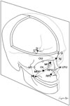
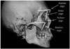
This study included a total of 223 Asians, all of whom were born and living in South Korea. For the evaluation of angular changes with aging, angular measurements of 4 bony regions (glabellar, orbital, maxillary, and pyriform aperture angles) were made using a method based on 3D vector mathematics. A line from the sella to the nasion was taken as the reference line. Each landmark as described below was identified, and a line was drawn to the reference line. Angular measurements were made as follows: the glabellar angle between the reference line and a line drawn from the maximal prominence the glabella to the nasofrontal suture; the orbital angle between the reference line and a line drawn from the most superior to the most inferior midportion of the orbit; the pyriform angle between the reference line and a line drawn from the nasal bone to the lateral inferior pyriform aperture; and the maxillary angle between the reference line and a line drawn from the most superior to the most inferior maxilla at the articulation of the inferior maxillary wing and alveolar arch. By employing the methods described in Fig. 1, 3D co-ordinates of each point on axial images were measured using a pixel lens cursor in a picture archiving and communication system (PACS) report viewer, software version 5.0 (INFINITT Co., Ltd., Seoul, Korea). Each point on an axial image was verified using reconstructed coronal and sagittal images in the PACS report viewer (Fig. 2). From given co-ordinates, the 2 vectors AB and AC were calculated. The dot product of the 2 vectors was defined as AB · AC= |AB| |AC| cos θ from geometric definition.10 This equation allows the angle θ, an angle between AB and AC to be precisely computed. The orbital aperture width (distance from the posterior lacrimal crest to the frontozygomatic suture) and the pyriform width (between both lateral margins of the pyriform aperture) were also measured using the PACS.
Statistical analysis was performed by the IBM SPSS software version 19.0 (International Business Machines Corp., Armonk, NY, USA), and a p value of <0.05 was considered statistically significant. The Pearson correlation test was used to find any changes associated with aging, and the independent sample t test was used to identify any trends in facial aging between the age groups. This study design and experimental protocol were approved by the Institutional Review Board of our hospital (IRB No. ECT13-41A-19).
RESULTS
A total of 223 facial CT scans were analyzed (108 men, 115 women). The age of the subjects ranged from 20 to 81 years. Male and female subjects each were divided into 3 groups: the young, middle, and old age groups (Table 1).
Correlations between age and midfacial anglular measurements
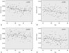
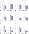
All 4 midfacial angles in females and the glabellar and maxillary angles in males showed statistically significant decreases with aging (p<0.05). LOESS regression curves were also created to better illustrate trends (Figs. 3 and 4). Each angular measurement was marked on the sample 3D reconstructed image for better understanding (Fig. 5).
Angular changes between individual age groups
For both men and women, the glabellar and maxillary angles showed statistically significant decreases with aging in the young, middle, and old age groups. For males, mean glabellar angles were 69.4±6.14, 67.4±6.26, and 65.0±4.77 degrees in the young, middle, and old age groups, respectively. For females, similar changes in the glabellar angle were observed with mean angles measuring 75.6±4.98, 72.0±5.88, and 71.1±4.51 degrees in the young, middle, and old age groups, respectively. For males, mean maxillary angles were 66.5±4.70, 64.3±4.27, and 63.0±4.10 degrees in the young, middle, and old age groups, respectively. For females, more prominent changes in the mean maxillary angle were observed with aging in the young and middle age groups; mean maxillary angles were 67.2±5.35, 63.6±5.90, and 61.9±5.73 degrees in the young, middle, and old age groups, respectively. For females, the orbital angle showed a statistically significant decrease between the middle (76.6±5.61 degrees) and old age groups (74.1±5.18 degrees), and the pyriform angle also showed a statistically significant decrease between the young (59.3±6.52 degrees) and middle age groups (56.7±7.21 degrees) (Fig. 6A-D).
Periorbital and perinasal changes
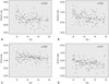
The orbital and pyriform widths did not show significant changes with aging. For males, mean pyriform widths were 22.2±2.43, 22.6±2.11, and 20.3±2.15 mm in the young, middle, and old age groups, respectively. For males, mean orbital widths were 39.1±3.51, 38.9±3.90, and 38.1±2.35 mm in the young, middle, and old age groups, respectively. For females, mean pyriform widths were 21.6±2.11, 21.7±1.93, and 19.9±1.83 mm in the young, middle, and old age groups, respectively; while mean orbital widths were 36.8±3.67, 37.4±2.59, and 36.7±2.61 mm in the respective groups (Fig. 6E and F).
DISCUSSION
In the present study, we demonstrated that Asian midfacial skeletons change continuously throughout life, mainly in a clockwise angular rotation around the orbit in a right-facing craniofacial skeleton. It can be described as Lambros's theory, summarized as a rotation of the maxilla relative to the cranial base.8 Shaw and Kahn9 suggested the trend of aging midfacial skeletal changes in 60 Caucasian subjects, and they expanded the study by increasing the sample size to 120 and by including measurements of upper and lower facial skeletons.11 The finding showed that glabellar and maxillary angles significantly decreased, whereas the pyriform aperture significantly increased with aging in both male and female subjects. In study of Richard, et al.,7 angular measurements of four bony regions (glabellar, orbital, maxillary, and pyriform aperture angles) were taken from 50 male and 50 female subjects, and all four the measurements were found to significantly decrease with aging. In our data, the orbital and maxillary angles showed less changes, the pyriform angle showed more prominent changes compared to those studies conducted on Caucasians.7911 These ethnic differences may be explained by characteristic features of Asian facial skeletons. Numerous studies revealed significant skeletal differences between Asians and Caucasians, demonstrated by cephalometric analysis.241213 The characteristics of Asian facial skeletons can be summarized as wide and flat midface contours, prominent zygomas, small nasal bones, and wide mandible angles. In particular, the relatively short midfacial skeleton and strong zygoma in Asians may be contributing factors to less prominent angular changes in orbital and maxillary regions in Caucasians.
There have been markedly different results among previous studies on Caucasians. One of the reasons is the different study design between studies. Shaw and Kahn9 and Richard, et al.7 used different reconstruction protocols and measurement programs. Furthermore, sample sizes were not large enough to compare each other.
In the present study, we increased the study population size for normal distribution and 3D vector mathematics using the PACS. Because of using these methods, we did not require 3D rendering and the angular measurement process in the reconstructed images. Compared to methods using the reconstruction program, much time were saved, and relatively small differences were observed between repetition of tests. Finally, therefore, we concluded these methods are quite effective and accurate, especially in a large study population.
The female subjects tended to exhibit more midfacial angular changes with aging than the male subjects. This gender difference in angular changes was observed in all four regions in varying degrees. In addition, female facial skeletal remodeling seemed to occur more rapidly between the young and middle age groups than between the middle and old age groups, whereas male subjects it did not. This difference may be due to bone remodeling by sex hormonal change during menopause.14
The orbital and pyriform widths showed no significant changes with aging. These results are quite different from those of previous studies on Caucasian subjects.91516 A plausible explanation of these results is differences in nutritional status and physique between generations. Since nutritional status has changed considerably in the Korean society over the past several decades, there have been large differences in facial skeletal size between generations.1718 This is a limitation of the cross-sectional design of our study. On the other hand, size differences between generations had little effect on angular changes of facial skeletons.
The growing desire to rejuvenate the aging faces in Asian countries is due to the increases of aging population and socioeconomic status.1 Especially, people living in the Far East, including Korea, Japan, and China, have greater interest in cosmetic surgeries, including facial rejuvenation. Understanding inherent ethnic differences and applying them to rejuvenation surgery would certainly lead to better surgical outcomes. Until now, there have been few studies on the facial skeletal aging process in Asians. This is the first study to show midfacial skeletal changes in Asians, which is based on a larger sample size than previous similar studies performed in Caucasians.
In conclusion, the results of this study suggest that changes in Asian midfacial skeletons may occur continuously throughout life, and show different rates and degrees depending on the regions of the face and sex, and that the most striking changes could occur in young female subjects.


 XML Download