

 PDF
PDF ePub
ePub Citation
 Print
Print


INTRODUCTION
Nonalcoholic fatty liver disease (NAFLD) is one of the most common causes of liver disease in the Western population and is becoming more prevalent. The presentation of NAFLD varies from simple fatty liver to nonalcoholic steatohepatitis that potentially progress to cirrhosis. Several studies have indicated that NAFLD is closely associated with several metabolic disorders including obesity, diabetes mellitus, dyslipidemia, and hypertension,123 which have all been recognized as risk factors for cardiovascular disease, and a recent study associated NAFLD with coronary artery disease (CAD).4 Moreover, NAFLD is associated with greater overall mortality and independent predictors of the risk of future cardiovascular disease (CVD) events.5 There are few cross-sectional studies on the association between NAFLD and CAD, and the studies that were performed evaluated only coronary calcification or the presence of coronary artery stenosis in individuals of clinically suspected CAD.46 The CAD risk of NAFLD is worth particular attention to the screening and surveillance strategies from the perspective of the increase in patients with NAFLD.
Atherosclerosis, an asymptomatic process that begins early in life, eventually leads to overt coronary heart disease (CHD). Multidetector computed tomography (MDCT) coronary angiography is a non-invasive method that provides a direct anatomic assessment of coronary arteries to allow detailed examination of coronary atherosclerosis, and it can also give additional information regarding plaque composition and the severity of coronary atherosclerosis.789
In the present study, we aimed to assess the relationship of NAFLD, as determined by ultrasonography, with subclinical coronary stenosis and plaque morphology, as detected by dual-source 64-slice MDCT, in asymptomatic subjects without a history of CVD.
MATERIALS AND METHODS
Study subjects
A retrospective review was performed using data from 1034 self-referred subjects who had undergone both 64-slice MDCT coronary angiography and hepatic ultrasonography as part of a general routine health evaluation at CHA Bundang Medical Center between March 2007 and December 2010. Participants were excluded if they had a history of myocardial infarction or stroke (n=1), coronary artery bypass surgery (n=1), malignancy (n=1), positive hepatitis B surface antigen or anti-hepatitis C antibody (n=30), habitual consumption of >20 g/day of alcohol (n=208), or insufficient medical records (n=21). Ultimately, 772 subjects were enrolled in this study. The Institutional Review Board of CHA Bundang Medical Center approved the study protocol.
Basic demographic data were acquired from a database maintained by the Health Promotion Center at CHA Bundang Medical Center. Medical history of myocardial infarction, angina, hypertension, stroke, diabetes mellitus, and tobacco use, as well as each patient's current medication profile, was collected from a systemized questionnaire prior to a general health examination. Body weight, height, waist circumference, and blood pressure were also measured during their visit. Hypertension was defined as a self-reported history of hypertension, the use of antihypertensive medication, or a blood pressure of ≥140/90 mm Hg. Total cholesterol, triglycerides, high-density lipoprotein (HDL) cholesterol, low-density lipoprotein (LDL) cholesterol, fasting plasma glucose, serum uric acid, alkaline phosphatase (ALP), aminotransferase, total bilirubin, and gamma glutamyl transferase (GGT) were measured after at least a 12-hour fasting period on the same day of the study. Diabetes was defined as a self-reported history of diabetes, the use of antidiabetic treatment, or a fasting plasma glucose level of ≥126 mg/dL. The mean age of the subjects was 49.2±9.8 years, and the mean body mass index (BMI) was 24.1±3.1 kg/m2. In addition, 68.0% of subjects were males, 215 (27.8%) were hypertensive, and 73 (9.5%) had diabetes. There were 202 current smokers (26.2%), of whom 97.5% were males.
Metabolic syndrome was defined based on the National Cholesterol Education Program Adult Treatment Panel Phase III (NCEP-ATP III) criteria, with a modification for the cutoff of waist circumference. Subjects with at least three of the following criteria were considered to have metabolic syndrome: 1) central obesity by waist circumference (>90 cm in men, >80 cm in women), 2) hypertriglyceridemia (≥150 mg/dL), 3) low HDLcholesterol (<40 mg/dL in men, <50 mg/dL in women), 4) high blood pressure (≥130/85 mm Hg), and 5) elevated fasting plasma glucose (≥100 mg/dL). Framingham risk scores (FRS), as used by the National Cholesterol Education Program (NCEP) guidelines, were calculated, and the 10-year risks of coronary events were estimated.10 All subjects were assigned to three different CHD risk groups according to the revised NCEP guidelines: a high-risk CHD group (CHD equivalents or a 10-year risk >20%), a moderate-risk group (more than two risk factors and a 10-year risk ≤20%), and a low-risk group (0 to 1 risk factors).
Hepatic ultrasonography
A liver ultrasound was performed to assess the presence and severity of NAFLD using a high-resolution B-mode ultrasound machine (LOGIQ S6; Siemens, Munich, Germany). The degree of steatosis was assessed semiquantitatively (absent, mild, moderate, or severe) on the basis of hepatorenal echogenicity contrast, liver brightness, deep attenuation, and vascular blurring.1112
Multidetector computed tomography
Non-enhanced CT was performed to measure coronary artery calcium (CAC) score on a 64-slice MDCT scanner (Light-speed VCT, GE Healthcare Systems, Waukesha, WI, USA). The scanning parameters of CAC CT were prospective electocardiography (ECG) gating, 120 kV tube voltage, 200 mA tube current, 175 ms temporal resolution, and 3 mm slice thickness. Calcification in the coronary artery was deemed present if there was a high density corresponding to the coronary arterial wall of >130 Hounsfield units (HU) using Agatston's method. The presence or absence of descending thoracic aortic calcification was also analyzed using the same method. Z-axis coverage of CAC CT was from the tracheal carina to the base of the heart. Contrast-enhanced coronary CTA was performed using variable kV and mA based on each patient's BMI. Tube voltage and current were 120 kV and 550 mA in patients with a high BMI (≥25 kg/m2) and 100 kV and 500 mA in patients with a low BMI (<25 kg/m2), respectively. Other scanning parameters for coronary CTA included 0.625 mm slice thickness, 32 cm field of view, 165 ms temporal resolution, 0.625 mm reconstruction interval, and 7-11 s scanning time. Retrospectively-ECG-gated contrast enhancement was performed after intravenous injection of 50-80 mL Optiray (Ioversol 300 mg/mL, Tyco Healthcare, Montreal, Canada). The volume of contrast media was adapted to match each body weight with a constant dose of iodine per body weight (0.5 g iodine per kilogram). To achieve a constant injection duration of 30 seconds, the flow rate was adjusted accordingly. A saline chaser bolus of 50 mL was subsequently injected at the same flow rate. A trigger delay of 5 s and bolus tracking of the contrast material were used. Scanning commenced 5 s after the CT attenuation reached 100 HU in the ascending aorta. Tube current modulation was used in patients to reduce the radiation dose. Subjects with a heart rate >65 beats/min were given beta-blocker (Propranolol 40-80 mg, Hyundae Pharmaceutical Co., Seoul, Korea) before MDCT imaging. Each subject was also given nitroglycerin (0.6 mg, Myungmun Parmaceutical Co., Seoul, Korea) sublingually 1 min before MDCT.
MDCT coronary angiography images were analyzed on an Advantage Workstation 4.3 (GE Healthcare Systems) by two experienced radiologists who were unaware of the clinical information. Each lesion was identified with a multiplanar reconstruction technique and maximum intensity projections of short-axis, 2-chamber, and 4-chamber views. The contrastenhanced portion of the coronary lumen was semi-automatically traced at the maximal stenotic site, and the degree of stenosis in the maximal stenotic site was compared with the average diameters of proximal and distal reference sites. Nonsignificant coronary atherosclerotic plaque was defined as lesions causing ≤50% luminal narrowing, and significant coronary atherosclerotic plaque was defined as lesions causing >50% luminal narrowing.
We analyzed plaque characteristics according to the modified American Heart Association classification.13 Plaques were defined as structures >1 mm2 within or adjacent to the vessel lumen, which could clearly be distinguished from the lumen and surrounding pericardial tissue. Plaques occupied by calcified tissue comprising more than 50% of the plaque area (density >130 HU in native scans) were classified as calcified, plaques with <50% calcium were classified as mixed, and plaques without any calcium were classified as non-calcified lesions. Subjects were stratified into subgroups having primarily mixed plaque (defined as a mixed component >70% of all plaque area), non-calcified plaque (defined as a non-calcified component >70% of all plaque area), and calcified plaque (defined as a calcified component >70% of all plaque area).
Coronary artery calcium scores (CACS) were measured with the scoring system previously described by Agatston, et al.14 Participants were categorized on the basis of the CACS in the following manner: no calcification, 0; mild calcification, 0.1 to 100; and moderate-to-severe calcification, >100.
Statistical analysis
Statistical analyses were performed using the Statistical Package for the Social Sciences software (SPSS Inc., version 19.0; Chicago, IL, USA). The categorical variables were reported as numbers with percentage, and the continuous variables were reported as the mean±standard deviation. Student's t-test, the chi-square test, and ANOVA were used for assessing the intergroup comparison. Logistic regression analysis was performed as appropriate to decide the factors related to coronary atherosclerosis, with consideration of cardiovascular risk factors such as increased age (men over 45 years old, women over 55 years old), tobacco use, hypertension, diabetes mellitus, high LDL cholesterol (>160 mg/dL in the low-risk group, >130 mg/dL in the moderate-risk group, and >100 mg/dL in the high-risk group), low HDL-cholesterol (<40 mg/dL in men, <50 mg/dL in women), metabolic syndrome, and NAFLD. Each odds ratio (OR) was presented together with its 95% confidence interval (CI). A p-value of <0.05 was considered statistically significant.
RESULTS
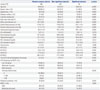
Significant coronary plaques were detected in 76 subjects (9.8 %). The percentage of patients with non-significant plaques was 31.1% (n=240), and normal coronary arteries were found in 456 patients (59.1%). The baseline characteristics of subjects with normal coronary arteries, non-significant stenosis, and significant stenosis detected by MDCT are shown in Table 1.
The prevalence of hypertension (18.6%, 38.8%, and 48.7%, respectively; p<0.001), diabetes mellitus (4.6%, 14.2%, and 23.7%, respectively; p<0.001), metabolic syndrome (20.6%, 33.8%, and 42.1%, respectively; p<0.001), and NAFLD (37.9%, 54.6%, and 55.3%, respectively; p=0.001) increased significantly with the severity of coronary atherosclerosis. Significant differences between groups were noted regarding the distribution of males and current smoking history, age, BMI, waist circumference, systolic blood pressure, diastolic blood pressure, fasting plasma glucose, serum uric acid, triglycerides, HDL cholesterol, aspartate aminotransferase (AST), alanine aminotransferase (ALT), GGT, ALP, and 10-year CHD risk by FRS. However, total cholesterol, LDL cholesterol, and total bilirubin did not change with the severity of coronary atherosclerosis.
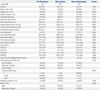
Among 772 patients, 316 (40.9%) had atherosclerotic plaques in coronary arteries. Of them, 122 (15.8%) patients had calcified plaques, 82 (10.6%) had mixed plaques, and 112 (14.5%) had non-calcified plaques (Table 2). The prevalences of diabetes, age, systolic/diastolic blood pressure, and 10-year CHD risk by FRS were different between groups. There was no significant difference in the prevalence of NAFLD according to plaque morphology between the primarily calcified, mixed, and non-calcified coronary atherosclerotic plaque groups (59.0%, 50.0%, and 53.6%, respectively; p=0.392).
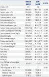
Table 3 shows the associations of NAFLD with clinical and biological variables in subjects. Age, gender distribution, the prevalences of hypertension and diabetes, smoking history, BMI, waist circumference, systolic/diastolic blood pressure, fasting plasma glucose, serum uric acid, triglyceride levels, HDL cholesterol, AST, ALT, GGT, ALP, and 10-year CHD risk by FRS were significantly different between subjects with and without NAFLD. However, the levels of total cholesterol, LDL cholesterol, and total bilirubin were similar between the two groups.
Coronary atherosclerotic plaques were detected in 143 patients (33.6%) among those without NAFLD and in 173 (50.0%) among those with NAFLD. NAFLD subjects had a higher prevalence of calcified, mixed, and non-calcified plaque than the subjects without NAFLD. There was no significant difference in the prevalence of significantly-stenosed coronary arteries according to the presence of NAFLD (8.0% vs. 12.1%; p=0.054) (Table 4).
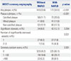
The results of binary logistic regression analysis to determine the presence of coronary atherosclerotic plaque are represented in Table 5. Increased age (OR: 3.46; 95% CI: 2.48-4.84; p< 0.001), diabetes mellitus (OR: 2.33; 95% CI: 1.30-4.17; p=0.005), hypertension (OR: 1.89; 95% CI: 1.28-2.79; p=0.001), low HDL cholesterol (OR: 0.58; 95% CI: 0.38-0.89; p=0.013), and NAFLD (OR: 1.48; 95% CI: 1.05-2.08; p=0.025) remained significant predictors of the presence of coronary atherosclerotic plaque after adjusting for major risk factors for cardiovascular disease. In contrast, the other binary logistic regression analysis showed no association between NAFLD and the presence of significantly-stenosed coronary artery segments determined by MDCT coronary angiography; however, increased age (OR: 4.84; 95% CI: 2.48-9.44; p<0.001) and diabetes (OR: 2.07; 95% CI: 1.09-3.94; p=0.027) remained significant predictors.
In the multinomial logistic regression analysis, increased age (OR: 2.23; 95% CI: 1.41-3.51; p=0.001), low HDL cholesterol (OR: 0.52; 95% CI: 0.29-0.92; p=0.026), and high LDL cholesterol (OR: 1.63; 95% CI: 1.01-2.64; p=0.046) seemed significant predictors of primarily non-calcified plaques after adjustment for other risk factors. NAFLD was not a significant predictor of non-calcified plaques in multinomial logistic regression (p= 0.053). Increased age (OR: 4.88; 95% CI: 2.74-8.70; p<0.001) and hypertension (OR: 2.24; 95% CI: 1.27-3.98; p=0.006) remained significant predictors of primarily mixed plaques. NAFLD was not a significant predictor of mixed plaque in multinomial logistic regression, with similar results for non-calcified plaque (p=0.691). In contrast to non-calcified and mixed plaques, calcified plaque appeared to be significantly predicted by NAFLD (OR: 1.70; 95% CI: 1.07-2.70; p=0.03). Increased age (OR: 4.47; 95% CI: 2.73-7.31; p<0.001), diabetes mellitus (OR: 3.60; 95% CI: 1.85-7.04; p<0.001), and hypertension (OR: 2.04; 95% CI: 1.22-3.40; p=0.006) remained significant predictors of primarily calcified coronary plaque (Table 6).
DISCUSSION
To the best of our knowledge, our study is the first to report on the association of NAFLD with subclinical coronary stenosis and plaque morphology using MDCT coronary angiography in a large middle-aged sample of asymptomatic individuals without a history of CVD. Our results demonstrated that NAFLD was associated with the presence and calcific morphology of coronary atherosclerotic plaque.
Few studies have documented the independent relation between NAFLD and cardiovascular disease.1516 An association between NAFLD and inflammatory markers including tumor necrosis factor (TNF)-α, interleukin (IL)-6, and C-reactive protein (CRP) was found, which indicates that NAFLD may contribute to the inflammatory CVD milieu, predisposing to atherosclerosis and CVD.171819 In addition to a direct predisposition to atherosclerosis, cytokines may have an indirect effect as well, as cytokines may be involved in insulin resistance. The pleiotropic cytokine TNF-α interferes with the hepatic insulin receptor and the intrahepatocellular insulin signaling, causing both hepatic and systemic insulin resistance.2021
Recent studies have demonstrated that NAFLD is correlated with circulatory endothelial dysfunction, which is accepted as an early process in the development of atherosclerosis.2223 It has been shown that non-diabetic patients with NAFLD have a significant decrease in flow-mediated vasodilation of brachial arteries (a surrogate measure of endothelial dysfunction) when compared with matched healthy controls, and this decrease correlated to histological features of NAFLD, independent of age, sex, BMI, homeostatic model assessment-insulin resistance (HOMA)-insulin resistance, and other metabolic syndrome components.24 Further accumulating evidence also exists demonstrating that patients with NAFLD had a marked increase in carotid artery intima-media thickness (IMT), a reliable index of subclinical atherosclerosis.25262728 In our study, NAFLD was found to be associated with the presence of CAD, though not with the severity. Increased age and diabetes mellitus appeared to be significant predictors of the severity of coronary atherosclerotic plaque after adjustment of other risk factors. The present study investigated asymptomatic subjects without a history of CVD; therefore, most of the study population was extremely low-risk (63.9% CACS=0), and the majority of coronary atherosclerosis was insignificant coronary stenosis.
MDCT coronary angiography has a high diagnostic accuracy in appropriate patients and has been proposed as an important non-invasive modality for detecting CAD. The cumulative adverse cardiac events rates over 21 months in patients with normal coronary arteries, non-significant CAD, and significant CAD have been reported as 0.5%, 3.5%, and 16%, respectively.8 Beyond the degree of luminal narrowing of the coronary artery, MDCT coronary angiography can characterize plaque composition, which can give valuable data about the various clinical manifestations of CAD. A recent study reported that younger age, hyperlipidemia, and a family history of CAD are all predictors of the extent of non-calcified plaque and that older age was significantly associated with the presence of mixed and calcified plaque.9 Our study revealed that, in addition to older age, diabetes mellitus, high LDL cholesterol, and NAFLD were associated with the presence of calcified plaques according to the multivariate regression analysis. NAFLD was not associated with non-calcified or mixed plaques. However, calcified plaques do not represent the entire atherosclerotic burden of coronary arteries, and non-calcified plaques, indicative of lipid-rich and vulnerable plaques, are associated with acute coronary syndrome more than calcified plaques.7 In a prospective study by Wong, et al.,29 fatty liver was associated with CAD independently in patients with clinical indications for coronary angiogram. However, in patients with CAD, fatty liver cannot predict adverse clinical outcomes such as cardiovascular mortality, non-fatal myocardial infarction, and the need for further coronary intervention. Our study results were in line with such previous studies, revealing an independent association between NAFLD and CAD.
A number of limitations of this study must be acknowledged, including the cross-sectional observational design with the lack of randomization and the retrospective descriptive nature of the study. All participants underwent a single measurement of all values. Although hepatitis B and C viruses and suspicious alcoholic hepatitis were excluded, we did not completely exclude other types of liver disease, such as autoimmune hepatitis, although the prevalence is low in Korea. Due to racial differences between the prevalence, extent, and morphology of coronary atherosclerosis, these findings cannot be generalized to all populations. All participants took part in a general routine health evaluation, and MDCT coronary angiography is a costly examination. Hence, a selection bias could be inherent.
In conclusion, our study revealed that NAFLD was associated with both the presence and the calcified-plaque morphology of subclinical coronary atherosclerosis detected by MDCT. Thus, in addition to traditional risk factors of CAD and subclinical atherosclerosis, NAFLD seems to be independently associated with the presence of coronary atherosclerotic plaque. Moreover, NAFLD is associated with calcific coronary plaque, which is an independent predictor of CAD and cardiovascular outcome. Further prospective clinical studies are needed to clarify the exact physiopathologic role of NAFLD in coronary atherosclerosis.


 XML Download