

 PDF
PDF ePub
ePub Citation
 Print
Print


INTRODUCTION
Primary percutaneous coronary intervention (PCI) is an established treatment for patients with ST-segment elevation myocardial infarction (STEMI). However, after successful PCI many patients continue to have microvascular dysfunction documented by various invasive and non-invasive tests.123 Impaired microcirculation after successful reperfusion therapy correlates strongly with a poor prognosis.4567
The index of microcirculatory resistance (IMR) is a simple and standard method of assessing microvascular integrity by using a pressure sensor/thermistor-tipped guidewire, and it is a strong predictor of microvascular damage, especially 3 months after STEMI.8 Using contrast-cardiac magnetic resonance imaging, it has also been shown that IMR can independently predict left ventricular function and infarct volume following STEMI.910 In a recent study, an elevated IMR measured immediately after primary PCI was a strong predictor of poor long-term outcomes.11
Multiple clinical-, angiographic-, and procedure-related factors could affect microcirculation in STEMI patients prior to or during primary PCI; however, data on which of these factors has the most influence on microvascular integrity are limited. In the present study, we used IMR to assess microvascular integrity in STEMI patients who underwent primary PCI. We aimed to assess the clinical and angiographic predictors of microvascular dysfunction in STEMI patients.
MATERIALS AND METHODS
Study design
The present study is a retrospective cohort study designed to find out the clinical and angiographic determinants of microvascular dysfunction in STEMI patients. We enrolled STEMI patients who underwent primary PCI and coronary physiologic measurements from 2011 to 2014 at Inha University Hospital. IMR, a parameter of hyperemic microvascular resistance, was measured with a pressure sensor/thermistor-tipped guidewire immediately after successful primary PCI. A transthoracic echocardiogram (TTE) was obtained within 24 hours. This study was approved by Inha University Hospital's Institutional Review Board, and written informed consent was obtained from each patient.
Study population
We enrolled 113 patients with STEMI who underwent primary PCI. The IMR was measured immediately after successful primary PCI. The patients were divided into three groups based on IMR values as tertiles: Low IMR (<18 U, n=38), Mid IMR (18-31 U, n=38), and High IMR (>31 U, n=37). Patients with STEMI were diagnosed based on symptoms of myocardial ischemia in association with ST-segment elevation and subsequent release of cardiac biomarkers. ST elevation was defined as new ST elevation at J point of ≥2 mm (0.2 mV) in men or ≥1.5 mm (0.15 mV) in women in at least two contiguous leads on electrocardiography.12 We excluded patients with prior myocardial infarction in order to focus on de novo coronary microvascular resistance, and also excluded patients with cardiogenic shock, Killip Class IV, and atrioventricular block, as they were contraindicated to an invasive coronary physiologic study using adenosine.13
Angiographic analysis
Lesion length and reference diameter (RD) were measured using an automated edge-detection algorithm (CASS 5.7.1, Pie Medical Imaging Systems, Maastricht, the Netherlands). Automated distance calibration was used to determine pixel size. Angiographic views with the least foreshortening and the best depiction of the stenosis were used. Thrombolysis in Myocardial Infarction (TIMI) grade and TIMI myocardial perfusion grade (TMPG) were obtained using a grading scale of 0-3. Lesions of the coronary artery were classified by the American College of Cardiology/American Heart Association (ACC/AHA) grading system as type A, B1, B2, and C.
Measurement of IMR
After successful primary PCI, a pressure sensor/thermistortipped guidewire (Radi Medical System, Uppsala, Sweden) was calibrated outside the body, equalized to the guiding catheter, and advanced to the distal two-thirds of the culprit vessel. Three bolus injections of 3 mL room temperature saline were administrated at the culprit vessel, and the mean transit time was obtained using a thermodilution technique.14 After intravenous adenosine (140 µg/kg/min) was administered to induce maximal hyperemia; the hyperemic mean transit time (hTmn) was measured again using the same method above. Simultaneously, mean aortic pressure (Pa) and mean distal pressure (Pd) were measured during the resting and maximal hyperemic state. The IMR value was calculated as Pd×hTmn.2 Fractional flow reserve (FFR) was derived from the ratio of Pd to Pa during maximal hyperemia.15 In addition, thermodilution coronary flow reserve (CFR) was calculated by dividing the resting mean transit time by the hTmn.16
Echocardiographic analysis
A TTE was obtained within 24 hours after the index PCI. Left ventricular ejection fraction (LVEF) was measured using the modified Simpson method. As recommended by the American Society of Echocardiography, the wall motion score index (WMSI) was assessed in a 16-segment model.17 An experienced cardiologist blinded to the IMR values scored segmental wall motion and the WMSI.
Statistical analysis
Statistical analysis was performed using SPSS 21.0 statistical software (SPSS Inc., Chicago, IL, USA). Data are presented as mean±SD for continuous variables and as proportions for categorical variables. Continuous variables were compared using the Student's t-test. Analysis of categorical variables was performed using the chi-square test. Continuous variables were compared using one-way analysis of variance (ANOVA) and Fisher's exact test as a post hoc test for each IMR group. Univariate correlations between variables were assessed by Pearson's correlation coefficients (r). Linear regression analyses were performed to assess the relationships between IMR and clinical, angiographic, and anatomical factors. Univariate regression analysis was used to identify relationships between each clinical and angiographic factor and increased IMR. The clinical and angiographic predictors of impaired microvascular function were assessed using multivariate logistic regression analysis. Figures were created by using GraphPad Prism v.5.01 (GraphPad Software, Inc., San Diego, CA, USA).
RESULTS
Patient characteristics between IMR groups
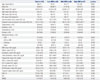
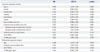
The mean age of the 113 study population was 56±11 years; 95 patients (84.1%) were men. The mean IMR in the study population was 28.2±17.8 U (range, 7.3-98.4 U). To determine the predictive factors for microvascular dysfunction, the study population was classified into three groups based on IMR values: Low IMR [<18 U (12.9±2.6 U), n=38], Mid IMR [18-31 U (23.9±4.0 U), n=38], and High IMR [>31 U (48.1±17.1 U), n=37] (Table 1).
Clinical and laboratory findings
The mean age of the Low IMR group was significantly lower than that of the Mid and High IMR groups (51±9 vs. 57±11 vs. 61±10, p=0.031) (Table 1). There were no significant differences in the presence of hypertension, diabetes, dyslipidemia, or smoking history between the three IMR groups.
The door-to-balloon time was <90 minutes for all patients. The High IMR group tended to have longer door-to-balloon times, compared to the Low and Mid IMR groups, although the difference was not statistically significant (65.7±14.6 min vs. 74.8±19.8 min vs. 82.0±44.6 min, p=0.068). However, the symptom-onset-to-balloon time of the high IMR group was significantly longer than that of the Low IMR and Mid IMR groups (172.2±80.1 min vs. 197.4±104.1 min vs. 299.8±195.1 min, p<0.001) (Table 1). The levels of cardiac enzymes, including creatine kinase (CK), creatine kinase-myocardial band (CK-MB), and troponin I, were significantly higher in the high IMR group, compared to the other groups (Table 1). The baseline LVEF of the High IMR group was lower than that of the Low and Mid IMR groups, although the difference was not significant (p=0.246). The high IMR group tended to have higher WMSI values than the Low and Mid IMR groups, although the difference in the values was not significant (p=0.162) (Table 1).
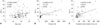
In correlation analysis, significant association was found between IMR values and age (r=0.219, p=0.020), CK level (r=0.342, p=0.010), and the symptom-onset-to-balloon time (r=0.463, p<0.001) (Fig. 1).
Angiographic findings
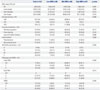
The culprit artery and coronary intervention including thrombus aspiration, direct stenting, the use of glycoprotein IIb/IIIa inhibitors, and lesion characteristics, including length, RD, and ACC/AHA lesion classification, were not significantly different between the IMR groups (Table 2). Culprit lesions located in the proximal portion of the target vessel were more frequent in the High IMR group than the Low IMR group (28.9% vs. 59.5%, p=0.008). The initial TIMI 0/1 flow before PCI was seen more frequently in the High than in the Low IMR group (55.3% vs. 81.1%, p=0.008). The achievement of a TIMI 3 (86.8% vs. 56.8%, p=0.015) and TMPG 3 (68.4% vs. 27.0%, p=0.008) after primary PCI was observed more frequently in the Low IMR than the High IMR group (Table 2).
Physiologic parameters
The baseline Pa and Pd showed no differences between the IMR groups. There were significant differences in Tmn at rest (0.37±0.18 vs. 0.59±0.34 vs. 1.01±0.54, p<0.001) and hTmn (0.18±0.04 vs. 0.31±0.09 vs. 0.66±0.27, p<0.001) (Table 3). The High IMR group tended to have higher FFR and lower CFR than the Low IMR and Mid IMR groups; however, the differences were not significant (Table 3).
Comparison of IMR according to clinical and angiographic parameters
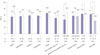
To determine whether certain parameters could influence microvascular dysfunction, we compared the IMR groups according to the presence or criteria of various parameters. As shown in Fig. 2, there were no significant differences in IMR regardless of major cardiovascular risk factors. However, our data did not show significant differences in IMR according to culprit arteries, the IMR values were significantly higher in proximal locations of the culprit lesion, as compared with non-proximal lesion (31.8±19.1 U vs. 24.6±15.8 U, p=0.033) and initial TIMI 0/1, as compared with initial TIMI 2/3 (23.0±12.8 U vs. 20.6±11.5 U, p=0.031) (Fig. 2). Since the door-to-balloon times were <90 minutes for all patients, we divided the patients into two groups (>60 minutes and ≤60 minutes). The two groups did not show a significant difference in IMR values (Fig. 2). Thereafter, the patients were divided according to a symptom-onset-to-balloon time of 180 minutes, and we observed that the IMR of those with a symptom-onset-to-balloon time of >180 minutes was significantly higher than the IMR of those with a symptom-onset-to-balloon time of ≤180 minutes (Fig. 2).
Predictors of microvascular dysfunction
As shown in Table 4, age, symptom-onset-to-balloon time, cardiac biomarker, such as CK-MB and final TMPG level, showed a significant correlation with increasing IMR. Our data showed that a symptom-onset-to-balloon time of >180 minutes had a significant odds ratio (OR) for higher IMR between those with a symptom-onset-to-balloon time of >180 minutes and those with a symptom-onset-to-balloon time of ≤180 minutes [OR, 4.376; 95% confidence interval (CI): 1.851-10.347; p=0.001]. Cardiac biomarkers, such as CK-MB, was also significantly correlated to increasing IMR (Table 4). In multivariate regression analysis, age and symptom-onset-to-balloon time remained independent determinants for High IMR [Exp (β)=1.085, 95% CI: 1.016-1.158, p=0.016; Exp (β)=1.007, 95% CI: 1.001-1.014, p=0.018, respectively] (Table 4).
DISCUSSION
The main findings of the present study are as follows: First, the High IMR group was older, had a longer symptom-onset-to-balloon time, and more frequently had a culprit lesion in a proximal location. Second, age and symptom-onset-to-balloon time were independent predictors of high IMR, reflecting the presence of microvascular dysfunction in patients with STEMI.
Aging is an important factor contributing to atherosclerosis and arteriosclerosis.18 It has been reported that arterial aging is associated not only macrovascular resistance but also with impaired microcirculation due to the diminution of the capillary bed, which accompanies atrophy of subcutaneous and other tissues.1819 The presence of structural alterations in the microcirculation may be an important link to ischemic heart disease.1820 Most studies about IMR do not describe the relationships between age and IMR well. Our study suggests that aging may be related to impaired microcirculatory resistance, as estimated by IMR in patients with STEMI.
Coronary microvascular dysfunction in diabetes mellitus has been explained by various mechanisms of endothelial dysfunction including insulin resistance, hyperglycemia, impaired vasodilatation, autonomic dysfunction, and inflammation.2122 Our data did not show a significant difference between individual IMR groups and diabetes mellitus. This may be due to the lack of adjusting for confounding factors such as medication, patient characteristics, and major risk factors, including hypertension, dyslipidemia, and smoking. The present data indicated that the IMR values do not significantly differ according to the presence of diabetes, as well as according to the presence of major cardiovascular risk factors, such as hypertension, dyslipidemia, and smoking. These results may be explained by the relatively small sample size of our study. In addition, as the IMR was measured shortly after the primary PCI, the angiographic- and procedure-related factors may have a greater effect on the IMR than classic cardiovascular risk factors.
Door-to-balloon time is associated with mortality in patients undergoing primary PCI for STEMI. Previous studies have shown a strong correlation between door-to-balloon time and clinical outcome.232425 The ACC/AHA guidelines for the management of patients with STEMI recommend a door-to-balloon time of ≤90 minutes.26 However, it has recently been reported that, despite reducing the door-to-balloon time from 83 minutes to 67 minutes, there has been no significant change in inhospital mortality and 30-day mortality.27 In our data, the door-to-balloon times were <90 minutes in all patients, and no significant differences in door-to-balloon time were noted between the IMR groups. However, the symptom-onset-to-balloon time in the present study showed significant differences between individual IMR groups. Therefore, the IMR correlated well with the symptom-onset-to-balloon time but not the door-to-balloon time of <90 minutes. In multivariate analysis, age and symptom-onset-to-balloon time remained independent determinants for impaired microvascular resistance. This suggests that, in patients with a door-to-balloon time of <90 minutes, decreasing the absolute door-to-balloon time does not affect the microvascular resistance in patients with STEMI. Therefore, the symptom-onset-to-balloon time might be more important for determining microcirculatory resistance in STEMI patients with a door-to-balloon time of <90 minutes.
Angiographic findings have been used as the classical parameter for microvascular dysfunction. Coronary blood flow and microvascular integrity have been estimated by using TIMI and TMPG in many previous studies,42829 and previous studies have shown that the final TIMI or TMPG score is related to mortality in patients with acute myocardial infarction.5 However, there are some limitations due to inter-observer variation since it is graded by visual estimation. In the present study, IMR was strongly correlated with TIMI and TMPG, as well as with the levels of cardiac biomarkers, such as peak CK, peak CK-MB, and peak troponin I. The achievement of the final TIMI 3 or TMPG 3 was much less frequent in the High IMR group than in the Low IMR group. These findings are consistent with previous studies that suggest that IMR predicts clinical outcome and prognosis.8911303132 In contrast, the IMR requires less inter- and intra-observer variability than the visual estimation of the TIMI or TMPG system; therefore, IMR may be a more objective method for assessing microvascular dysfunction.
Some recent studies have shown that increased microvascular resistance and dysfunction in patients with STEMI might lead to an alteration in flow dynamics associated with a smaller pressure drop through the culprit lesion. FFR would be overestimated in this setting.333435 Our data showed that the High IMR group tended to have a higher FFR than the Low IMR and Mid IMR groups, although the difference was not statistically significant.
To find out the relevant factors for microvascular dysfunction in STEMI, our study population was classified into three groups based on IMR value as tertiles. A definite cut-off value of IMR to determine LV recovery and clinical outcomes in STEMI patients has not yet been established. However, it is interesting that our cut-off value (>31 U) for the High IMR group was similar to suggested IMR values in previous studies on predicting myocardial dysfunction in STEMI patients.830 To apply the cutoff value of IMR for predicting a clinical outcome in STEMI patients, further large-scaled studies may be needed in the future.
Murai, et al.36 reported that right coronary artery lesion location was significantly associated with increased IMR in patients with intermediate coronary artery lesions. Our data did not show significant differences in IMR according to culprit arteries. The proximal location of the culprit lesion in STEMI is associated with greater myocardial damage. It has been reported that patients with proximal culprit lesions are more likely to have a poorer clinical outcome and prognosis than patients with non-proximal culprit lesions.37 Proximal culprit lesions were found more frequently in the High IMR group than in the Low IMR group in the present study, and the mean IMR in patients with proximal culprit lesions was significantly higher than that in patients with non-proximal culprit lesions. This suggests that a proximal location of culprit lesion might play an important role in deteriorating microcirculatory resistance in patients with STEMI.
The Thrombus Aspiration during Percutaneous coronary intervention in Acute myocardial infarction Study (TAPAS) demonstrated that aspiration of the thrombus before stenting seemed to improve 1-year clinical outcomes following primary PCI for STEMI.38 However, a recent study showed that, compared with PCI alone, routine thrombus aspiration before PCI did not reduce the 30-day mortality in patients with STEMI.39 In a recent randomized study, it was reported that thrombus aspiration as an adjunctive therapy to primary PCI for STEMI may reduce IMR and have beneficial effects on myocardial microcirculation.40 Our data showed that thrombus aspiration was more frequent in the High IMR group, compared with the Low IMR group; however, the difference was not significant, although there may be an operator-dependent bias in the thrombus aspiration, which was determined by operator at the time of the primary PCI. Although the use of glycoprotein IIb/IIIa inhibitor may reduce mortality in high-risk patients with STEMI,41 the use of glycoprotein IIb/IIIa inhibitors did not differ between the IMR groups in the present study.
Study limitations
Our study consisted of a relatively small number of STEMI patients who underwent primary PCI. Because patients with cardiogenic shock, prior myocardial infarction, and hemodynamic instability were excluded, our results may not represent all patients with STEMI. Moreover, this study is a retrospective analysis and was performed at a single center. We need more data and randomized control study to better understand determinants of microvascular dysfunction in STEMI patients. The 113 study subjects were relatively small as a study group and most of our enrolled STEMI patients had mild LV systolic dysfunction or preserved LV function with a LVEF of approximately 50%. Therefore, there were no significant differences in LVEF and WMSI among the IMR groups, although the High IMR group tended to have lower LVEF and higher WMSI than the Low and Mid IMR groups. If more patients presenting severe LV systolic dysfunction in STEMI patients were included, the differences of LVEF and WMSI might be clarified among the IMR groups. Furthermore, the present study was not performed using a randomization study protocol for interventional techniques and medications such as direct stenting, thrombus aspiration, and glycoprotein IIb/IIIa inhibitors; therefore, it is possible that an operation-dependent bias may be present in these variables. These variables might play a role as confounding factors. In the multivariate analysis, it would have been ideal to exclude all factors in order to avoid selection bias and confounding factors; however, this was not ensured in the present study, except for age, gender, comorbidities, lesion location, and symptom-onset-to-balloon time.
In conclusion, our study suggests that age, proximal location of the culprit vessel, and symptom-to-onset-balloon time are correlated with microvascular dysfunction estimated using IMR. In STEMI patients undergoing primary PCI with a door-to-balloon time of <90 minutes, age and symptom-onset-to-balloon time might be major predictors of microvascular dysfunction.

 XML Download