

 PDF
PDF ePub
ePub Citation
 Print
Print


INTRODUCTION
Breast cancer is the most common malignancy among women globally. In the United States, more than 200000 new cases of invasive breast cancer and more than 60000 new cases of in situ breast cancer were expected among US women in 2013.12 Approximately 40000 US breast cancer patients were expected to die in 2013; however, breast cancer death rates decreased by 34% from 1990 to 2010.2 Similarly, the incidence rate of breast cancer is continuously increasing in Korea, and it is the second most common cancer among Korean women.3 The Korean Breast Cancer Society reported that the number of newly-diagnosed breast cancer patients was more than 16000 and that the crude age-specific incidence rate of women aged 40 to 49 years was the highest, with approximately 148 cases per 100000 women in 2010.4 The 5-year relative survival rate of female breast cancer patients diagnosed in 2006-2010 improved from 78.0% to 91.0% compared to those diagnosed in 1993-1995.3
Increasing survival rates in breast cancer patients are attributed to both early detection of malignancies and improvements in treatment.23 With increasing incidence and survival rates of such malignancies, the number of cancer survivors continues to increase in Korea. Although many cancer survivors return to normal daily activities after the completion of primary treatment, the cancer itself and related treatments may also result in a wide range of chronic, long-term physical and psychological problems.5 Of these issues, second or multiple primary cancers (MPCs) are both disastrous and lethal in this patient population. An increased risk of MPCs in cancer survivors compared to the general population has been associated with complex factors including genetic predisposition, host factors, environmental determinants, gene-environment interactions, shared lifestyle factors (e.g., tobacco use or excessive alcohol intake), and the late effects of cancer treatments (e.g., cytotoxic, radiation, or hormonal therapies).6
MPCs are generally defined according to the criteria of Warren and Gates7 as follows: 1) each tumor must have clear evidence of malignancy on histologic examination, 2) each tumor must be geographically separate and distinct, and 3) the possibility of a metastatic lesion having spread from a prior cancer must be excluded. Recent rules for classifying MPCs have been modified based on the cancer site of origin, date of diagnosis, histology, tumor behavior (i.e., in situ versus invasive), and laterality of the paired organ.8 International coding rules for MPCs are more restrictive, and MPCs occurring at the same site or on different sides of a paired organ are usually considered to be first primary unless the two tumors are of completely different histology.89
Trends of cancer incidence and mortality vary by age and between nations. Considering that the crude age-specific incidence rates of Korean breast cancer patients are different from those of Western countries,4 risks for and survival of MPCs should be clarified in Korean breast cancer patients. However, limited information is available on this subject.510 This study aims to investigate patterns of MPCs in Korean breast cancer patients treated at a single institution and to examine the characteristics and survival rates of these patients after diagnosis according to the presence or absence of MPCs.
MATERIALS AND METHODS
A total of 8204 patients with locoregional breast cancer were retrospectively selected from the Severance Hospital breast cancer registry. The Severance Hospital registry prospectively records clinicopathological information including past histories of cancer and details on survival outcomes. Additional information for MPCs was obtained from the Yonsei Cancer Registry of Yonsei University Health System in Seoul, Korea. All patients underwent surgery for primary breast cancer between January 1990 and December 2012. Patients with stage IV disease at the time of diagnosis were excluded. In this study, patients with bilateral breast cancer at initial diagnosis or during follow-up periods were not considered to have MPCs. In cases of simultaneous bilateral breast cancer, the side with the more advanced stage was selected, and in cases of metachronous bilateral breast cancers, the initial index tumor was considered for analysis.
In this study, the criteria of Warren and Gates7 were used to define MPCs. Synchronous MPCs were defined as a tumor diagnosed simultaneously with breast cancer or within a time interval of 6 months. Metachronous MPCs were considered to be a tumor detected more than 6 months before or after diagnosis of breast cancer. For survival analysis according to follow-up time period and time of diagnosis of MPCs, the patient cohort was subdivided into two groups as follows: patients with or without MPCs at a time point within a follow-up duration of 5 years or less (time period ≤5 years, n=8204) and those with or without MPCs among patients with a follow-up duration of more than 5 years (time period >5 years, n=3745). Therefore, patients who died after >5 years of being diagnosed with breast cancer were considered to be alive at ≤5 years. Similarly, patients diagnosed with metachronous MPCs at >5 years after being diagnosed with breast cancer were placed in the breast-cancer-alone group with a time period ≤5 years. Patients who died or were lost to follow-up within 5 years of being diagnosed with breast cancer were excluded from the analysis of the time period >5 years.
Clinical follow-up included a patient interview, physical examination, laboratory tests, and breast imaging every 6-12 months. If necessary, an abdominopelvic ultrasound, bone scan, computed tomography (CT) scan, or fluorin-18 fluorodeoxyglucose positron emission tomography/CT scan was performed. Tumor-node-metastasis (TNM) staging was based on the 6th American Joint Committee on Cancer criteria.11 Tumors with ≥1% nuclear-stained cells were considered positive for estrogen receptor (ER) and progesterone receptor (PR) according to the American Society of Clinical Oncology/College of American Pathologists guidelines.12 Human epidermal growth factor receptor 2 (HER2) staining was not available during the early 1990s, and HER2 3+ scores were considered positive.
Differences between groups were evaluated by the chi-square test. Continuous variables were compared using the independent two-sample t-test. Overall survival (OS) was calculated from the date of surgery for breast cancer to the date of the last follow-up or death from any cause. The date of the last follow-up was used as censored data in this analysis. Survival curves were plotted using the Kaplan-Meier method, and group differences in survival curve were investigated by the log-rank test. A Cox proportional hazard model was used to identify variables that were independently associated with OS. All statistical tests were two-sided and a p-value<0.05 was considered statistically significant. Categorical variables were expressed as a frequency and percentage. SPSS version 20.0 (IBM Inc., Armonk, NY, USA) was used for all statistical analyses.
RESULTS
Of the 8204 breast cancer patients with stage 0 to III disease, 858 patients (10.5%) had MPCs: 268 patients (31.2%) had metachronous MPCs alone ≥6 months before diagnosis of breast cancer, 180 patients (21.0%) had synchronous MPCs alone, 370 patients (43.1%) had metachronous MPCs alone ≥6 months after diagnosis of breast cancer, 10 patients (1.2%) had both metachronous MPCs ≥6 months before the diagnosis of breast cancer and synchronous MPCs, 14 patients (1.6%) had both synchronous and metachronous MPCs ≥6 months after the diagnosis of breast cancer, and 16 patients (1.9%) had metachronous MPCs ≥6 months both before and after the diagnosis of breast cancer. Synchronous MPCs were noted in 204 patients (23.8%), and metachronous MPCs were identified in 678 patients (79.0%). There were also 24 patients (2.8%) with both synchronous and metachronous MPCs.
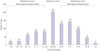
In the 858 patients with MPCs, a total of 962 primary malignancies were detected. Second primary cancer alone was noted in 762 patients (88.8%), and 96 patients (11.2%) had two or more primary malignancies other than breast cancer. Disease sites and numbers of cases are presented in Table 1. Cancers of the endocrine system, which mainly included the thyroid gland, were the most prevalent malignancy in Korean breast cancer patients, and more than two-thirds of synchronous MPCs were thyroid cancer. Subsequently, primary tumors of the gynecologic system including the ovary, cervix, and uterus were the second-most prevalent cancer. Interestingly, in patients with metachronous MPCs, primary cancers more frequently developed in respiratory and hematologic systems after, rather than before, the diagnosis of breast cancer. Fig. 1 shows the time intervals between breast cancer and MPCs. When excluding 53 metachronous cases (5.5%) that were diagnosed as MPCs before diagnosis of breast cancer and did not have information available to identify the date of cancer diagnosis, 166 cases (18.3%) had a past history of malignancy between 6 months and 5 years before diagnosis of breast cancer, and 275 cases (30.5%) developed MPCs between 6 months and 5 years after diagnosis of breast cancer.
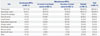
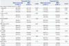
Table 2 summarizes the clinicopathologic characteristics associated with breast cancer according to the presence or absence of MPCs and stratified by a cutoff follow-up duration of 5 years. At ≤5 years, the mean age of patients with MPCs was 52.7 years, which was significantly older than those with breast cancer alone. Patients with MPCs showed smaller tumor size, higher node-negative disease, and lower TNM staging at the time of diagnosis of breast cancer. ER, PR, and HER2 expressions were not significantly different between groups. Similarly, at >5 years, patients with MPCs were older in age at diagnosis of breast cancer and showed smaller tumor sizes. However, node status was not significantly different between patients with and without MPCs; differences in TNM stage showed borderline statistical significance.
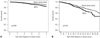
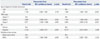
During a mean follow-up duration of 67.3 months for the whole population, the OS of patients with and without MPCs are shown in Fig. 2. Although patients with MPCs demonstrated a higher proportion of stage 0-I disease, they also showed worse survival than the breast-cancer-alone group at both ≤5 and >5 years. Table 3 shows causes of death. Of the 978 patients who died during this study, 53 (5.4%) did not have an identifiable cause of death, and most were in the breast-cancer-alone group. At ≤5 years, a significantly higher number of patients with MPCs died due to reasons not associated with breast cancer, and at >5 years, causes of death were similar to those at ≤5 years.
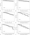
When adjusting for age, TNM stage, and ER expression at diagnosis of breast cancer, patients with MPCs were significantly associated with an increased risk of death at ≤5 years (Table 4). However, a slightly increased risk of death in patients with MPCs did not reach statistical significance at >5 years. Older age, advanced TNM stage, and ER-positive disease were independent prognostic factors at >5 years. A stage-matched subgroup analysis showed that patients with MPCs had significantly worse OS than the breast-cancer-alone group for stage 0-I disease at both ≤5 and >5 years and for stage II disease at ≤5 years (Fig. 3). Nevertheless, no statistical difference in OS was noted between stage II disease at >5 years and stage III disease irrespective of follow-up duration.
DISCUSSION
It is occasionally clinically difficult to distinguish new second primary malignancies from a metastatic neoplasm during the follow-up of cancer survivors. MPCs are generally considered new primary cancers when detected in a patient with a prior history of malignancy that is in a new site or tissue and subsequent to the initial cancer.5 Cancer survivors have a higher risk of developing MPCs than the general population, and studies using population-based registry datasets have demonstrated standardized incidence ratios for subsequent MPCs of 1.17 to 1.6 in female cancer survivors.581314 Recently, these relative risks for developing MPCs among women with breast cancer were reported differently, with ratios as high as 1.96 [95% confidence interval (CI), 1.48-2.44] according to age at diagnosis of breast cancer.1516 However, there has been no official study on MPCs from the nationwide Korean Central Cancer Registry.
In Korea, incidence rates of female thyroid cancer have increased sharply since the early 2000s, and this was found to be the most common malignancy of Korean women, with an age-standardized incidence rate of 87.4 in 2010.3 In our study, thyroid cancer was the most prevalent malignancy among breast cancer patients. Interestingly, more than two-thirds of patients with synchronous MPCs had a thyroid malignancy with breast cancer. Globally, incidence rates of thyroid cancer have increased, with the exception of countries such as Sweden, Norway, and Spain. The cause of such changing trends in the incidence of thyroid cancer may be multifactorial, although it remains unclear.17 It also remains to be determined whether our results reflect a true increase in the development of Korean thyroid cancer, an increased identification of previously undetectable subclinical thyroid disease along with improved diagnostic techniques and screening rates, or close biological connections between female breast and thyroid cancers.318
It has been suggested that certain types of anticancer treatments are closely linked to an increased risk of developing MPCs in cancer survivors.192021 Examples include findings that cytotoxic chemotherapy was associated with an increased risk for leukemia, chest irradiation for Hodgkin's disease was related to an increased risk of breast cancer, and tamoxifen treatment for breast cancer was connected to a higher risk of endometrial cancer.15182022 By analyzing patients with MPCs among our study cohort, the proportions of patients with malignancy in the thyroid gland, lung, thorax, and hematologic system were elevated after, rather than before, the diagnosis and treatment of breast cancer. In this study, we were unable to confirm whether certain types of cancer were developed in association with anticancer therapies, and in the future, it will be necessary to further investigate this topic. In addition, given that MPCs in the gynecologic system were the second-most common malignancy and development of breast and ovarian cancer shares a close genetic linkage in deleterious BRCA 1 & 2 mutation carriers, results of the Korean Hereditary Breast Cancer (KOH-BRA) Study for the Korean population may shed further light on this topic.23
In the present study, patients with MPCs showed a higher proportion of early-stage breast cancer, especially at ≤5 years. Recent meta-analyses suggested that cancer survivors were more likely to be screened for breast, cervical, colorectal, and prostate cancer than non-cancer controls.2425 Cross-sectional surveys involving the Korean population have shown that screening rates for breast cancer within 2 years were 46.4% (95% CI, 36.2-56.7) in cancer survivors, 36.0% (95% CI, 33.2-38.9) in non-cancer chronic disease controls, and 30.0% (95% CI, 27.8-32.2) in non-cancer non-chronic disease controls when adjusted for gender, age, marital status, education, income, working status, insurance status, smoking and drinking status, self-reported health status, and survey year.26 Higher screening rates and interest in personal health among patients with metachronous MPCs could partly explain our results; however, further investigation of the association of health screening with the first primary cancer and subsequent MPCs is required.
There have been limited data regarding the impact of MPCs on the survival of breast cancer patients. One early study of Korean breast cancer patients reported no difference in survival according to MPCs.10 However, in a recent study by the M.D. Anderson Cancer Center on 4198 patients treated with breast conservation therapy, patients with MPCs showed a worse OS than those without MPCs after excluding patients who had a past history of malignancy prior to the diagnosis of breast cancer. 27 Similarly, patients with MPCs in this study demonstrated a worse survival than those without MPCs, and most died due to diseases other than breast cancer. Although statistical significance in the multivariate analysis was not maintained at >5 years as more than half of the patients were lost to follow-up, further long-term study is needed. Stage-matched subgroup analysis revealed that the implications of MPCs on breast cancancer survival were more significantly evident among patients with early stage 0-I disease, yet not among those with advanced stage II-III disease.
Many cancer survivors do not perceive their risk of a subsequent second primary malignancy, and appropriate screening rates among Korean cancer survivors are suboptimal compared to the United States.5 Therefore, awareness and education regarding the development of MPCs and importance of screening programs are required for both cancer survivors and surgical and medical oncologists.2829 In addition, as there are no evidence-based guidelines on screening programs for cancer survivors, it is accepted that cancer survivors should, at minimum, follow the screening guidelines for the general population until the establishment of screening guidelines for cancer survivors. Limitations of the present study include the retrospective nature of the survival analysis over a short duration of follow-up and the use of a hospital-based registry database at a single institution. More importantly, it was not possible to incorporate details of TNM stage, treatment patterns of MPCs, or environmental risk factors in the analysis.
In conclusion, Korean breast cancer patients are at a risk of second or multiple primary malignancies. Cancers most commonly occurred in the thyroid gland and gynecologic system. Breast cancer patients with MPCs exhibited worse survival, and many died due to causes unrelated to breast cancer. However, these patients were older at diagnosis and had lower breast cancer staging. The implications of MPCs on survival were more evident when patients had early-stage breast disease. Therefore, further efforts are needed to investigate the nationwide incidence of MPCs in order to discover the causes of increased risk for MPCs, to develop preventive methods for MPCs, and to increase awareness of and enrollment in screening programs for breast cancer survivors.

 XML Download