

 PDF
PDF ePub
ePub Citation
 Print
Print


Oxidative stress plays an important role in proliferation, apoptosis, and inflammation in gastric epithelial cells infected with Helicobacter pylori (H. pylori).1 High levels of reactive oxygen species (ROS) were observed in gastric mucosa of patients infected with H. pylori.1,2 The activated leukocytes recruited to gastric mucosa during infection produce ROS.2 However, gastric epithelial cells themselves also produce ROS in gastric epithelial cells with H. pylori infection.3,4
NADPH oxidase, which is a complex of the membrane-bound subunits (gp91phox, p22phox) and the cytosolic subunits (p67phox, p47phox), produces superoxide (O2-) upon activation of the enzyme. Guinea pig gastric mucosal cells express Nox1, p22phox, p67phox, NOX1, and Rac1 constitutively.5 Previous study showed that H. pylori activates NADPH oxidase by translocation of heat shock protein (Hsp) 90β from cytosol to membrane in gastric epithelial cells.6 An NADPH oxidase inhibitor dipehenylene iodonium (DPI) inhibited inflammatory signaling such as activation of mitogen-activated protein kinases and induction of monocyte chemoattractant protein-1 in H. pylori-infected gastric epithelial cells by suppressing the production of ROS.7 Since ROS mediate inflammation by activating inflammatory signaling and DNA damage to induce apoptosis,8 NADPH oxidase may be involved in H. pylori-induced apoptosis of the infected gastric epithelial cells and tissues. In addition, the clinical outcome of the infection may be different depending on H. pylori-virulence-associated genes such as cagA, vagA, and ice A genes.9,10 Therefore, it is essential to study using the predominant genotype of H. pylori (cagA+, vacA s1b, m2, iceA genotype)11 to understand the pathogenesis of H. pylori-associated gastric disorders in Korea.
The purpose of the present study is to investigate whether NADPH oxidase-generated ROS mediate apoptotic cell death by increasing the ratio of Bax/Bcl-2, induction of p53, and DNA fragmentation in gastric epithelial AGS cells infected with H. pylori in a Korean isolate (HP99) by treating cells with an NADPH oxidase inhibitor DPI. Cell viability, hydrogen peroxide levels in the medium, DNA fragmentation, and protein levels of p53, Bcl-2, and Bax were determined in the cells treated with or without DPI and cultured in the presence of H. pylori.
A human gastric epithelial cell line AGS (gastric adenocarcinoma, ATCC CRL 1739) was purchased from the American Type Culture Collection (Manassas, VA, USA) and cultured as previously described.7 H. pylori strain HP99 in a Korean isolate were inoculated onto chocolate agar plates (Becton Dickinson Microbiology Systems, Co-ckeysville, MD, USA) at 37℃ under microaerophilic conditions using an anaerobic chamber (BBL Campy Pouch® System, Becton Dickinson Microbiology Systems, Franklin Lakes, NJ, USA).7 Prior to the experiment, the cells (1×10⁵/mL/well) were cultured in the presence of H. pylori at a bacterium/cell ratio of 100:1, 300:1, and 500:1 for 24 h. For the study on DPI, the cells were pre-treated with DPI (2.5 or 5 μM) for 2 h and infected with H. pylori at a bacterium/cell ratio of 300:1 for 2 h (to determine hydrogen peroxide level), 12 h (protein levels of p53, Bcl-2, and Bax), and 24 h (DNA fragmentation and cell viability). Hydrogen peroxide levels in the medium were determined by the modified ferrithiocyanate method.12 Viable cell numbers were determined using trypan blue exclusion test (0.2% trypan blue). DNA fragmentation was assessed according to the amount of oligonucleosome-bound DNA in the cell extracts using a Cell Death Detection ELISA plus kit (Roche, Mannheim, Germany). For determination of protein levels of p53, Bcl-2, and Bax, the cells were extracted with lysis buffer containing 10 mM Tris, pH 7.4, 15 mM NaCl, 1% NP-40, and complete protease inhibitor complex (Roche, Mannheim, Germany), and centrifuged at 13000×g for 15 min. The supernatants were used as whole cell extracts. The protein concentration was determined by Bradford assay (Bio-Rad Laboratories, Hercules, CA, USA). Whole cell extracts (50 μg protein) were subjected to 10-12% SDS-PAGE under reducing condition. After transfer to nitrocellulose membranes, the proteins were detected with polyclonal antibodies against p53, Bcl-2, and Bax (Santa Cruz Biotechnology, Santa Cruz, CA, USA) followed by goat anti-rabbit secondary antibodies (1:2000, Cat. No. sc-2004, Santa Cruz Biotechnology) conjugated to horseradish peroxidase, which was followed by enhanced chemiluminescence (Santa Cruz Biotechnology). Actin was used for protein loading control. For the ratio of Bax/Bcl-2, the protein bands of Bax and Bcl-2 were scanned using a GS-700 scanner (Bio-Rad Laboratories). Band intensities were quantified using the Scion image program (Scion Corporation, Frederick, MD, USA). The statistical differences were determined using one-way ANOVA and Newman-Keul's test. All values are expressed as mean±SE of four different experiments. A value of p<0.05 was considered statistically significant.
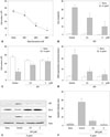
At 24 h-culture, cell viability was decreased with the density of H. pylori added to the cells (Fig. 1A). At the ratio of 100:1, 300:1, and 500:1 (H. pyori:AGS cells), cell viability of H. pylori-infected cells was 82%, 34%, and 14% of non-treated cells, respectively. Therefore, for following study on DPI, the cells were pre-treated with DPI and infected with H. pylori at a bacterium/cell ratio of 300:1. H. pylori increased hydrogen peroxide levels in the medium (Fig. 1B) and induced cell death (Fig. 1C) and DNA fragmentation (Fig. 1D) of the infected cells. In addition, H. pylori increased the protein levels of p53 and Bax, but decreased Bcl-2 levels of the infected cells (Fig. 1E), which increased the ratio of Bax/Bcl-2, the marker of apoptosis (Fig. 1F). DPI inhibited H. pylori-induced alterations in hydrogen peroxide levels in the medium, cell viability, DNA fragmentation, and apoptotic indices (increment of p53 and Bax, decrease in Bcl-2) of AGS cells dose-dependently. In non-infected cells, DPI did not affect cell viability, hydrogen peroxide levels, and DNA fragmentation.
Our study shows that DPI, an inhibitor of NADPH oxidase, suppresses H. pylori-induced cell death, hydrogen peroxide production, DNA fragmentation, increase in the ratio of Bax/Bcl-2, and p53 induction in AGS cells. The results demonstrate that NADPH oxidase mediates H. pylori-induced apoptosis in gastric epithelial cells. We previously reported that H. pylori-induced expression of integrin alpha 5/beta1 was inhibited by DPI, suggesting that NADPH oxidase mediates the induction of integrin in AGS cells.13 NADPH oxidase is present in gastric mucosal tissues of humans14 and mice.15 Which may contribute to gastric inflammation, apoptosis, and carcinogenesis associated with H. pylori-infection. DPI is a non-specific inhibitor of NADPH oxidase.16 Therefore, transfection of siRNA for NADPH oxidase subunits may be a good approach to definitely confirm the mediation of NADPH oxidase in apoptotic cell death of H. pylori-infected gastric epithelial cells.
Previous study showed that ROS play a critical role in the development of H. pylori-induced gastric damage. Although H. pylori possesses antioxidants as a constitutive compartment, SOD and catalase may not play a significant role for scavenging ROS from injured gastric mucosa.17 In the infected children in Turkey, SOD levels both in gastric tissue and erythrocytes were found to be not different between H. pylori (+) and H. pylori (-) patients.18 However, H. pylori SOD activity was higher in the cancer group than in the non-cancer group in Japan.19 These results may be related to high oxidative stress in cancer patients compared to healthy subjects.20
Recent study showed that H. pylori catalase decomposes cell-derived hydrogen peroxide and prevents HOCl synthesis. H. pylori-associated SOD inhibits interactions between superoxide anions and HOCl, as well as superoxide anions and NO. Therefore, H. pylori-associated antioxidants (catalase, SOD) inhibit HOCl and NO/peroxynitrite signaling pathways which is the control step in oncogenesis. These results suggest that H. pylori antioxidant enzymes play a role in the survival of transformed cells.21
In the present study, H. pylori infection contributes to the production of ROS in the infected cells, which induces apoptotic cell death of the infected cells. Since abnormal apoptosis may stimulate abnormal proliferation of the adjacent cells, H. pylori may induce hyper-proliferation of the adjacent cells with DNA damage. We previously showed that H. pylori infection results in oxidative DNA damage of the infected cells.8 Therefore, both H. pylori-associated antioxidant enzymes and H. pylori infection-induced ROS may contribute to the pathogenesis of H. pylori-induced carcinogenesis.
Even though H. pylori activate NADPH oxidase and ROS contribute to apoptosis, other mediators or antioxidant enzymes, either secreted by H. pylori or activated by H. pylori in the infected cells, may be involved in H. pylori-induced apoptosis of the infected cells. To investigate direct effect of hydrogen peroxide on apoptosis, apoptotic indices should be determined in gastric epithelial cells treated with various concentrations of hydrogen peroxide in the absence of H. pylori.
In conclusion, NADPH oxidase may mediate apoptosis by increasing the levels of p53 and the ratio of Bax/Bcl-2 as well as DNA fragmentation in H. pylori-infected gastric epithelial cells. DPI, an inhibitor of NADPH oxidase, may prevent H. pylori-associated inflammation and may suppress abnormal apoptosis of gastric epithelial cells.
 XML Download