

 PDF
PDF ePub
ePub Citation
 Print
Print


INTRODUCTION
Despite recent technological and therapeutic advances in neonatal medicine, bronchopulmonary dysplasia (BPD) remains a leading cause of respiratory morbidity in very low-birth weight infants. However, there are no effective therapeutic options for BPD currently.1 Although it has been demonstrated that BPD is the result of prolonged inflammation, oxidative stress and impaired healing of immature lungs, the mediators that are involved in the lung injury and repair are not completely understood.2-4 In recent years, there has been increasing evidence that the transdifferentiation of pulmonary lipofibroblasts to myofibroblasts may be an important element in the pathogenesis of BPD.5,6 Interactions between lung epithelium and mesenchyme mediated by peroxisome proliferator-activated receptor-γ (PPARγ) are critical for normal lung development and homeostasis. PPARγ, the main target of the parathyroid hormone-related protein (PTHrP) signaling pathway, prevents excessive differentiation of fibroblasts into myofibroblasts and induces differentiation into lipofibroblasts, thus contributing to normal alveolarization.7 Notably, PPARγ is the key nuclear transcription factor that supports the lipofibroblastic phenotype, which provides alveolar type II cell growth and differentiation. Recently, it has been suggested that exposure to hyperoxia impairs paracrine communications between lung fibroblasts and epithelial cells, and subsequently induces and accelerates pulmonary lipofibroblast transdifferentiation into myofibroblasts.8 Although the role of PPARγ in the lung development is well known, its role during lung injury induced by intra-amniotic lipopolysaccharide (LPS) exposure and postnatal hyperoxia is not yet known.9 The aim of our study was to determine the effects of a rosiglitazone (RGZ), a PPAR-γ agonist, on alveolar and vascular development in a rat model of BPD induced by intra-amniotic LPS administration and postnatal hyperoxia.
MATERIALS AND METHODS
Materials
Timed pregnancy Sprague-Dawley rats (term, 22.5 d), weighing 300-360 g, were used. One to three pregnant rats were used per group. The litter sizes ranged from 8-12. The male : female ratio in the study groups ranged from 0.8 to 1.2, and did not differ between treatment groups.
Animal exposures
The animal experimental procedures were approved by the Seoul National University Bundang Hospital Animal Care and Use Committee. On gestation day 20, the pregnant rats were anesthetized with isoflurane inhalation. After a midline abdominal incision, 1 µg of LPS (Escherichia coli 0111:B4; Chemicon International, Temecula, CA, USA) dissolved in 0.05 mL of saline or same volume of vehicle (saline) was injected into the amniotic sacs. After recovery, the pups were delivered spontaneously 24-48 hours after the injections. At 10-12 hours after birth, pups from each treatment group were randomly re-assigned to dams from the same treatment group to balance the litter sizes (5-8 pups/dam). The rat dams reared pups which were allocated to a single group only. The newborn rat pups which were exposed to intra-amniotic LPS were then exposed to 80% O2 in a Plexiglas chamber with adaptors to supply and vent gas (BPD groups), and pups which were exposed to vehicle were kept in room air (No BPD groups) with their dams for 7 days beginning on the day of birth (P1), as previously described.10 To the newborn rat pups in the BPD groups, 80% O2 was constantly administered using mass flow controllers (TSC-220; MFC Korea, Suwon, Korea). Nursing rat dams were switched between 80% O2 and air every 24 hour to avoid oxygen toxicity. The duration of exposure to ambient air in the rat pups in the BPD groups was <10 min/day for drug administration, weighing, and cage cleaning. After 7 day-exposure to 80% O2 or room air, all the pups were subsequently kept in room air for another 7 days until sacrifice on D14. As the main intervention of the study, RGZ or vehicle was administered to the rat pups. RGZ (3 mg/kg/d; Santa Cruz Biotechnology, Santa Cruz, CA, USA) dissolved in 1 : 3 solution of dimethyl sulfoxide (DMSO): phosphate buffered saline (PBS) or same volume of vehicle (1 : 3 DMSO : PBS solution) was injected intraperitoneally once daily for 14 days beginning on P1. These experimental protocols led to four treatment groups: No BPD+vehicle (n=11); No BPD+RGZ (n=9); BPD+vehicle (n=8); BPD+RGZ (n=8).
Tissue preparation
Rat pups were anesthetized with an intraperitoneal injection of ketamine (50 mg/kg; Yuhan Corp., Seoul, Korea) and xylazine (50 mg/kg; Bayer AG, Leverkusen, Germany) on D14. Lungs were prepared for immunoassays and histologic analyses on D14 as previously described.11 Briefly, for immunoassays, after removing the lungs and hearts as an en-bloc, the right lower bronchi were ligated and the right lower lobes were resected and stored at -80℃. For histological analyses, after removing the right lower lobes, the tracheas were cannulated, and buffered formaldehyde was infused at 25 cmH2O for 5 min. The tracheas were then closed with a suture, and the lungs were fixed in buffered formaldehyde for 24 hours at 4℃. Paraffin sections (4 µm) cut from the right upper and left lobes were mounted onto Super Frost Plus slides (VWR Scientific, West Chester, PA, USA). The slides were then deparaffinized and stained with hematoxylin and eosin (H&E).
Immunohistochemistry
The slides with 5 µm-paraffin sections were deparaffinized in xylene and rehydrated via serial immersions in 100% ethanol, 95% ethanol, 80% ethanol, and deionized water. Endogenous peroxidase activity was quenched by immersion in 3% hydrogen peroxide in methanol, followed by a rinse with distilled water. For platelet endothelial cell adhesion molecule (PECAM)-1 immunohistochemistry, the sections were incubated with primary goat anti-PECAM-1 polyclonal antibody (Abcam, Cambridge, MA, USA) for 1 hour at room temperature. The sections were then incubated with biotin-labeled goat anti-mouse secondary antibody (Jackson ImmunoResearch Laboratories, West Grove, PA, USA) diluted 1 : 500 for 10 min at room temperature. Following the secondary antibody incubation, the sections were incubated with streptavidin peroxidase (Abcam) for 10 min at room temperature, rinsed in Tris-buffered saline, and developed with diaminobenzidine (DAB; Vector Laboratories, Burlingame, CA, USA) and hydrogen peroxide. A wash with water stopped the DAB reaction. A light H&E counterstain was applied. The sections were dehydrated by sequential immersion in 80% ethanol, 95% ethanol, 100% ethanol, and then xylene before placing a coverslip on the section.
Lung morphometry and pulmonary vascular development
Four random non-overlapping fields per pup in two distal lung sections were used for the morphometric examinations. The lung sections were photographed using a digital camera (Axioskop MRc5; Carl Zeiss, Oberkochen, Germany) attached to an Axioskop 40 microscope (Carl Zeiss, Oberkochen, Germany) at 100× magnification and saved as JPEG files. Morphometric analysis was performed as previously described.11 The objective assessment of the extent of alveolarization was determined by the mean cord length (Lm) and alveolar surface area (SA). Lm which is an estimate of the mean alveolar size, was determined by counting intersections of alveolar wall with an array of 84 lines, each -24 µm long. SA was calculated as 4×volume density of tissue×lung volume/Lm. Volume density of tissue was determined using a 10×10 grid (grid element side length -29 µm). These morphometric analyses of the photographed lung section images were performed using Image-Pro Plus software (Media Cybernetics, Rockville, MD, USA).
For pulmonary vascular development assessment, images of PECAM-1-stained slides were captured at 400× magnification. The number of PECAM-1-positive vessels (<100 µm in size) was counted per each high-powered field (400×). The vascular density was expressed as the ratio of area of blood vessels (PECAM-1-postive area) to the total area of lung parenchyme. Color thresholding of the gray-black DAB was performed using the differential interference contrast image analysis function of Image-Pro Plus software. At least five counts from 5 fields were performed for each animal, and the fields were chosen randomly in areas not containing large airways or vessels. Four to six animals were examined per group.
Western blotting of vascular endothelial growth factor (VEGF) and VEGF receptor-2
Frozen lung tissue was homogenized in ice-cold buffer containing 25 mM Tris-HCl, 1 mM EDTA, 1 mM EGTA, 0.1% 2-mercaptoethanol, 1 mM phenylmethylsulfonyl fluoride, 2 mM leupeptin, and 1 mM pepstatin A. Homogenates were centrifuged at 1500 g for 20 min at 4℃ to remove cell debris, and proteins in the supernatants were resolved by SDS-PAGE and then transferred to nitrocellulose membranes by electroblotting. Blots were blocked overnight in 5% of nonfat dry milk in PBS. These blots were incubated for 1 hour at room temperature with a rabbit polyclonal VEGF antibody (1 : 500; Santa Cruz Biotechnology, Dallas, TX, USA) or rabbit anti-human polyclonal VEGFR-2 antibody (1 : 500; Santa Cruz Biotechnology, Dallas, TX, USA) in 5% of nonfat dry milk in PBS with 0.1% Tween 20. After the blots were washed to remove unbound antibody, a secondary antibody, goat anti-rabbit HRP (Enzo Biochem, New York, NY, USA) diluted in blocking buffer (1 : 2000), was applied for 30-60 min. After the blots were washed, pico-enhanced peroxidase detection (ELPIS-Biotech, Daejeon, Korea) was performed. We initially determined the accuracy and consistency of the protein loads for each gel by Ponceau S (Sigma-Aldrich, St. Louis, MO, USA) staining before applying different antibodies. For each blot, β-actin was used as a housekeeping protein to compare expression between experimental groups. The experiments were performed with at least four animals per study group. The density of the protein bands was quantified using an imaging densitometer (ChemiDoc XRS; Bio-Rad, Hercules, CA, USA), and the results were normalized for β-actin expression.
Right ventricular hypertrophy
After removing hearts as an en-bloc with the lungs on D14, the right ventricle (RV) and left ventricle plus interventricular septum (LV+IVS) were dissected and weighed, and Fulton's index (the ratio of RV weight to LV+IVS weight), an indicator of right ventricular hypertrophy, was measured.12
RESULTS
Light microscopic findings of lung tissue
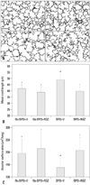
Photomicrographs of lung tissue sections from the animals which were exposed to intra-amniotic LPS and postnatal hyperoxia (BPD+V group) showed large and simple distal air spaces, a hallmark of disrupted alveolarization, compared to the animals which were not exposed to either (No BPD groups). The RGZ treatment of the animals which were exposed to intra-amniotic LPS and postnatal hyperoxia obviously restored alveolarization as indicated by small and complex distal air spaces (BPD+RGZ group). However, in the No BPD groups, the RGZ treatment did not significantly alter alveolar development (Fig. 1A).
Morphometric evaluation of the alveolar development
As expected from the light microscopic findings of the lung tissue, Lm, an indicator of average alveolar size, was significantly increased and SA was significantly decreased in the animals which were exposed to intra-amniotic LPS and postnatal hyperoxia (BPD+V group) compared to the animals which were not exposed to either (No BPD groups). The RGZ treatment significantly decreased Lm and increased SA in the animals which were exposed to intra-amniotic LPS and postnatal hyperoxia. However, in the animals in the No BPD groups, the RGZ treatment did not cause significant alteration of either Lm or SA (Fig. 1B and C).
Pulmonary vascular development
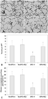
Photomicrographs of the lung tissue sections immunostained with PECAM-1 showed significantly decreased number and density of the vessels in the animals exposed to intra-amniotic LPS and postnatal hyperoxia (BPD+V group). The RGZ treatment significantly increased the number and density of the vessels in the animals which were exposed to intra-amniotic LPS and postnatal hyperoxia. However, in the animals which were not exposed to intra-amniotic LPS or postnatal hyperoxia, the RGZ treatment did not cause significant alternation in the number and density of the vessels (Fig. 2).
Western blotting of VEGF and VEGFR-2
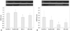
The levels of VEGF and VEGFR-2 in the lungs were significantly decreased in the animals which were exposed to intra-amniotic LPS and postnatal hyperoxia (BPD+V group). The RGZ treatment restored VEGF and VEGFR-2 levels to comparable levels in the animals which were not exposed to intra-amniotic LPS or postnatal hyperoxia (No BPD groups). However, the RGZ treatment did not significantly affect VEGF and VEGFR-2 levels in the animals in the No BPD groups (Fig. 3).
Right ventricular hypertrophy
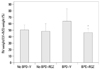
The RGZ treatment significantly decreased the Fulton's index (RV weight/LV+IVS weight) in the animals which were exposed to intra-amniotic LPS and postnatal hyperoxia. However, in the animals which were not exposed to intra-amniotic LPS or postnatal hyperoxia, RGZ treatment did not significantly alter the Fulton's index (Fig. 4).
DISCUSSION
In our previous studies, intra-amniotic LPS and subsequent postnatal hyperoxia caused an arrest in alveolar and pulmonary vascular development, which is the hallmark of BPD, in a newborn rat model.10,11,13,14 In the present study, the RGZ treatment was found to preserve alveolar and pulmonary vascular development against the detrimental influence of intra-amniotic inflammation and postnatal hyperoxia. Increased pulmonary vascular density and restored VEGF and its receptor VEGFR-2 levels in the lungs by the RGZ treatment of the animals exposed to intra-amniotic LPS and postnatal hyperoxia suggest that this protective effect of RGZ might be associated with enhanced angiogenic activity. VEGF plays a central role in the microvascular development of the lungs.15 VEGF signaling was reduced by hyperoxia and enhanced angiogenesis by VEGF treatment improved both alveolar and pulmonary vascular development in neonatal rats.16,17 In our previous study with the same animal model, sildenafil improved alveolar development by activating angiogenic factor VEGF.18 Taken together, it is suggested that enhanced angiogenic activity induced by RGZ is involved in the improvement of not only pulmonary vascular development but also alveolar development.
PPARγ-induced angiogenesis can occur through direct stimulation of endothelial cells.19 However, increasing evidence suggests that PPARγ promotes angiogenesis through the actions of angiogenic growth factors and cytokines that stimulate endothelial cells.19 Impaired distal microvascular development has been linked to poor alveolar development.20 Many studies have investigated the contribution of lung vasculature and angiogenic growth factors to the maintenance of alveolar structures.15,21 Pulmonary vasculature is disrupted in human infants dying of BPD and VEGF and VEGF receptors are reduced in their lungs.22 The disruption of VEGF signaling leads to the arrest of alveolar and pulmonary vascular development, which is the hallmark of BPD.23 The clinical importance of PPARγ originates from its insulin-sensitizing action. PPARγ has been known to ameliorate the progression of cardiovascular disease in diabetic patients.24 PPARγ stimulates the expression of genes involved in the regulation of vascular tone, thus improving endothelial function and decreasing vascular inflammation. Results of the present study correspond well with the results of several studies that have identified a potential role of PPARγ agonists in the microvascular development in the lungs.9,25 Yamakawa, et al.25 suggested that PPARγ agonists can promote VEGF secretion in vascular smooth muscle cells and modulate angiogenesis, and another study has shown that systemically administered RGZ, a PPARγ agonist, significantly enhances lung vascular maturation during normal lung development in rat pups;9 RGZ increased the expression of VEGF and its receptor. In our present study, we observed a striking decrease in vascular growth and the expression of angiogenic growth factor in the animals which were exposed to intra-amniotic LPS and postnatal hyperoxia. Furthermore, these deleterious effects of intra-amniotic inflammation and postnatal hyperoxia on vascular development and VEGF expression were alleviated by RGZ.
RGZ is an anti-diabetic drug in the thiazolidinedione class. It binds to PPARs in fat cells and makes the cells more responsive to insulin. Among several PPARs, PPARγ is expressed mainly in fat tissue, where it regulates genes involved in adipocyte differentiation, fatty acid uptake and storage, and glucose uptake. It has recently been known that PPARγ plays an important role in the normal lung development via epithelial-mesenchymal signaling.26 Stretching stimuli on alveolar type II cell lead to the expression of PTHrP. PTHrP binds to PTHrP receptor which is expressed on the lipofibroblast and upregulates PPARγ. PPARγ induces lung myofibroblast-to-lipofibroblast trans-differentiation, and lipofibroblasts support alveolar type II cells proliferation and differentiation, thus contributing to alveolarization.27 Hyperoxia, volutrauma and infection, which are factors implicated in the pathogenesis of BPD, have been known to decrease PTHrP expression in the alveolar type II cells and consequently lead to downregulation of alveolar lipofibroblast PPARγ expression.7,28,29 Most importantly, all of these injurious factors have been shown to be prevented by PPARγ agonist.26 RGZ is one of the well known PPARγ agonists. Wang, et al.9 demonstrated that systematically administered RGZ significantly increased the expression of PTHrP receptor, PPARγ and surfactant protein-B in the lungs in rat pups without affecting their blood biochemical and metabolic profiles including serum electrolytes, blood glucose, blood gases and plasma cholesterol and triglycerides levels. In our present study, RGZ alleviated the inhibitory effect of intra-amniotic LPS and postnatal hyperoxia on alveolarization. Experimental data cited above together suggest that the protective effect of RGZ on alveolarization observed in the present study might be mediated by increased PPARγ expression in the alveolar lipofibroblasts. However, we did not measure PPARγ level or its expression, or mediators involved in PTHrP signaling pathway, which might unravel underlying mechanism associated with the findings. This is our major limitation and further investigation on these mediators should be carried out to delineate the mechanism involved in the protection of developing lungs from intra-amniotic inflammation and postnatal oxidative stress.
Although there is considerable evidence to suggest that PPARγ agonists alleviate hyperoxic lung injury, the effects of PPARγ agonists on the double lung injury induced by antenatal inflammation and subsequent hyperoxia remain unknown. We have already demonstrated that when developing lungs are exposed to both intra-amniotic inflammation and postnatal hyperoxia, the inhibition of alveolarization is amplified compared to when only hyperoxia is present.11 The priming effect of intra-amniotic LPS administration on hyperoxic lung injury is associated with increased expression of inflammatory cytokines in the lungs.14 Premature infants are born with an underdeveloped microvasculature, and inflammatory cytokines may be toxic to their immature endothelial cells. PPARγ seems to participate in the control of inflammation at the transcriptional level by inhibiting NF-κB, signal transducer and activator of transcription 1 and activator protein 1 signaling.30-32 Cuzzocrea, et al.33 investigated the anti-inflammatory effects of RGZ in animal models of acute inflammation, and found that RGZ attenuated the infiltration of the lung with neutrophils and down-regulated the expression of intercellular adhesion molecule-1 and P-selectin in the lungs of carrageenan-treated rats. Consistent with these results, RGZ has been found to have protective effects against inflammation in fetal rat lung explants exposed to the LPS.34 Rehan, et al.34 suggested that LPS affects PTHrP-driven epithelial-mesenchymal interactions, and thus agonists of PPARγ signaling have the potential to prevent inflammation-induced molecular injury that is implicated in BPD. However, inflammatory markers or cytokines were not studied in the present study, and this is also another limitation of the present study. Therefore, the effects of RGZ on the inflammatory responses in the lungs which might have been induced by intra-amniotic LPS cannot be delineated here.
In conclusion, RGZ, a PPARγ agonist, restored alveolar and pulmonary vascular development and alleviated pulmonary hypertension in a neonatal rat model of lung injury, which was induced by intra-amniotic LPS administration and postnatal hyperoxia exposure. Moreover, the lung vascular growth was accompanied by increased angiogenic growth factor expression in the lungs. We speculate that augmented angiogenesis through RGZ administration can be one of the potential therapeutic approaches to improve the lung growth in extremely premature infants who are at greater risk of disrupted microvascular development.
 XML Download