

 PDF
PDF ePub
ePub Citation
 Print
Print


INTRODUCTION
Cardiovascular disease is the leading cause of mortality throughout the world, with studies showing cardiovascular disease to be the cause of death in 30% of all mortalities reported in the USA.1 Atherosclerosis is a disease process that begins at an early age and is progressive in nature. Atherosclerotic lesions do not arouse any symptoms in the early stage, but its initial presentation may result in catastrophic cardiovascular events resulting from plaque rupture.2 As such, it is imperative to identify subjects at increased risk of cardiovascular disease and modify their risk factors early on. Also, the treatment of advanced atherosclerosis is less effective than the inhibition of atherosclerosis progression.3 However, traditional risk factors for predicting cardiovascular disease, such as the Framingham Risk Score (FRS) and the European Systematic Coronary Risk Evaluation, comprise modest predictive value for future cardiovascular events.4 For this reason, the treatment of atherosclerosis at an earlier stage is crucial with more precise patient selection for preventive treatment. Landmark studies have demonstrated that measurements of subclinical atherosclerosis, such as carotid ultrasound, ankle brachial index and coronary calcium score, offer significant benefit in improving cardiovascular risk prediction beyond that with traditional risk factors.5,6,7 Among these measurements, ultrasonographic measurement of carotid intima-media thickness (cIMT) and carotid plaque have been widely applied to detect early atherosclerotic lesions. This review aims to discuss up to date evidence for the clinical usefulness of cIMT and plaque measurement for cardiovascular risk stratification.
LIMITATIONS OF TRADITIONAL RISK FACTORS FOR CARDIOVASCULAR RISK STRATIFICATION
In spite of recent advances in treatment strategies, the incidence of cardiovascular disease is expected to increase as a result of ageing populations.8 In addition, the socioeconomic cost thereof is high due to the chronic nature of cardiovascular disease. Therefore, the establishment of a prevention plan is essential, and research regarding biomarkers, imaging studies, and risk classification models for predicting cardiovascular events have been studied extensively.
Although FRS is the standard score system for predicting cardiovascular risk using traditional risk factors, it is modest at best in predicting cardiovascular risk. This is not surprising since studies have shown that the majority of patients with cardiovascular disease would have been classified as low risk by traditional risk scores. A 26-year follow-up data of the Framingham Heart Study revealed that 35% of subjects with total cholesterol level below 200 mg/dL, which is considered a desirable cholesterol level, were predisposed to cardiovascular disease. Moreover, the total cholesterol levels were the same in 80% of individuals who experienced myocardial infarction versus those who did not experience the event.9 The National Cholesterol Education Program Adult Treatment Panel III (NCEP-ATPIII) guidelines, which are based on FRS, suggest that the target goal of low density lipoprotein (LDL) cholesterol lowering, the primary target for reduction of cardiovascular disease, differs according to risk category, as determined using traditional cardiovascular risk factors.10 However, some studies have revealed some limitations with the NCEP-ATPIII guidelines on risk stratification. Akosah, et al. showed that the only 25% of young patients with acute myocardial infarction would have been recommended for statin drug therapy criteria according to NCEP-ATPIII guidelines.11 In addition, this guideline did not categorize 69% of acute myocardial infarction patients to the drug therapy group in a large cohort without a history of coronary artery disease.12
High blood pressure is one of the most important causes of cardiovascular disease and there is no argument that treatment of hypertension lowers cardiovascular risk. However, in the British United Provident Association (BUPA) study, which included 21520 subjects of ages 35-64 years, blood pressure was not a strong indicator for prediction of future cardiovascular events. The study demonstrated that the distribution of blood pressures among men who died of ischemic heart disease and men who did not exhibited noticeable overlap.13
Risk factors, like those above, are closely related with the incidence and the progression of atherosclerosis. However, these are indirect rather than direct markers for the current status of atherosclerosis and have limitations for evaluating atherosclerosis. Thus, more direct markers with anatomical delineation through imaging technology or functional studies have been developed and widely used. Among them, many researchers or clinicians have utilized cIMT measurement via ultrasonography.
ADVANTAGES OF cIMT MEASUREMENT WITH CAROTID ULTRASOUND
Carotid ultrasonography can measure cIMT and detect focal atherosclerotic plaque using ultrasound. The measurement of cIMT has several advantages for monitoring of atherosclerosis. First, cIMT can be performed with no adverse effects on subjects. Second, cIMT can be carried out at relatively low cost. Third, cIMT gives better visualization of atherosclerotic changes on arterial wall than other imaging modalities. In addition, ultrasound B-mode imaging of cIMT has been shown to be well correlated with IMT measured on microscopic examination.14 Therefore, the measurement of cIMT can provide precise information about atherosclerotic burden. Data from several sources have identified that traditional risk prediction models could not accurately reflect the presence of atherosclerosis as measured by carotid ultrasonography. Naqvi, et al. studied the prevalence of subclinical atherosclerosis in the groups of low, intermediate, and high FRS in 136 asymptomatic subjects using carotid ultrasonography. In the 103 low-risk (FRS<10%) subjects, 66% had cIMT>75th percentile or plaque >1.5 mm.15 In another study, when carotid ultrasonography was performed in 336 asymptomatic healthy young subjects with FRS of <5%, nearly 38% had high risk carotid ultrasound findings, defined as the cIMT>75th percentile. These studies demonstrated that traditional risk factors do not accurately reflect the actual plaque burden in the arteries, and as such, cIMT may be of use for risk stratification.16 The recently published European Society of Hypertension/European Society of Cardiology have defined either a mean cIMT of over 0.9 mm or the presence of carotid plaque as a marker of target organ damage in hypertension.17
THE ASSOCIATION BETWEEN cIMT AND CARDIOVASCULAR RISK FACTORS
Studies have demonstrated significant correlations between the degree of cIMT with other cardiovascular risk factors. Age and high blood pressure are known to be major determinants of cIMT in the general population.18 This is consistent with the fact that atherosclerosis progresses with age and high blood pressure. Moreover, other proatherogenic factors, like total cholesterol, LDL-cholesterol and insulin resistance, have been shown to be significantly associated with cIMT.19,20 The Bougalusa Heart Study showed that the progression of cIMT was significantly associated with fasting blood glucose and systolic blood pressure in young adults.21 When the lifetime risk for cardiovascular disease was estimated using an algorithm including total cholesterol, blood pressure, smoking, and diabetes mellitus (DM), individuals with higher life time risk experienced a greater subclinical atherosclerosis burden.22 These associations between traditional risk factors for cardiovascular disease and cIMT suggest that cIMT exhibits potential as a surrogate marker for predicting cardiovascular events.
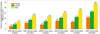
Many prospective studies have evaluated the relationship between cIMT and cardiovascular clinical events.23 Although each study set a differing cutoff point of high risk, the studies demonstrated an absolute yearly risk for cardiovascular disease, ranging from 1.6% to 3.2%, with increase in cIMT. Additionally, the relative risk of high cIMT ranged from 2.2 to 3.2 for coronary heart disease.24,25,26 The Atherosclerosis Risk In Communities (ARIC) study, which was an epidemiologic study of cardiovascular incidence in the general population, revealed that a higher coronary heart disease incidence was significantly associated with the both the increasing degree of cIMT and the presence of plaque (Fig. 1).27 Baldassarre, et al. demonstrated that the progression rate of maximum cIMT was significantly associated with cardiovascular risk.28 The results from the aforementioned epidemiologic studies clearly demonstrated that cIMT is associated with increased cardiovascular risk.
IMPROVEMENT IN RISK STRATIFICATION USING A COMBINATION OF TRADITIONAL RISK FAVTORS AND cIMT
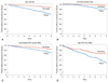
Numerous studies have demonstrated that cIMT is a significantly predictive for cardiovascular events. However, what is still not centain is the additional predictive value of cIMT beyond traditional risk score. Lorenz, et al. studied the additive value of cIMT using the 10-year follow-up data of 4904 subjects from the Carotid Atherosclerosis Progression Study (CAPS).29 Subjects were reclassified to new risk strata using traditional risk prediction models with cIMT, and were compared to classifications based on traditional risk scores to determine the improvement in cardiovascular risk prediction with the addition of cIMT. Surprisingly, the risk prediction with cIMT did not demonstrate an improvement in cardiovascular risk prediction, compared to the traditional risk prediction model. However, a study done in 13415 subjects enrolled in the ARIC study showed that adding cIMT to traditional risk factors improves predictability. Also, this study showed that the addition of carotid plaque further improved the prediction strength.27 Likewise, Polak, et al. demonstrated that cIMT is able to contribute to improving traditional risk prediction models.5 They carried out a study in 2965 subjects of the Framingham Offspring Study Cohort and found that the prediction of cardiovascular disease became better after addition of maximum intima-media thickness of internal carotid artery, with an improvement of the net reclassification index by 7.6% (p<0.001); however, no improvement in net reclassification was demonstrated when mean intima-media thickness of common carotid artery was added to the traditional risk factors. In this study, the presence of plaque, defined as internal cIMT of more than 1.5 mm, was associated with significant increase in both C statistics [0.014, 95% confidence interval (CI): 0.003-0.025, p=0.02] and improvement in the net reclassification index by 7.3% (p<0.01). Also, the presence of plaque significantly improved the prediction of new-onset cardiovascular disease (Fig. 2). Therefore, the assessment of both common carotid and internal carotid plaque add incremental value in predicting cardiovascular events.
Notwithstanding, cIMT may not be useful for risk stratification in all spectrums of risk categories. Among 10-year follow-up results from the CAPS, which included a cohort of relatively young and low risk subjects (n=4904) in a primary healthcare setting, cIMT did not improve the risk classification of individuals.29 Likewise, a recent meta-analysis of 14 population cohorts of 45828 subjects by Den Ruijter, et al. demonstrated that addition of common cIMT to FRS was associated with a small improvement in 10-year risk prediction of first time myocardial infarction or stroke, but without significant improvement in the net reclassification (0.8%, 95% CI: 0.1-1.6%). However, the net reclassification improvement was 3.6% (95% CI: 2.7-4.6%) in subjects with intermediate risk of cardiovascular disease.30 Therefore, cIMT may be useful for risk stratification in subjects with intermediate risk of cardiovascular disease as determined by traditional risk factors.
MEASUREMENT OF cIMT: IS THE PROGRESSION OF cIMT A PREDICTOR OF CARDIOVASCULAR DISEASE?
With cIMT being accepted as a measure of subclinical atherosclerosis and organ damage, it has been presumed that the progression of cIMT, as a marker of atherosclerosis progression, will be associated with adverse cardiovascular prognosis. However, recent publications are contradictory to these assumptions. Analysis of the European Lacidipine Study on Atherosclerosis showed that both baseline IMT and on-treatment IMT, but not treatment induced changes, were associated with incident cardiovascular events in treated hypertensive patients.31 In the PROG-IMT collaborative project, a meta-analysis was done on 16 studies with 36984 participants who had at least two measurements of cIMT and outcome data regarding myocardial infarction, stroke, and death.32 The results showed that for mean cIMT progression the hazard ratio was not significantly increased (hazard ratio=0.98, 95% CI: 0.95-1.01) when adjusted for age, gender, mean cIMT, and vascular risk factors. As such, recent European Society of Hypertension (ESH) and European Society of Cardiology (ESC) guidelines did not endorse the usefulness of follow-up cIMT for assessing vascular organ damage in hypertensive subjects.17 From the evidence available at this time, it is uncertain as to whether or not change in cIMT during the follow-up carotid ultrasound is associated with increased risk of cardiovascular disease. As such, follow-up carotid ultrasound should not be performed for the sole purpose of cardiovascular risk stratification.
LIMITATIONS OF cIMT AND ISSUES THAT NEED TO BE RESOLVED
Despite the widespread use of cIMT, it still has limitations. Above all, the threshold value of cIMT, above which the risk is increased, needs to be clearly defined. cIMT is affected by multiple factors. Age is the most influential factor on cIMT and common cIMT increases 0.01 mm annually.33 Thus, it is hard to apply one absolute value to the general population as abnormal IMT. Although the ESH/ESC guidelines define a high risk cIMT finding as a mean cIMT of more than 0.9 mm, this may not be generalizable to all populations, especially Asians. In addition, as cIMT is a continuous variable, a definition to distinguish between plaque and vessel wall hypertrophy is arbitrary. The 34th Bethesda conference suggested the use of nomograms or ratios of observed to predicted IMT based on age and other factors affecting cIMT, such as the approximate age-adjusted 75th percentile values for cIMT.34 The Manheim cIMT Consensus defines plaque as a focal structure encroaching into the arterial lumen by at least 0.5 mm or 50% of the surrounding wall or demonstrates a plaque thickness of >1.5 mm.35 However, each of these studies still used an arbitrary cutoff point for abnormal IMT, and more research is needed to further standardize a protocol.
Another key question that needs to be resolved is how the presence of high risk cIMT findings in a patient affects management decisions. When abnormal results are found on carotid ultrasound screening, physicians are more likely to prescribe aspirin and lipid-lowering medication.36 However, recent meta-analyses suggested that aspirin may not offer benefits for primary prevention of cardiovascular disease when considering the increase in bleeding risk.37,38 Therefore, a prospective randomized study to determine whether the addition of antiplatelet agents, based on the presence of high risk cIMT finding alone, may have significant benefit needs to be performed. Also, while several clinical trials reported that statin reduced the progression of carotid atherosclerosis and cardiovascular disease risk, there are no prospective studies to show the cardiovascular benefit of prescribing statin based on the presence of high risk cIMT. Although the ESC/EAS guidelines for the management of dyslipidemia grouped subjects with carotid plaque as very high risk and recommended an LDL-cholesterol treatment goal of 70 mg/dL in these patients, this is based more on expert recommendation rather than clinical evidence.39 As such, prospective randomized study to determine the LDL-cholesterol treatment goal for obtaining maximal efficacy in subjects whose risk classification has changed according to cIMT needs to be performed. In conclusion, although there are still unresolved issues regarding the usefulness of cIMT in primary prevention, cIMT is considered to have additive value in cardiovascular risk prediction, especially in subjects with intermediate risk of cardiovascular disease by traditional risk factors. However, further studies are needed to determine treatment strategies to possibly reduce cardiovascular risk in patients with high risk cIMT.
 XML Download