

 PDF
PDF ePub
ePub Citation
 Print
Print


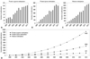
The term protein methylation in reference to a posttranslational modification reaction first appeared in scientific journals in the mid-1960's as a potential epigenetic regulator.1 However, the initial enthusiasm ended by the mid-1980's due to the lack of recognizable progress in understanding its biological significance. Fortunately, with the introduction of modern molecular biotechnology, the last decade witnessed a tremendous upsurge of interest and accomplishment in protein methylation research. Figure 1 depicts the result of a PubMed search using arbitrary keywords related to protein arginine methylation (Fig. 1A), protein lysine methylation (Fig. 1B), and histone methylation (Fig. 1C) for articles published over the past thirteen years (2001-2013), and we found 78%, 89% and 98% of all articles published in each field (Fig. 1D), respectively (Footnote 1). An average of six papers per day are published in the field of protein methylation. Indeed, protein methylation constitutes one of the most hotly pursued topics, covering many fronts of modern biological science. In the present article, we describe the historical background and general status of such endeavors, followed by a brief review of the interaction of protein arginine methyltransferase and antiproliferative genes and BTG/Tob families. We also focus on cell differentiation, growth arrest and apoptosis, and DNA damage signal pathway to suggest the regulation of genomic DNA stability.
BEFORE THE TERM PROTEIN METHYLATION APPEARED1
In 1939, Schoenheimer, et al.2 observed that all the amino acids, except threonine and lysine, in the acid-hydrolysate of body proteins isolated from a rat that had previously been injected with isotope-labelled NH3 contained N.15 This cleared any uncertainty of whether body proteins remain unchanged during the life span of animal and indicated that lysine and threonine had to be supplemented exogenously to maintain life. Subsequently, in the mid-1940s, Neuberger and Sanger3 found that the administration of ε-N-acetyl-L-lysine or ε-N-methyl-L-lysine (Fig. 2A) to weaning rats on a basal gliadin diet (a diet sufficient to maintain but not increase body weight) resulted in weight gain similar to that which results from the addition of L-lysine. However, they failed to demonstrate the production of free L-lysine from ε-acetyl-L-lysine in vitro. Therefore, as described below, we sought to identify two separate enzymes to produce L-lysine from these compounds.
Meanwhile, protein-bound ε-N-methyl-lysine was first found in 1959 in the flagella protein of Salmonella typhimurium4 and five years later in histones from various sources.5 The discovery of the presence of ε-N-methyl-lysine in histone molecules generated a great deal of excitement. In 1962, Huang and Bonner6 observed that histones stoichiometrically inhibited DNA-dependent RNA synthesis, and in 1964, Allfrey, et al.7 found that the formation of ε-N-methyl-lysine in histone molecules was insensitive to a protein biosynthesis inhibitor puromycin. These two observations suggested the possibility that posttranslational methylation might allow a histone with a wide range of specificity to control DNA-dependent RNA synthesis.
Until now, only two kinds of proteins, histone and flagella proteins, were known to contain ε-N-methyl-lysine. Furthermore, methyllysine was only known as a single entity of ε-N-methyl-lysine, and a single experiment with an antibiotic suggested the possibility of a posttranslational reaction. However, enzymes involved in the reactions had never been mentioned.
OUR INVOLVEMENT IN THE FIELD OF PROTEIN METHYLATION
In support of the aforementioned observation by Neuberger and Sanger,3 Paik, et al.8 identified an enzyme ε-lysine acylase that hydrolyzes ε-N-acetyl-L-lysine to result in free L-lysine and acetate (Footnote 2). Afterwards, it was postulated that an additional enzyme different from ε-lysine acylase to metabolize ε-N-methyl-lysine exists since the bond in ε-methyl-L-lysine is an amide type, unlike that of ε-N-acetyl-L-lysine. We discovered an enzyme, ε-alkyllysinase, in 1963 that oxidatively demethylated ε-N-methyl-L-lysine resulting in the formation gof free L-lysine and formaldehyde.9,10 This enzyme was also found to demethylate protein-bound ε-N-methyl-lysine residues,11 and became the forerunner of many demethylases that were subsequently identified.12
Even though their conclusion was later proven false, the observation by Huang and Bonner6 that histones stoichiometrically inhibited the DNA-dependent RNA synthesis (Footnote 3) together with the fact that various histones contained ε-N-methyl-lysine5 and that the formation of ε-N-methyl-lysine in histones was insensitive to puromycin,7 made us focus our attention on methylation at the protein level instead of free methylysine. As the first step toward this goal, we decided that it was necessary to find out if ε-lysine methylation was a posttranslational process, even though Allfrey, et al.7 showed that the reaction was insensitive to the recently discovered antibiotic puromycin; however, this result was based on only one experiment. Thus, we explored this problem by systematically examining various steps of reactions involved in protein biosynthesis, including the activation of amino acid, formation of aminoacyl t-RNA, and transfer of amino acid from aminoacyl t-RNA to ribosomal protein as well as evidence that indicated histone methylation was a posttranslational reaction.1 For the first time, we also observed that S-adenosyl-L-methionine (AdoMet) could serve as a methyl donor for the reaction.1
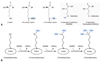
When isolated calf thymus nuclei were incubated with [methyl-14C]-AdoMet, and the acid-hydrolysate of the protein was analyzed by paper chromatography, two radioactive ninhydrin spots were detected whose retention factor (RF) values corresponded to those of ε-N-monomethyllysine and ε-N-dimethyllysine.1 Although ε-N-methyllysine was present in the flagella proteins of Salmonella typhimurium and in histones of higher animals, the presence of ε-N-dimethyllysine in these sources had not yet been reported. These findings prompted us to reexamine the resolution of the analytical methods employed by others, particularly column chromatography with a Beckman automatic amino acid analyzer. By increasing the pH of the elution buffer (from 5.28 to 5.84) and decreasing the elution temperature (from 50℃ to 28℃), we were able to resolve ε-N-mono and ε-N-dimethyllysine using column chromatography, thus confirming the presence of ε-N-dimethyllysine in the histone.13 One year later using the modified elution condition, Hempel, et al.14 established the presence of ε-N-trimethyllysine in histones, and this completed the discovery of all the theoretically possible ε-N-methyl substituted lysine derivatives (Fig. 2B). ε-N-trimethyllysine, which is also called laminine, had previously been found in kelp (laminariaces) as a free amino acid in 1964.15 The resolution of these three ε-N-methyl substituted lysine derivatives and confirmation of their presence in histones constitute one of the milestones in the field of protein methylation.
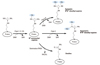
Thus, by 1968, it was firmly established that the ε-NH2 group of lysines in histones was mono-, di- or trimethyl-substituted. It was for this reason that we pursued the identification and purification of an enzyme responsible for such a methylation reaction. Using [methyl-14C]-AdoMet as the methyl donor, we purified such an enzyme from the cytosol of a calf thymus (Supplementary Table 1, only online). However, much to our dismay at the time, the enzyme (called protein methylase I-EC 2.1.1.2316) could not methylate the ε-NH2 group of lysine residue, rather some unknown amino acid. In hindsight, this turned out to be a turning point in our research; nevertheless, it was one of the most discouraging times in our scientific career. After more than half a year's effort, we came to the conclusion that the methylated amino acid was an arginine derivative, since (methyl-14C)-labelled derivatives were sensitive to NaOH, giving rise to ornithine and radiolabelled methylamines.16 However, due to a lack of standard compounds and minute amounts of radiolabelled materials available at that time, we were unable to precisely determine the structure. In 1970, Kakimoto and Akazawa17 chemically synthesized various guanidino-methyl substituted arginine derivatives, and finally identified the structures of the enzymatically formed [methyl-14C]-arginine derivatives as NG-monomethyl-arginine, symmetric NG,N'G-dimethylargininie, and asymmetric NG,NG-dimethylarginine (Fig. 3). The finding that the guanidino group of arginines could also be methylated greatly expanded the knowledge of protein methylation and marked another milestone in the field of protein methylation.
Protein methylase I was named because the enzyme was unexpectedly found. We had expected to find an another enzyme to methylate the ε-NH2 group of lysine residue. Although histones had been used as an in vitro substrate for the enzyme early on, myelin basic protein (MBP) was the first protein that was clearly shown to contain NG-methyl-arginine.18,19 The protein methylase I, which methylated MBP, was later found to be different from that of the methylating histone, and this foretold the diversity of the enzyme.20,21 At present, protein methylase I is known as "PRMT" (protein-arginine N-methyltransferase), and numerous natural proteins are shown to be methylated by nine different PRMTs22 (Supplementary Table 1, only online).
In the course of attempts to further purify protein methylase I from a calf thymus cytosol, we observed two protein peaks on column chromatography.23 One of the enzyme peaks had protein methylase I activity, as expected, which methylated the guanidino group of arginine, and the product was stable on acid-hydrolysis. By contrast, the second peak enzyme produced a product that was incorporated (methyl-14C) into the substrate protein (in this case histones), but was volatile on acid-hydrolysis. This led us to rediscover the enzyme that had previously been identified by Liss and Edelstein in 1967.24 This enzyme was capable of esterifying the dicarboxylic amino acid residues of proteins. We designated this enzyme as protein methylase II (SAM: carboxyl-O-methyltransferase; EC 2.1.1.24) because this was the second enzyme discovered that transfers a methyl group to a protein side chain. However, protein methylase II was later proven to be identical to what was then referred to the "methanol-forming enzyme" from the posterior lobe of the bovine pituitary gland.25 However, at the time, the authors of the study were unaware of the strict requirement of the enzyme for a protein substrate. Interestingly, there are several new concepts regarding the function of O-methyltransferase enzymes. These include effects related to bacterial chemotaxis26 and protein repair during aging.27
By the 1960s, while our research on protein methylase I and II was in progress, we had accumulated enough evidence to indicate the presence of an enzyme involved in protein-lysine methylation. However, in light of the enzymatic methylation of arginine and dicarboxylic amino acid residues, it was necessary to devise an assay method specific for lysine methylation. By treating the incubation mixture with 0.1 M NaOH after allowing the enzyme to react, we were able to remove the methylated arginines (Fig. 3) and methylated dicarboxylic amino acids from our enzyme reaction products while preserving methylated lysines (Footnote 4). With this assay method, a protein lysine methyltransferase (PKMT) from a calf thymus was readily characterized and designated as protein methylase III in 1970 (SAM: protein-lysine N-methyltransferase; EC2.1.1.43).28,29 This enzyme and others similar to it comprise what is now known as a family of lysine-specific histone methyltransferases.30
The identification of protein methylase III marked the period during which various naturally occurring methylated amino acids and enzymes responsible for their methylation were identified. Therefore, the scope of this field was greatly expanded and most of the ground work was laid for future studies on protein methylation.29 However, until the end of the last century, there has been a hiatus from numerous efforts made to substantiate the biological function of these reactions. It was not until the application of molecular biology techniques to the field of protein methylation in the mid-1990s that the field began to advance.
PROTEIN METHYLATION TODAY
In the following section, we summarize some of the highlights of current protein-methylation research to provide more perspective on this field.
Protein-arginine methylation
Before the advent of modern molecular biology and its associated experimental techniques, the enzyme known to methylate the guanidino group of arginine residues (i.e. protein methylase I, PM-I) was believed to have only two subtypes: histone or heterogenous nuclear ribonucleoproteins (hnRNP)-specific PM-I and myelin basic protein (MBP)-specific PM-I21 (Footnote 5). These subtypes were identified using classical enzymological techniquesas described earlier.
PRMT, formerly known as PM-I, is not only specific for Gly-Arg-Gly or Gly-Ala-Arg primary sequences but also highly specific for the tertiary structure of proteins. The PRMT family has been shown to include at least nine methyltransferases31-33 designated as PRMT1-9, based on differences in primary sequence and substrate specificity. These enzymes can also be broadly divided into three types based on its methylation activity: type I PRMT can form NG-monomethyl-L-arginine and asymmetric dimethyl-L-arginine, type II PRMT forms NG-monomethyl-L-arginine and symmetric dimethyl-L-arginine, whereas type III forms only NG-monomethyl-L-arginine (Fig. 3). Furthermore, an ever expanding list of proteins are known to be methylated both in vivo and in vitro, which includes histones, fibrillarin, nucleolin, scaffold-attachment factor A, signal transducer and activator of transcription, Ewing sarcoma in addition to myelin basic protein and hnRNP proteins.33,34 As in the case of ε-N-methyl-L-lysine residue,11,35 ε-NG-methyl-L-arginine residues are now known to be demethylated with the formation of citrulline and releasing methylamine via the arginine deimination reaction by PADI (Fig. 3).36,37 The resulting citrulline-containing protein could be destined for degradation or regenerated by a uncharacterized mechanism.
In conjunction with the expanding number of proteins that are known to be arginine-methylated, a growing list of biological process has been shown to involve arginine methylation, such as signal transduction, cellular proliferation, transcriptional processes, and splicing of mRNA. In particular, some recent review articles describe a specific area of interest and discuss protein-arginine methylation in cancer,38,39 the function of RNA-binding protein,40 plant development,41 parasitic protozoa42 and T-lymphocyte as well as its function.43 Earlier studies suggested an association of MBP-arginine methylation with myelin formation and myelin-related diseases such as multiple sclerosis.44 Furthermore, evidence also indicates that arginine-specific histone methylation has a role in association with other histone modifications such as acetylation, phosphorylation and ubiquitination.45
One of the direct outgrowths of protein-arginine methylation research has been the identification of methyl-arginines as naturally occurring competitive inhibitors of nitric oxide synthases, the enzymes responsible for the production of nitric oxide. Because nitric oxide is involved in so many important physiological and pathophysiological processes, NG-methyl-L-arginines (asymmetric NG,NG-dimethyl-L-arginine in particular) and their associated enzymes have provided targets for pharmacotherapy.46 Furthermore, the use of NG-methylarginine (and other arginine derivatives) has provided a means for studying nitric oxide-mediated processes such as smooth-muscle relaxation and inflammation. Indeed, some of the earliest studies delineating the biological importance of the L-arginine-nitric oxide pathway in the cardiovascular system used NG-methyl-L-arginines as an indispensable tool for blocking nitric oxide production.47 Thus, it is noteworthy that the preliminary work in the field of protein-arginine methylation facilitated the development of the field of nitric oxide biology.
Protein-lysine methylation
Regarding protein-lysine methylation, the bulk of recent efforts have primarily been focused on histone methylation (Fig. 1C). This is undoubtedly a result of the lure that histones possess control of genetic expression since they are components of the nucleosome. When first identified protein methylase III, which methylates the ε-amino group of lysine residues in protein, from isolated calf thymus nuclei it was found to be tightly bound to the particulate fraction (most likely chromatin) and highly labile once solubilized. Consequently, we could purify it only three-fold.28 Nevertheless, the 'crude' nuclear protein mixture methylated exogenous and endogenous histones and gave rise to ε-N-mono, ε-N-di-, and ε-N-trimethyl-lysine (Fig. 2B). In hindsight, we know that protein methlyase III was a mixture of histone methyltransferases. Furthermore, we now know from studies involving several plant and animal species that histone subunits H3 and H4 are the primary substrate targets, and multiple different lysine residues are methylated by more than a dozen highly specialized histone methyltransferases.33 These enzymes share a SET [Su(var)3-9, suppressor of position effect variegation; E(z), enhancer of zeste; and Trx, trithorax] domain that is responsible for catalysis and binding of the methyl donor AdoMet.
The chemical mechanisms and biological importance of methylation at each particular lysine residue in the H3 and H4 histone subunits continue to be unraveled. Methylation of several lysine residues in the H3 subunit [i.e. Lys36 (H3K36) and Lys79 (H3K79)] is associated with euchromatin and transcriptional activation, whereas methylation of other residues in H3 and H4 [i.e. H3Lys4 (H3K4), H3Lys9 (H3K9), H3Lys27 (H3K27) and H4Lys20 (H3K20)] is associated with heterochromatin and transcriptional repression.33 Furthermore, histone-lysine methylation is enzymatically reversible, indicating a dynamic process.
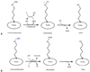
The biochemical mechanism of lysine demethylation can be explained by two different pathways. Lysine-specific demethylase 135 (LSD1, KDM1A) is a nuclear homolog of amine oxidase (also known as oxygen oxidoreductase), and is thought to be identical to the first enzyme that we discovered relating to protein methylation, ε-alkyllysinase.10 LSD1 demethylates H3K4 by the FAD-dependent amine oxidase and releases formaldehyde and free lysine (Fig. 4A). Importantly, the methylation or demethylation of H3K4 has been associated with the regulation of transcription. On the other hand, JmjC domain-containing histone demethylase 148 (JHDM1, KDM2A) demethylates H3K36me or H3K36me2 by the activity of dioxygenase, thus formaldehyde and free lysine can be released in the presence of Fe++ (Fig. 4B). In the reaction, α-ketoglutarate is metabolized to succinate along with decarboxylation. Here, we have summarized several other demethylases that have since been identified and the references associated with pathology were also marked (Supplementary Table 2, only online). In addition, JHDM2A demethylates H3K9me and H3K9me2,49 and JMJD2 (JmjC domain-containing protein 2) demethylates H3K9me3 or H3K36me3 to give rise to dimethylated counterparts.50 This dynamic nature of histone-lysine methylation and demethylation has an important role in regulating transcription and functions in conjunction with acetylation, phosphorylation, and ubiquitination. As mentioned previously, peptidyl arginine deiminase 4 (PADI4 or PAD4) converts methyl-arginine to citrulline and releases methylamine (Fig. 3) that leads to the antagonizing arginine methylation on the N-terminal tail of histone H3.36,37
In addition to histones, ε-amino groups of lysine residues of highly specialized proteins, such as calmodulin, cytochrome c, Rubisco (ribulose biphosphate carboxylase-oxygenase), heat-shock proteins, ribosomal proteins and actin, are known to be methylated in vivo.51 Some of these proteins have been associated with highly specific PKMTs.52 Recent review articles indicate an ever-expanding scope of involvement of protein-lysine methylation in many areas of biological sciences such as gene transcription and remodeling,53-55 life span,56 stem cell,57 DNA packaging in spermatozoa,58 alternate splicing,59 plant development,60,61 diabetes,62 AIDS,63 and psychiatric disorders.64
Finally, an additional area of scientific knowledge that was a direct outgrowth from protein methylation research is carnitine biosynthesis.65 Free ε-N-trimethy-L-lysine is formed by the intracellular hydrolysis of ε-N-trimethylated proteins and converted to betaine, the precursor of carnitine, which has important functions related to fatty acid transport. Once again, as in the case of nitric oxide biology and methyl-arginines, primary work in the field of protein methylation found fertile ground in a seemingly unrelated area and gained importance.
REGULATION OF BIOLOGICAL FUNCTIONS BY INTERACTION OF PRMT1 WITH ANTIPROLIFERATIVE PROTEINS
In the past, arginine methylation was mainly observed on abundant proteins such as RNA-binding proteins and histones, but recent advances have revealed a plethora of methylated proteins implicated in a variety of cellular processes including RNA metabolism, epigenetic regulation of chromatin remodeling to regulate signal integration, and DNA damage response pathways. Herein, we briefly review recent advances in the understanding of mutual interaction between protein arginine methyltransferases and antiproliferative genes, BTG/Tob family, focusing on their roles in cell differentiation, growth arrest and cell death, the DNA damage signal pathway, and its importance in the maintenance of genomic stability. The antiproliferative gene, BTG/Tob, includes at least six distinct members in vertebrates,70,71 namely BTG1, BTG2/TIS21/Pc3, BTG3, BTG4/Pc3B, Tob1/Tob and Tob2.66,67 This family of proteins are structurally related and characterized by the presence of three conserved domains in their amino-terminal region called BTG Box-A, B, and C.68 Mouse TIS2169 and rat PC370 are homologs of human B-cell translocation gene-2 (BTG2). Data accumulated during the past two decades with both cells and animal models implicate that BTG2/TIS21/PC3 (BTG2) plays roles in various biological processes such as mediation of stage specific expansion of developing thymocytes,71 regulation of the hematopoietic progenitor cell expansion in response to estradiol,72 cell cycle arrest at G2/M73 and G1/S phases,74,75 enhancement of cancer cell death via interaction with Pin-1 in response to growth factor stimulation76 or via accumulation of hydrogen peroxide after doxorubicin treatment,77 and regulation of neuronal differentiation.78,79 Moreover, BTG2 is involved in the differentiation of myelocytic leukemia cells and CD34+ hematopoietic precursor cells,80,81 DNA repair,82,83 inhibition of cancer cell migration84 as a transcriptional co-regulator in different model systems, and in the antioxidant defenses through the antioxidant transcription factor NFE2L2.80,85 Murine BTG2 gene, TIS21, has originally been identified as a primary response gene86 induced by stimulations with either growth factors, tumor promoters, a high concentration of serum addition, Ca++ flux changes, or depolarization. Under the oxidative stress generated by serum deprivation or exogenous treatments, however, BTG2 expression is regulated via NFκB activation.87
In 1996, Herschman's group cloned protein methyltransferase, which interacts with mammalian immediate-early gene, TIS21/BTG2/Pc3 and leukemia-associated B-cell translocation gene (BTG1), through the yeast two hybrid system and renamed it as PRMT (protein-arginine N-methyltransferase),88 which corresponds to our protein methylase I. PRMT1 was found to be bound to Box C domain of BTG1 and BTG2,89 and the interaction of PRMT1 with BTG2 significantly increased the activity of PRMT1,83 strongly suggesting BTG2 as a regulating factor of PRMTs. Meanwhile, we observed the in vitro methylation of recombinant TIS21/BTG2/Pc3 protein by protein methylase I,90 indicating TIS21 is one of the PRMT substrates.
Regulation of erythroid differentiation
The expression of BTG1 can be directly regulated by PI3K-controlled Forkhead box class O (FoxO) subfamily, FoxO3a. BTG1 and BTG2 can be the direct target of FoxO3a, and expression of BTG1 down regulates the outgrowth of erythroid colonies during erythroid differentiation.91 Inhibition of methyl transferase activity blocks erythroid maturation without affecting expansion of progenitor cells. Therefore, FoxO3a-controlled expression of BTG1 and the subsequent regulation of PRMT activity have been considered a novel mechanism controlling erythroid expansion and differentiation. However, the expression of BTG2 inhibits uncontrollable proliferation of bone marrow precursor cells (Lin-Sca1+cKit+) in mouse via downregulation of mTOR activation and phosphorylation of Akt.72
Differentiation of myeloid leukemia cells and CD34+ hematopoietic progenitors
The promoter region of retinoic acid receptor alpha (RARα) provides the binding site for PRMT1, BTG2 and Sin3A. Upon retionoic acid treatment, Sin3A, BTG2, and PRMT1 are detached from RARα promoter to the cytoplasm and prime histone H4 demethylation and acetylation. Retinoic acid induces BTG2 overexpression and increases RARα transcriptional activity along with the differentiation of HL-60 promyeloid leukemia cells via degradation of c-Myc protein.81 The overexpressed BTG2 increases PRMT1 participation in the RARα protein complex on the RARβ promoter and enhances gene-specific histone H4 arginine methylation, and this contributes to retinoic acid activity by favoring differentiation through a gene-specific modification of histone H4 arginine methylation and acetylation levels.80 BTG2 enhances retinoic acid-induced differentiation by modulating histone H4 methylation and acetylation.
Regulation of neurite outgrowth in Neuro2a cells by PRMT1 and Btg2
Neurite outgrowth is an important event in the formation of neural circuits, and the signal transduction processes are usually based on the phosphorylation and acetylation. When PRMT1 was depleted, neurite outgrowth, but not cell growth and differentiation, was significantly affected. Moreover, depletion of BTG2 expression, an activator of PRMT1, downregulates arginine methylation in the nucleus and inhibits neurite outgrowth, suggesting a possibility of regulation of nuclear proteins by PRMT1 during neuritogenesis.92
Regulation of growth arrest and apoptosis
Engagement of membrane immunoglobulin on WEHI-231 murine B lymphoma cells upregulates BTG1 and BTG2 expression and induces growth arrest at the G1 phase and subsequent apoptosis via interaction of anti-immunoglobulin M with BTG1 and BTG2 bound to PRMT1. This was shown by employing PRMT1-deficient cells via a small interference in RNA and by treatment of WEHI 231 cells with the arginine methyltransferase inhibitor, S-adenosyl-L-homocysteine (AdoHcy). Methylation, detected by a monoclonal antibody specific for asymmetric (not symmetric methyl residues), can be observed as early as 1 h to 2 h after stimulation with anti-membrane immunoglobulin and sustained for up to 24 h. These results indicate that anti-immunoglobulin M-induced growth inhibition is mediated via the upregulation of BTG1 and BTG2, resulting in the activation of arginine methyltransferase activity and culminating in growth inhibition of WEHI-231 cells.93
The RGG/RG motif of nucleolin, a RNA binding protein, physically interacts with poly-A-binding protein (PABP), and the association stabilizes Bcl-XL mRNA by inhibiting the action of poly-A-specific ribonuclease.94 Thus, BTG2/TIS21/Pc3, which is a general activator of mRNA deadenylation,95 and its physical interaction with PRMT1 protein,59 forming BTG2-PRMT1-nucleolin-PABP interaction, might play an important role in the Bcl2 family-mediated apoptosis in response to various cellular damages (UV, IR) and chemical agents.
Regulation of PRMT1-mediated crosstalk between transcription and RNA processing
As mentioned above, BTG1 and BTG2 interact with PRM-T188 and regulate its activity. In addition, hCAF1 (CCR4-associated factor 1), which interacts with B-box of BTG1 and BTG2,96 has recently been characterized as a new regulator of PRMT1.97 Co-immunoprecipitation and immunofluorescence experiments demonstrated in vivo interaction of hCAF1 and PRMT1 in the nuclear speckles, a sub-nuclear compartment enriched in small nuclear ribonucleoproteins (snRNPs) and splicing factors. In vitro methylation assays also revealed that hCAF1 regulates methylations of Sam68 and histone H4 proteins. These results imply that the hCAF1 and PRMT1-regulated transcription and RNA metabolism contribute to the crosstalk between transcription and RNA processing. Interaction between BTG proteins and hCAF1 may recruit the Ccr4-not complex to target mRNA.98
DNA damage signaling and regulation of protein methylation
MRE11 and 53BP1, proteins regulating the repair of a DNA double-strand break (DSB), contain RGG/RG motifs that are methylated in a PRMT1-dependent manner.99 MRE11, part of the MRE11/Rad50/NBS1 complex, is one of the initial proteins recruited for DNA double strand breaks (DSBs)100 as well as the RGG/RG motif deleted mutant renders MRE11 unable to efficiently localize to DSBs.101 The mouse model harboring R-to-K mutations of the nine arginines in the RGG/RG motif of MRE11 and its mouse embryo fibroblasts cells exhibit defects in the DNA end resection, in the ATR kinase activation in response to ionizing radiation, the G2/M checkpoint defect similar to ATR-deficient Seckel syndrome, and the phenotypes observed in the ATR- and Fanconi-deficient cells.102 This suggests arginine-methylation has an essential function in the maintenance of genomic stability.
Chromosome instability syndromes such as ataxia telangectasia, Nijmegen breakage syndrome, and ataxia telangenctasia-like disease are strongly associated with defects in the recombinational DNA repair proteins during cell cycle regulation after DNA damage,103 and mostly involved in the regulation of Mre11/Rad50/Nbs1 (MRN) complex activity. The physiological functions of the MRE11/Rad50/NBS1 complex and their interactions with other molecules might explain the wide clinical presentation observed in patients with chromosome instability syndrome. We previously reported regulations of Mre11 and PRMT1 activity by BTG2 using TIS21KO mouse embryonic fibroblasts and adenoviral transduction of the TIS21 gene into Huh7 cancer cells.83 Expression of BTG2 increased methylation of Mre11 and PRMT1 activity, leading to1 Mre11 activation in vitro and in vivo.
CLOSING REMARKS
Although remarkable progress has been made in protein methylation research during the past decade, clear outcomes still cannot be predicted. The late Mahlon Hoagland, who is credited for the discovery of tRNA and amino acid activation, is quoted to have said, "Fundamental research can predict no outcomes."
Last, we would like to quote the words of the late Lehninger,104 which can be found in the preface of the first edition of his well-known textbook, "living organisms normally contain no functionless compounds, although there are some biomolecules whose functions are not yet understood."
 XML Download