

 PDF
PDF ePub
ePub Citation
 Print
Print


INTRODUCTION
During DNA replication, the linear ends of chromosomes are eroded at each cell division due to the end replication problem.1 Telomeres, the very ends of linear chromosomes, are predominantly composed of tandem repeats of short sequences; in vertebrates, the repeats consist of the TTAGGG hexanucleotide.2 Telomere lengths are also remarkably heterogeneous among individuals and vary according to the origin, age, and proliferative history of cells.3,4 Telomere length variations among individuals of the same age are, therefore, thought to be related to variations in ageing and longevity.5 As a ribonucleoprotein complex that is composed of telomerase reverse transcriptase (TERT) and telomerase RNA component (TERC),6 telomerase is responsible for elongation of the telomeres, and thus maintains genome stability.7,8 The enzymatic activity of telomerase is not detected in normal somatic cells, but is detected in embryonic and highly proliferative adult tissues.9 Furthermore, telomerase is re-activated in most cancers,10 thus suggesting the possibility that telomerase is a potential therapeutic target in cancers.
Recently, a number of reports have shown extra-telomeric functions of TERT. The following observations pose questions regarding the rationality of telomere length-dependent singular role of telomerase:
1) Although tissue stem and progenitor cells have sufficient telomere reserves, they highly express TERT. Notably, laboratory mice have significantly longer telomeres than humans (40-60 kb vs. 5-15 kb);11-14 however, no apparent role for long telomeres has been found in the survival of mice.
2) TERT overexpression promotes tumor development without further telomere elongation.15
3) The reconstitution ability of hematopoietic stem cells (HSCs) is essentially linked with TERT, although there is no evidence that HSC activities are fully dependent on the telomere-elongation function of TERT.16,17
4) Transgenic mice overexpressing murine TERT show significant resistance to ischemic brain injury and N-methyl-D-aspartic acid (NMDA) receptor-mediated excitotoxicity without any detectable change in telomere length.18 Interestingly, ischemic injury induces TERT expression in the wild-type brain.18
5) Suppression of TERT expression decreases cell growth rate and induces apoptosis prior to measurable telomere shortening,19 and the expression of specific TERT mutants lacking telomerase activity prevents apoptotic cell death.20
6) Ectopic expression of TERT in the hair follicle stem cells of mouse epidermis activates stem cell capacities.21 The phenotype appears to be independent of telomerase activity.
7) Cancers without functional telomerase (10-15% of all cases) maintain their telomere lengths by adopting an alternative lengthening of telomeres (ALT) pathway.22
Furthermore, indirect evidence also indicates extra-telomeric functions of telomerase: TERT may have additional functions because the reverse transcriptase (RT) domain of TERT is only 15 kDa, which is less than 10% of the total molecular weight. Thus, it is quite possible that other regions may mediate distinct activities other than telomerase activity.23 In support of this hypothesis, alternatively spliced forms of TERT devoid of the RT domain have been identified in humans.24 Although TERT is known as a nuclear protein, it is exported from the nucleus and delays replicative senescence in endothelial cells dependent on reactive oxygen species (ROS).25
Several lines of evidence obtained using mouse models have strengthened the idea that TERT contributes to preventing ageing and cell death, as well as promoting carcinogenesis, cell signaling, and transcriptional regulation, which are unrelated to its role in telomere lengthening. Here, we review the studies encompassing the extra-telomeric functions of telomerase conducted using transgenic or knockout mouse models.
SURVIVAL-PROMOTING FUNCTIONS OF TELOMERASE
Apoptosis induced by critically short telomeres has been extensively documented. Dysfunctional telomeres increase apoptosis in highly proliferative tissues including intestine,26,27 male germ cells,26,28 and splenocytes (B cells) from immunized fifth and sixth generation mice after mitogen treatment.29 These dysfunctional telomeres can be generated in early generations, and can increase apoptosis in these tissues as well. Protection of Telomeres 1 (POT1) is a single-stranded telomere binding protein that is essential for proper maintenance of telomere length. When Pot1 is deficient, abnormal apoptosis is induced in proliferative tissues as well as cells derived from Terc-/- mice, including male germ cells, hematopoietic cells, and intestinal cells.30 Critically short telomeres also affect highly proliferative developmental processes. Terc-/- embryos at embryonic day 10.5 (E10.5) with dysfunctional telomeres frequently exhibit neural tube closure defects, suggesting that this is one of the most sensitive developmental processes to telomere loss and chromosomal instability.31 Although cardiac tissues are not highly proliferative, the balance between cell growth and cell death is critical for maintaining normal heart function. Consistently, dysfunctional telomeres lead to abnormal apoptosis in cardiomyocytes, resulting in cardiac dilatation and heart failure in the late generation of Terc-/- mice.32 Additionally, cardiomyocyte survival is promoted in transgenic mice overexpressing wild-type TERT, but not expressing mutant TERT.33 Tert deficiency also induces apoptotic phenotypes; Tert-/- and Terc-/- mice show frequent apoptosis in intestinal crypt cells34-36 and male germ cells37 respectively in their late generations. Furthermore, the deleterious effects can be rescued by turning on telomerase activity in the late generation of homozygous ER-Tert knock-in mice by treating with tamoxifen.37 These results clearly demonstrate that telomerase deficiency elicits telomere erosion, resulting in abnormal apoptotic phenotypes in vivo.
Telomerase-deficient mouse models have provided opportunities for unraveling the mechanisms which induce these apoptotic phenotypes in late generations. Rajaraman, et al.36 showed using Tert-/- mice that apoptosis is dependent on S phase, and thus is primarily triggered by newly uncapped (or critically short) telomeres. It is not triggered by chromosome fusion-bridge breakage because mitotic blockade did not alter the apoptotic pattern.36 It is also dependent on p53 that is activated by genotoxic stresses, including critically short telomeres.36,38,39 In fact, growth arrest and/or apoptosis in late generations of Terc-/- mice are dependent on proper p53 activation.32,40 However, p53-mediated regulation of the phenotypic manifestations in telomerase knockout mice is complicated by its negative effect on TERT gene expression.41 Rahman, et al.20 evaluated the effect of human TERT (hTERT) overexpression on p53-dependent apoptosis. In HCT116 colon carcinoma cells carrying endogenous p53, genotoxic stress-induced apoptosis that is p53-dependent is suppressed by constitutive hTERT expression. Indeed, a telomerase-inactive hTERT mutant equally antagonizes p53-induced apoptosis.20 Similarly, ectopic mouse TERT expression in mouse embryonic stem cells that exhibit high levels of telomerase activity and maintain sufficiently long telomeres confers resistance to p53-dependent apoptosis.42 In addition to telomere-associated functions, these results indicate that TERT exerts antiapoptotic activity beyond telomeres.
In addition to the phenotypes induced by critically short telomeres, emerging evidence has indicated the existence of extra-telomeric functions of telomerase. The first clue for an extra-telomeric role of TERT was obtained by revealing the neuroprotective effect of TERT on neuronal cell death induced by the neurotoxic protein amyloid β-peptide, a protein believed to promote neuronal degeneration in Alzheimer's disease.43 Using transgenic mice ubiquitously overexpressing TERT, we also have provided evidence that TERT prevents NMDA neurotoxicity through the transfer of cytosolic free Ca2+ into the mitochondria, thereby playing a protective role in ameliorating ischemic neuronal cell death.18 Because TERT is induced in postmitotic neurons by ischemic brain injury and its overexpression confers resistance against NMDA neurotoxicity, these protective phenotypes are considered to be independent of telomerase activity. Similarly, first generation (G1) Tert-deficient mouse embryonic fibroblasts (MEFs) displayed increased sensitivity to staurosporine (STS), whereas Tert transgenic MEFs were more resistant to STS-induced apoptosis than wild-type.44 Consistent phenotypes were also observed upon NMDA treatment of Tert-deficient and Tert transgenic mice, respectively.44 Although extensive studies were conducted, it remains unclear whether the protective function is dependent on telomerase activity.44 In fact, Terc deficiency does not alter the sensitivity of Tert transgenic MEFs to STS treatment, and NMDA-induced excitotoxic cell death of primary neurons was suppressed by TERT, but not by Terc deficiency, in vitro and in vivo.44 Furthermore, although telomerase activity is evidently suppressed in transgenic mice overexpressing hTERT, hTERT transgenic MEFs still show resistance to STS-induced apoptosis.44 Based on these lines of evidence, telomerase activity must not be essential for the protective function of TERT. Therefore, independent of its roles in telomere maintenance, diverse telomerase mouse models have demonstrated that TERT-mediated antiapoptotic functions may contribute to tumorigenesis.
ONCOGENIC ROLES OF TELOMERASE IN TUMORIGENESIS
Telomerase knockout or transgenic mouse models have been extensively employed to elucidate the in vivo roles of telomerase and dysfunctional telomeres in tumorigenesis.
Telomeres are dedicated to the maintenance of linear chromosomes and thus prevent chromosomal abnormalities. In cultivated cells from late generations of Terc-/- mice, critically short or dysfunctional telomeres induce aneuploidy and chromosomal abnormalities, including end-to-end fusions.45 These phenomena are prevalent in cancer, and spontaneous tumors are more frequently induced in late generation Terc-/- mice,46 indicating that dysfunctional telomeres are genotoxic and possess mutagenic effects in mice. Additionally, it is plausible that p53 deficiency significantly attenuates genotoxic stresses triggered by telomere dysfunction. In fact, p53 deficiency contributes to the neoplastic transformation of cells with critically short telomeres from late generation Terc-/- mice40 and promotes non-reciprocal translocations and epithelial cancers.47 In contrast, these critically short telomeres also suppress tumor formation in cancer-prone Ink4a/Arf-deficient mice that still possess intact DNA damage responses.48 The phenotypes obtained from studies using Terc-/- mice support the tumor suppressive role of intact telomeres in maintaining genomic integrity, and prove the intimate genetic interaction between telomere regulation and p53-governed genomic surveillance.
Tert deficiency also results in overtly similar phenotypes to Terc deficiency in tumorigenesis, but the phenotypic manifestations are not completely identical, thus revealing the extra-telomeric role of TERT in tumorigenesis. The seminal observation was obtained from in vitro experiments employing human cell lines. Immortalized human cells are frequently transformed by introducing an oncogene such as ras; however, oncogenic ras cannot fully transform immortalized human cells that are TERT-deficient ALT cells.49 Interestingly, hTERT overexpression confers fully malignant traits to cells expressing oncogenic ras.49 A hemagglutinin (HA) epitope-tagged hTERT (hTERT-HA) that is defective in maintaining telomeres in vivo also exhibits comparable effects on cellular transformation.49 Similarly, hTERT overexpression in human mammary epithelial cells with epigenetically silenced p16INK4a resulted in increased resistance to growth arrest mediated by transforming growth factor β (TGF-β).50 Because resistance to TGF-β-induced growth inhibition is independent of telomere length,50 TERT possesses telomere-independent roles that cooperate with p16INK4a inactivation to promote tumor development. These results clearly demonstrate an oncogenic role of TERT beyond telomeres.
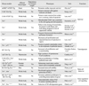
Extensive studies adopting diverse models have recently revealed the extra-telomeric roles of oncogenic TERT at an organismal level (Table 1). For example, telomere dysfunction in late generation Terc-/- mice enhances the initiation of hepatocellular carcinogenesis, but suppresses progression into fully malignant carcinomas.51 In contrast, enhanced tumor initiation does not occur in late generations of Tert-/- mice,35 indicating a possible oncogenic effect of TERT, other than telomeres, in tumorigenesis. Strong induction of TERT expression in hepatic neoplasms may also support its procarcinogenic effect on hepatic tumorigenesis.35 Consistently, transgenic overexpression of Tert promotes the development of spontaneous cancers in ageing mice.52 When TERT overexpression is targeted to basal keratinocytes using the bovine keratin 5 promoter, these transgenic mice show normal telomere length in their stratified epithelia even with high levels of telomerase activity.15 Interestingly, these mice are more susceptible to experimental skin carcinogenesis employing 7,12-dimethylbenz[a]anthracene and 12-o-tetradecanoylphorbol 13-acetate than wild-type mice.15 In addition, TERT overexpression actively promotes proliferation in epidermal tissues without telomere elongation.15 These results from mouse models suggest extra-telomeric roles of TERT, particularly in promoting tumor progression.
Telomerase mouse models have been also extensively used to validate telomerase as an important target for anticancer therapies. Since telomere dysfunction increases the chemo-sensitivity of Terc-deficient transformed MEFs, the combination of chemotherapy and telomerase inhibition may be an effective anticancer approach.53 Recently, Ding, et al.34 showed that telomerase reactivation by conditional rescue of Tert expression in mice with dysfunctional telomeres resulted in bone metastases of prostate tumors. Although the authors did not discuss the extra-telomeric roles of TERT in their study, this report is reminiscent of ALT cell transformation by hTERT overexpression.49 From this standpoint, anti-telomeric drugs are considered as an effective strategy for curing cancers. However, anti-telomerase therapy certainly provokes ALT and mitochondrial adaptive mechanisms in cancer,54 and with respect to the extra-telomeric functions of telomerase, anti-telomeric drugs may not be the best drug candidates.55
Taken together, telomerase exerts pleiotropic effects in cancer both dependent on and independent from its roles in telomeres. As described in Table 1, there are complex genetic interactions of telomerase with diverse genes. In conjunction with the currently emerging mechanisms of extra-telomeric roles, telomerase mouse models will expedite the invention of anti-telomerase strategies for cancer treatment.
REGULATION OF STEM CELLS BY TELOMERASE
Stem cells support tissue homeostasis and regeneration after certain types of damage. Because stem cells possess self-renewal potential and indefinitely propagate, high levels of telomerase activity should be essential for telomere maintenance. Therefore, extensive studies have been conducted to identify the patho-physiological consequences of telomerase deficiency or overexpression in stem cell function using diverse telomerase mouse models. However, telomere dysfunction is likely to affect stem cell functions in a context-dependent manner. Indeed, late-generation Terc-/- HSCs with short telomeres exhibit reduced proliferation capacity, but still possess long-term repopulating ability.56 Interestingly, when serially transplanted into recipient mice, the telomeres are considerably shortened even in wild-type HSCs, which is accelerated by approximately 2-fold in both Terc-/- and Tert-/- mice.16 Consistently, these telomerase-deficient HSCs exhibit considerably reduced replicative capacity compared to wild-type HSCs.16 However, although the telomere length of HSCs is constantly maintained by TERT overexpression in the transgenic mice, the long-term transplantation capacity of HSCs is not enhanced.57 Furthermore, Tert deficiency exacerbates senescence and the sensitivity of ataxia-telangiectasia mutated deficient murine HSCs against ROS-induced apoptosis, which does not accompany telomere shortening or dysfunction.17 These results suggest that telomerase may regulate the long-term replicative capacity of HSCs independently of telomere length.
Epidermal stem cells are also regulated by telomerase both dependent and independent of telomeres. In late generation Terc-/- mice, epidermal stem cell functions are significantly suppressed by critically short telomeres.21,58 However, epidermal overexpression of TERT under the control of the K5 promoter does not alter telomere length, but promotes stem cell mobilization, hair growth, and stem cell proliferation in vitro.18 Similarly, transgenic mice conditionally overexpressing TERT show robust hair growth via proliferation of quiescent, multipotent stem cells in the hair follicles.13 These phenotypes are also reproduced in a Terc-deficient genetic background without telomere dysfunction,13 thereby indicating the extra-telomeric activity of TERT.
In addition to the critical roles of p53 in mediating phenotypic manifestations against critically short telomeres,59 clues for the molecular mechanisms governing the extra-telomeric roles of telomerase have been obtained by identifying the positive effect of TERT on gene expression. Choi, et al.60 found that TERT triggers a rapid change in gene expression in the skin and hair follicles. This gene expression pattern significantly overlaps those controlling natural hair follicle cycling in wild-type mice. TERT affects the developmental program mediated by Myc and Wnt, which is intimately associated with stem cell function and cancer.60 Furthermore, TERT binds BRG1 (also called SMARCA4), a SWI/SNF-related chromatin remodeling protein, and directly modulates Wnt/β-catenin signaling as a cofactor in the β-catenin transcriptional complex.61 Therefore, independently of telomeres, TERT can act as a transcriptional regulator that is directly involved in stem cell functions, including in mouse epidermal tissues.
CONCLUSIONS
For more than a decade, diverse telomerase mouse models have provided us with precious opportunities for evaluating the patho-physiological significance of telomerase in genetically defined environments and at an organismal level. With an emphasis on defective telomeres, these mouse models have considerably contributed to understanding a broad spectrum of phenomena associated with cancer and ageing. Furthermore, growing evidence has indicated that defective telomerase functions are involved in distinct diseases other than human cancers including dyskeratosis congenita, atherosclerosis, and renal diseases.62-64 The list of disease-associated mutations has been expanding. To genetically define the pathological aspects and thus to establish animal models of these mutations, novel mouse models should be still generated and analyzed.
Despite the evident roles in telomeres, currently emerging extra-telomeric functions of telomerase are completely changing the scope of this enzyme. Notably, the direct roles of TERT in transcriptional regulation (e.g. Wnt/β-catenin and nuclear factor-κB or NFκB) provide good rationale for several phenotypes that cannot be explained by telomere dysfunction, and their physiological significance has been also confirmed using telomerase mouse models.62,64,65 As might be expected, these lines of evidence make us consider that diverse observations supporting extra-telomeric roles of telomerase should be scrutinized and validated in vivo by generating novel mouse models. For example, in addition to the effect of short telomeres on mitochondria,39 mitochondrial targeting of telomerase upon certain stressful conditions66 and the recently identified RNA-dependent RNA polymerase activity of TERT,67,68 indicates that telomerase has direct roles in mitochondria. Furthermore, considering the important roles of telomerase in cellular homeostasis, telomerase may be a critical factor for regulating the subcellular organelle homeostasis. Undoubtedly, we believe that these extra-telomeric functions of telomerase should be intimately associated with life span regulation, and that some regions of TERT, other than the RT domain, will be required for mediating protein-protein interactions with known functions in controlling the life span of an organism. In this context, we cannot rule out speculations for divergent mechanisms of telomerase function regulating survival, tumor progression, development/differentiation, and stress responses. These extra-telomeric functions have inevitably complicated the phenotypic manifestations elicited by dysfunctional telomeres and vice versa; thus, to separate these distinct functions of telomerase, more sophisticated genetic strategies should be developed in mice.
 XML Download