

 PDF
PDF ePub
ePub Citation
 Print
Print


INTRODUCTION
Femoral neck fractures are associated with femoral shaft fractures in 1% to 9% of cases.1,2 Some femoral neck fractures are undisplaced and can only be diagnosed by preoperative computed tomography (CT) (i.e., silent neck fractures). Such fractures are susceptible to displacement during preparation of the patient for surgery or during reaming or insertion of the nail.3 During an operation, we encountered what appeared to be a femoral neck fracture, which caused us to change our procedure from antegrade femoral nailing to reconstruction nailing. After the operation, however, we determined that it was a false femoral neck fracture, as no fracture could be detected on simple radiogram, fluoroscopy or CT scan.
CASE REPORT
A 57-year-old man sustained a motor vehicle accident and was transferred to our ER for femoral shaft comminuted fractures on the left side. We planned closed reduction and internal fixation with intramedullary nails for the left femoral shaft fracture. In the operating room, the patient was placed on a fracture table under general anesthesia. A skin incision was made proximal to the tip of the greater trochanter about 5 centimeters. A 3.2-mm guide pin was inserted at the tip of the greater trochanter. After trochanteric reaming along the 3.2-mm guide pin, a ball-tip guide was introduced into the medullary cavity of the proximal and distal fragments after reduction. A femoral nail (greater trochanter starting nail, 12 mm in diameter, 40 cm in length) was introduced after reaming a hole to 13 mm. The nail was inserted uneventfully without excessive force. At that time, we identified a vertical radiolucent line at the femoral neck, which was thought to be further displacement of a hidden silent fracture or an iatrogenic fracture that developed during nail insertion (Fig. 1). Consequently, we decided to perform reconstructive femoral nailing. We inserted two 6.0-mm partially threaded cancellous screws into the femoral neck and head. Afterwards, we observed that the radiolucent line at the femoral neck had decreased in size. Three distal interlocking screws were then inserted, and a nail cap was applied.
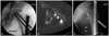
Postoperative hip imaging failed to show the femoral neck fracture that we saw in the operating room. Fourteen days after surgery, we took a CT scan of the proximal femur in order to analyze the radiolucent line observed during intraoperative fluoroscopy; the femoral neck was confirmed as intact. Reversal of the fluoroscopic image taken in the operating room showed a radiolucent line running from the femoral neck to the medial thigh (Fig. 2).
DISCUSSION
Mach bands are a perceptual phenomenon in which dark and bright lines are observed on the borders of structures of different radiographic densities, resulting in contrast-enhancing effects in various areas of the body. Pseudo-fractures (i.e., false fractures) are among a group of illusory phenomena that result from overlapping images, differences in background illumination, subjective contour formation and parallax. Pseudofracture at the base of the dens of the second cervical vertebra is a well-known artifact. Skin fold or fat tissue plane simulating trochanteric fracture as well as pseudofracture of the femoral neck secondary to osteophyte formation on the femoral head and acetabulum have also been reported.5
In our case, the pseudofracture was not detected during preparation of the entry point, but instead was found after final settlement of the femoral nail. Unfortunately, it is not uncommon to detect displacement of an undisplaced femoral neck fracture during antegrade shaft nailing. The pseudofracture was vertically oriented in the middle of the femoral neck, thus mimicking a typical femoral neck fracture associated with femoral shaft fracture. After the operation, we realized that it was a pseudofracture, so we attempted to determine the cause of the event. We put the patient on the fracture table and recreated the positions that we used for femoral nailing, but we were unable to make the false femoral neck fracture reappear. We then reversed the saved image that we took during the operation and determined that a radiolucent line was running from the femoral neck to the medial thigh. The boundary of the radiolucent band was a little smoother than that of fracture lines seen in real cases. Intraoperative CT scan or 3D fluoroscopy may also have allowed us to determine that it was a false femoral neck fracture; however, this would have been particularly impractical during femoral nailing with the patient positioned on the fracture table.
To our knowledge, this is the first report of a pseudofracture in the femoral neck associated with femoral shaft nailing. It is true that the pseudofracture line seemed to decrease after femoral neck screw fixation; however, we realize that rotation of the limb was different. The lesser trochanter was more prominent in the post-fixation image. We suspect a tiny difference in limb rotation caused the decrease in pseudofracture gap on the fluoroscopic image.5 In conclusion, we recommend that surgeons check the fracture line again in the reversed fluoroscopic image (tracing the adjacent fat plane and its connectivity with the fracture line) before converting to reconstructive nailing. However, if the presence of a pseudofracture cannot be definitively ascertained, then antegrade femoral nailing must be converted to reconstructive nailing or another method of fixation. Additionally, all supporting fluoroscopic images should be saved for further evaluation and documentation.

 XML Download