

 PDF
PDF ePub
ePub Citation
 Print
Print


INTRODUCTION
Unilateral plating for proximal tibial fractures with conventional plating system has often been blamed for secondary loss of reduction due to the lack of angular stability.1-3 Dual plating, however, is commonly associated with serious complications involving soft tissue breakdown.4,5 The introduction of anatomically pre-contoured locking plate such as less invasive stabilization system (LISS), Locking compression plates-proximal lateral tibia (LCP PLT, Synthes®, West Chester, PA, USA) and biological plating concept, has made unilateral plating for complex proximal tibial fractures very popular in recent years.6-19 With this new technique, union rate has increased without bone grafting, and good clinical results have been reported. The main problem with this technique, however, is malalignment.8,12 Although many technical tips have been described to assess the alignment intraoperatively,20 there is still a need for more methods which could be used to assess the alignment intraoperatively.
The purpose of this study is to evaluate whether the angle formed between the proximal most screw through the LCP PLT and the joint line has any relationship with the coronal plane alignment of the tibia.
With this objective in mind, we measured the angle between the proximal most locking screw through LCP PLT and the joint line in normal adult tibia of cadaver limbs [this angle was termed as the 'joint screw angle' (JSA)] and established a normal JSA range. Then, we evaluated our results in a clinical setting to assess if the JSA can be used as a radiological guideline intraoperatively, for the assessment of final coronal alignment.
Our hypothesis is that the position of the proximal most screw through LCP PLT in relation to the joint line can be used as a rough guideline to assess the alignment in the coronal plane.
MATERIALS AND METHODS
Our basic methodology was to measure the angle between the proximal most locking screw through LCP PLT and the joint line in normal adult tibia of cadaver limbs (this angle was termed as the 'JSA') and to establish a normal JSA range. Subsequently, we evaluated our results in a clinical setting to assess if the normal JSA has any constant relationship with the coronal alignment of tibia. This was done in two parts. In the first part, we conducted a cadaveric study using 30 adult tibial bones. In the second part, in a clinical set up, we retrospectively analyzed the relationship between the postoperative alignment and the radiological guideline (JSA). The study was approved by the Konkuk University Hospital Institutional review board.
Anatomical study
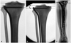
We retrieved 30 tibial shafts from adult cadavers of 24 males and 16 females. The mean age was 62 years (range 31-90 years). An 11-hole LCP PLT was applied to the lateral aspect of the proximal tibia. The LCP PLT was applied such that the plate nestled up with the lateral aspect of proximal metaphysis and was parallel to the axis of the tibial shaft. The two proximal most locking screws were then placed. One locking screw was placed on the shaft according to the manufacturer's recommendations. We grossly observed the plate and bone construct to see if there was any part where the plate did not fit to the bone surface well. The anteroposterior (AP) and lateral views of the proximal tibia were taken. On the AP image, we measured the angle between proximal tibial joint line and the locking screw, and termed the angle as 'JSA' (Fig. 1A). We designated a negative value to the JSA when the screw was tilted in a varus position to the joint line, such that the tip of the screw pointed towards the joint line, resulting in a lateral opening angle (Fig. 1B). If the screw was tilted in a valgus position, such that the tip pointed away from the joint line, resulting in a medial opening angle, we described it as a positive JSA value (Fig. 1C).
Clinical study
We retrospectively reviewed 56 cases of proximal tibial fractures that were treated with LCP PLT. They comprised 44 males and 12 females. The mean age was 52 years (range, 30-88 years). Forty-nine fractures were treated in an acute setting within 2 weeks after the injury. There were six nonunion and one malunion of the proximal metaphysis. The fresh fractures were classified according to the AO-OTA classification. They were of the following types: 41A1(2), 41A2(1), 41A3(7), 41B2(1), 41C2(1), 41C3(18), 42A1(1), 42A2(3), 42A3(1), 42B2(5), 42C1(1), 42C2(3), 42C3(5). All operations were performed by two experienced trauma surgeons or they were performed under their guidance. In all the fractures, the reduction was achieved first and then LCP PLT was applied via a lateral approach. The plate was applied over the lateral surface of the tibia, anterior to fibular articular surface of the tibia. The first screw was placed in the mid diaphysis and then the proximal screws were inserted. All efforts were made to align the plate well with the tibial surface proximally and with the shaft.
X-ray analysis
The radiologic analysis formed the backbone of our study. This was done based on the radiograms from 56 patients. In 29 of these patients, we could retrieve the intraoperative images as well as the postoperative radiographs. For the remaining 27 patients, we had the postoperative radiographs only.
1) Postoperative radiograms were taken at approximately 4-20 weeks postoperatively or as soon as patient was able to stand and bear weight on the operated leg. Postoperative radiograms were standing orthoradiograms of the lower extremity centering at the knee joint with the patient facing the radiographic tube and the patella pointing forward.
2) Intraoperative images were taken with the patient positioned supine, knee in full extension, patella pointing towards the ceiling, and the image intensifier aligned perpendicular to the knee joint. Following measurements were made on the radiograms. Two angles were measured:
(1) The medial proximal tibial angle (MPTA; angle measured on the medial side formed between the tibial plateau line and the line along the tibial axis) was measured in both the tibiae on the postoperative orthoradiogram and the difference was calculated. Malalignment was defined as a difference of more than 5° in the MPTA. In three cases with bilateral proximal tibial fractures, the average population MPTA (87°) was used as a reference angle.
(2) The JSA was measured simultaneously on both intraoperative and postoperative images in 29 cases in whom the C-arm images were also saved in addition to the postoperative X-rays. For the remaining 27 patients, the JSA was measured only on the postoperative orthoradiogram. The screw was considered acceptable if it is approximately parallel to the joint line and the JSA was found to be in the range of 0±5°.
In the 29 patients, we did not find a difference of more than 1 degree between the JSA on the intraoperative image and the JSA on the postoperative image. On the basis of this interim analysis, we decided to use the postoperative measurement of the JSA as an alternative to the JSA on the intraoperative image in the remaining 27 patients. We then statistically analyzed the relationship between the JSA and the postoperative malalignment (MPTA) in all 56 cases. All the radiographic measurements were made by two independent blinded observers (two orthopedic surgeons: first and corresponding authors).
Statistical analysis
Inter-observer variance was assessed using the paired 't' test. No difference was found between the observations made by the two observers (p<0.05).
Patients were categorized into two groups based on their postoperative coronal plane alignment: one group with normal alignment (MPTA within ±5° of the contralateral normal limb) and the other group with malalignment. Patients were also divided into two groups based on the degree of the JSA: one group with acceptable JSA (within ±5°) and the other group with unacceptable JSA. The kappa statistics was calculated to assess the agreement of alignment status between the MPTA and the JSA. We also conducted an exact test version of McNemar test to evaluate the agreement between MPTA and JSA. In this test, the null hypothesis was agreement between two variables while the alternative hypothesis was difference between two variables. All analyses were conducted using the SAS version 9.1.3 (SAS Institute Inc., Cary, NC, USA). The level of significance was set at 0.05.
RESULTS
Anatomical study
On gross observation, we found that the LCP PLT fits to the lateral aspect of the proximal tibiae fairly well. However, we also found that the head of the plate tends to stay apart from the lateral aspect of the lateral condyle by a few millimeters. In 30 specimens that we studied, the JSA was -2° on an average (ranged from 0° to -7°). In 22 cases (73%), the proximal locking screw was almost parallel to the joint line (JSA -2° to 0°). In 7 patients (24%), the JSA was between -5° and -2°, and the JSA was -7° in one specimen with severe osteoarthritis. This particular bone showed severe osteoarthritis with varus inclination of the joint line (MPTA 82). In this case, the unacceptable JSA was due to an abnormal inclination of the joint line. Two other specimens showed degenerative changes and a varus inclination of the joint line of 5° in the coronal plane and a JSA of 5°. None of the specimens showed a positive JSA.
Clinical and radiological study
Our clinical and radiographic retrospective study yielded the following results. In 49 of the 56 patients (87.5%), the MPTA was found to be acceptable and within the normal range (within 5° of the contralateral normal limb or average of the population). In the remaining 7 patients (12.5%), there was malalignment in the coronal plane and the MPTA was out of the normal range. Out of the 49 patients with normal alignment, the JSA was found to be in the acceptable range in 47 patients (95.92%), and in the unacceptable range in the remaining 2 patients. In the 7 patients with malalignment, the JSA was unacceptable in 4 patients (57.14%) and acceptable in the remaining 3 patients. Among the 50 patients with a normal JSA, 47 patients [94% (47/50×100)] were found to have a normal alignment. Out of the 6 patients with an abnormal JSA, 4 patients (67%) showed malalignment. The kappa value for the measure of agreement between JSA and MPTA was 0.565 (95% confidence interval: 0.225, 0.905) (Table 1).
The p-value from the exact test version of McNemar's test was 1.000, indicating a good agreement between JSA and MPTA and clearly stating that the association between the MPTA and the JSA was very relevant and not just coincidental.
DISCUSSION
Although the minimal invasive Locking plate fixation has shown promising results in reducing infections (4%) and achieving high rates of union, it has given rise to more troublesome, alignment related issues. The published data in literature show varied complication rates of this technique. According to Ricci, et al.,14 obtaining proper alignment with the use of LISS is technically demanding. Malalignment in the coronal plane after the use of LISS in treatment of various types of proximal tibial fractures has been reported in many papers.8,12,15,21
Our anatomical study was carried out to identify the relationship between the coronal plane alignment and the JSA. We found, the proximal most screw through LCP PLT to be parallel to the joint line or within ±5° in 29 of 30 cadaveric tibial bones. Only in 1 grossly arthritic specimen, the JSA was out of the acceptable range (-7°) which was due to the abnormal inclination of the joint line (MPTA 82°). Thus, the relationship established was that the JSA was 5° or less in nearly all the normal tibial bones, except in the severely arthritic bones with abnormal inclination of the joint line. The relationship is difficult to establish in severely arthritic bones, and thus we would generally limit the results of our study to bones which do not show severe arthritis.
We further set out to evaluate if this relationship holds true in a clinical setting and if it could be used intraoperatively to assess the alignment in the coronal plane.
On a retrospective analysis, the relationship that we found between the JSA and the MPTA had a high positive prediction of 94% (47/50); i.e. there is a great chance of having a normal alignment if the JSA is within the normal acceptable range. The results of our anatomic study and clinical correlation indicate that an acceptable JSA does point towards a normal MPTA and the relationship cannot be explained by chance alone. The relationship between the JSA and the MPTA could thus be used as an additional tool to evaluate the coronal alignment.
However, out of 6 patients with an unacceptable JSA, there were 4 cases of an abnormal MPTA, and 2 cases of a normal MPTA. These 2 cases of a normal MPTA with an unacceptable JSA can be explained by the fact that there are various ways in which the plate could be positioned over the tibia due to various kinds of possible mismatches between the proximal tibia and the pre-contoured plate. If the position is like that of an impingement fit with the distal part of the plate away from the shaft and the proximal part fitting well on the shaft as described by Goyal, et al.,22 the screw and the JSA will be in a valgus position over an anatomically well aligned tibia.
Preservation of the normal anatomic alignment is important in achieving a favorable outcome. Blokker, et al.6 found unsatisfactory results in 100% of the patients with inadequate anatomical reduction. In many series, the authors underscore the importance of good technical tricks to avoid malalignment. Cole, et al.8 emphasized the importance of good intraoperative radiographs to decrease the incidence of malreduction. They tried to underscore the importance of good radiography techniques; i.e. taking radiographs on a large flat plate before the placement of screws, using fluoroscopy & strategic bumps supporting the distal fragment in order to achieve a good postoperative alignment. Krettek, et al.20 described the cable technique to avoid the coronal plane malalignment. Although this technique is quite useful when the alignment is evaluated after the fixation is over, it is often not feasible to have two assistants holding the cable and moving the C-arm from the top to the bottom while the alignment is maintained. On the other hand, the JSA can be easily measured on a single C-arm AP image. We did not find any other study which specifically dealt with alignment issues and addressed them. We were also unable to find any mention of the JSA or of its relationship to the alignment in the manufacturer's recommendation. Hence, we are not certain whether or not they intended to orient the first screw parallel to the joint line.
An ideally placed plate, is likely to demonstrate the relationship between the MPTA and the JSA. In view of the above limitations, we can state that the JSA may not be used as a standalone assessment tool, however, it can rather be used as an adjunct to the other techniques of assessment of alignment, since it points very well towards the type of alignment we are likely to achieve and may also provide an indication on the occurrence of malalignment.
In conclusion, we can safely conclude from our observation that the JSA can be used intraoperatively as a rough guideline to assess the achievement of a final normal alignment in the coronal plane when applying a LCP PLT/LISS internal fixator for fractures of the proximal tibia.

 XML Download