

 PDF
PDF ePub
ePub Citation
 Print
Print


INTRODUCTION
Syncope is a frequent symptom in the general population. However, it is often difficult to diagnose the cause of syncope because diverse diseases and factors are involved in the occurrence of syncope.1 Although several diagnostic tests can be performed, the cause of syncope is unclear in 5-25.5% of patients with syncope.2,3 If the cause of syncope is not correctly diagnosed, patients are at risk of physical injury from recurrent syncope. In some patients with cardiac or arrhythmic causes, delay in correct diagnosis and treatment may lead to sudden cardiac death.2,4 Recently, use of an implantable loop recorder (ILR) and a standardized diagnostic pathway have led to breakthroughs in reducing the number of unknown causes of syncope.1,2,4,5 The ILR has become the diagnostic tool of choice in unexplained syncope even after negative initial evaluations. However, domestic adoption and implementation of the ILR has been slow in Korea. Thus, we analyzed our patients who underwent ILR implantation.
MATERIALS AND METHODS
Study population
According to guidelines,1 the ILR is indicated for early evaluation of 1) patients with recurrent syncope of uncertain origin absent of high risk criteria and a high likelihood of recurrence within battery longevity of the device, as well as 2) high-risk patients in whom a comprehensive evaluation did not demonstrate the cause of syncope or lead to a specific treatment. Our indication mirrored guidelines of European Society of Cardiology (ESC).
Between February 2006 and June 2011, 18 patients had an ILR implanted at our center due to an unknown cause of syncope even after undergoing several diagnostic tests. As an initial evaluation of syncope, careful medical history taking, physical examination, electrocardiography (ECG), Holter monitoring, head-up-tilt test (HUTT) and treadmill test were performed. Invasive studies such as coronary angiography (CAG) and electrophysiologic studies (EPSs) were performed prior to ILR implantation in some patients.
Event recording and follow-up after ILR implantation
The ILR (Reveal DX, Medtronic Inc., Minneapolis, MN, USA) is a rectangular device measuring 62×19×8 mm and weighing 15 grams. To implant the device, a 2.0-cm incision was made in the left chest area. Blunt dissection was then employed to create a 6-cm subcutaneous pocket. The device can be implanted under local anesthesia in 15-20 minutes.
The ILR stores representative rhythm strips when the heart rate exceeds preset limits (automatic activation) or when a patient presses a button on the accompanying actuator (patient activation). These records can be transmitted to a physician for review during subsequent office visits.
In all patients, the device was programmed with the following settings: 40 minutes of total electrocardiographic recording consisting of three patient-activation recordings of a 10 min duration (8 minutes before and 2 minutes after symptom onset) and five automatic activation recordings of a 2 min duration. The following parameters were set for automatic activation: heart rate <40 beats per minute (four consecutive measurements); heart rate >160 beats per minute (16 consecutive measurements) and asystole lasting more than 3 seconds. A follow-up visit was set up after symptomatic events or every 3 months in asymptomatic subjects in order to retrieve the times and dates of episodes of bradycardia or tachycardia as well as the corresponding electrocardiographic tracings from the memory of the ILR.
RESULTS
Clinical characteristics
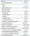
Eighteen consecutive patients were identified from February 15, 2006 to June 20, 2011. Thirteen patients (72.2%) were male. The mean age (±SD) of the patients was 61.2 (±15.0) years. Several underlying diseases were noted in more than one-fourth of the patients, among which hypertension was the most common (n=8, 44.4%). Coronary heart disease, diabetes mellitus, hyperlipidemia and cardiomyopathy were noted in 6 (33%), 5 (27.8%), 5 (27.8%) and 5 (27.8%) patients, respectively. The mean left ventricular ejection fraction (±SD) was 59.6% (±8.1). The mean estimated interval (±SD) between syncope or presyncope events was 164 (±219.5) days. The median number (interquartile ranges) of syncope or presyncope events was 5 (3-16). The mean estimated duration of syncope (±SD) was 145.5 (±182.2) seconds. The mean follow-up duration (±SD) of for all study subjects was 11.3 (±10.6) months. In addition, it took a mean of 5.6 (±9.2) months to diagnose patients (n=10, 55.6%) (Table 1).
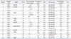
All but 3 patients (patients 15, 16 and 17) received EPSs before implantation of ILR. EPSs were performed after antiarrhythmic medications were stopped. Intervals were measured at first conduction; thereafter, antegrade and retrograde refractory periods were determined. Finally, for tachycardia induction, the high right atrium, right ventricular outflow tract and right ventricular apex were stimulated using programmed stimuli. The results were not diagnostic in 15 patients. CAG was performed on 11 patients. Ten (55.6%) of the 18 patients was successfully diagnosed using the ILR. Despite a short interval (3 days) between symptoms, patient 6 received ILR implantation because diagnosis was not possible using Holter monitoring and EPSs. Furthermore, the patient's pattern of symptoms was accompanied by shock. After 4.4 months using the ILR, syncope with seizure-like movements occurred while sleeping at night. The patient lost consciousness transiently due to ventricular tachycardia (VT) and ventricular fibrillation (VF) lasting for 90 seconds. The arrhythmia started with monomorphic VT, which was then changed into polymorphic VT, VF and monomorphic VT.6 He did not have any genetic disorder. Patient 16 also had a short interval (9 days) between symptoms. However, diagnosis could not be confirmed over several previous hospital visits despite a total 72 episodes of syncope. Holter monitoring and event recording at our hospital also could not confirm a diagnosis. Therefore, ILR was performed without EPSs and sick sinus syndrome (SSS) was diagnosed 8 days after ILR implantation (Table 2).
Documented arrhythmia and management
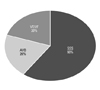
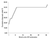
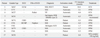
SSS (n=6, 60%) was the most common diagnosis among the 10 diagnosed patients. Two patients (20%) were diagnosed with VT and VF. Advanced atrioventricular block (AVB) was also diagnosed in 2 patients (20%) (Fig. 1). Nine patients (90%) were diagnosed successfully within 6.5 months. Only 1 patient (10%) required 30.8 months to arrive at a diagnosis (Fig. 2).
In SSS patients (n=6), diagnosis was confirmed more frequently through the patient-activation mode (n=4) than through the automatic activation mode (n=2). In cases of ventricular tachyarrhythmia, the patient activation and automatic activation modes were used in both cases. Both advanced AVB patients (n=2) were diagnosed through the automatic activation mode.
Although patient 1 was diagnosed with SSS, an implantable cardiac defibrillator (ICD) was implanted instead of a permanent pacemaker because of accompanied hypertrophic cardiomyopathy. In patient 17, non-sustained VT, which induced syncope, was diagnosed using the ILR through the patient activation mode without EPSs. Interestingly, syncope occurred during short runs of non-sustained VT. Right ventricular out tract VT was suspected based on the non-sustained VT, which was recorded by 2-channel ECG monitoring. However, no significant tachyarrhythmia was induced with programmed electrical stimulation. Therefore, we could not perform radiofrequency catheter ablation on the patient. Because of concerns about unnecessary defibrillation shock after recovery from the non-sustained VT which had already induced syncope, pharmacological therapy with sotalol was performed instead of ICD implantation. Fortunately, syncope has not recurred (Table 3).
DISCUSSION
The etiologies of syncope remain unknown in 21% to 49% of all patients even after comprehensive conventional work-ups.7,8 Among conventional tests, HUTT (19% to 61%) and postural blood pressure measurement (15% to 33%) show relatively high diagnostic yields.7,8 Our results showed that it was possible to diagnose more than half (55.6%) of the patients with unexplained syncope using the ILR. In previous reports, Boersma, et al.,9 Vitale, et al.10 and Lombardi, et al.5 reported of diagnosis rates of 28%, 32% and 50% using the ILR, respectively, in patients with recurrent unexplained syncope. Recently, in the PICTURE registry, a prospective, multicenter, observational study, ILR-guided diagnosis was established in 78% of unexplained syncope patients.4
Among the arrhythmic causes of the syncope diagnosed using the ILR, SSS was the most common (60%) in the present study, which was similar to the results of previous studies that reported the incidence of SSS to be between 54.5% and 58.3%.5,9 Advanced AVB was reported as the second most common cause, the occurrence of which ranged from 8.3% to 27.3%.5,9 However, in patients with bundle branch block and negative EPSs, the most commonly reported cause was prolonged asystolic pauses, mainly due to sudden-onset paroxysmal AVB.11
Furukawa, et al.12 reported that a quarter of patients who had unexplained syncope needed more than 18 months of follow-up using the ILR to establish a diagnosis. However, most patients (90%) were diagnosed within 18 months in our study. Furthermore, it took less than 6 months to arrive at a diagnosis in 8 of the 10 patients. There were significant differences between the results of previous reports and ours. These differences were probably due to a high recurrence rate among the selected patients. However, the actuarial curves showed similar patterns of steep rises during the first 6 months (Fig. 2). Furthermore, these patterns were prominent in cases of asystolic-type arrhythmias, such as SSS and significant AVB. On the other hand, none of the asystolic-type episodes showed a continuously increasing linear curve during the follow-up period up to 48 months.12 With greater advancements in technology, newly-developed devices offer a longer longevity of more than 3 years, allowing for more convenient and prolonged observation.
In the present study, 3 patients (patients 15, 16 and 17) underwent ILR implantation without EPS. They were all diagnosed successfully within 2 months. The updated version of the ESC guidelines highlights the use of ILR and recommends its early use in diagnostic workups.1
Krahn, et al.13 and Giada, et al.14 showed, in their randomized controlled trials, that a strategy of primary monitoring using an ILR is more cost-effective than conventional evaluation methods in establishing a diagnosis for recurrent unexplained syncope. These results may influence the use of ILR in Korea. However, indications for ILR implantation should be considered in terms of the frequency of syncopal episodes. For example, patient 16 had a short interval between symptoms (9 days) in this study. In addition, the patient was diagnosed just 8 days after ILR implantation. An event recorder was not available for this patient because her syncope occurred suddenly without prodromal symptoms. Even so, repeated Holter monitoring or prolonged ECG monitoring during hospitalization might also be a useful alternative. Giada, et al.14 limited target patients to those who had symptoms less frequently than once a month as part of the inclusion criteria. Further studies are needed to establish guidelines regarding symptom frequency in patients undergoing ILR implantation.
Newly developed ILR devices can detect cardiac arrest while its external receiver can alert bystanders to start resuscitation and to automatically call emergency medical services.15 However, wireless data transmission from the ILR is impossible in Korea because of radio frequency selection problems. It is clear that wireless data transmission offers the possibility of alerting care providers when cardiac arrests occur, decreasing response times and improving survival. Therefore, wireless data transmission should be instituted as soon as possible along with the use of the ILR.
In conclusion, the ILR may be a valuable and effective tool for determining arrhythmic causes of unexplained syncope. Furthermore, this selected Korean patient population behaved very much like patients from other parts of the world. Thus, it is believed that ILRs are equally helpful in evaluating Korean and Asian patients in clinical practice.
Study limitations of the present study
The small sample size and the potential selection bias of the population regarding patient economic status due to non-coverage by insurance in Korea are major limitations of our study. In addition, this study was conducted with a cross-sectional design. Further large-scale prospective studies with a larger sample size are needed to confirm our results.
 XML Download