Journal List > Yonsei Med J > v.54(3) > 1031247

Kim, Kim, Jo, Park, and Ahn: Modulation of the Transcriptional Activity of Peroxisome Proliferator-Activated Receptor Gamma by Protein-Protein Interactions and Post-Translational Modifications

Abstract

Peroxisome proliferator-activated receptor gamma (PPARγ) belongs to a nuclear receptor superfamily; members of which play key roles in the control of body metabolism principally by acting on adipose tissue. Ligands of PPARγ, such as thiazolidinediones, are widely used in the treatment of metabolic syndromes and type 2 diabetes mellitus (T2DM). Although these drugs have potential benefits in the treatment of T2DM, they also cause unwanted side effects. Thus, understanding the molecular mechanisms governing the transcriptional activity of PPARγ is of prime importance in the development of new selective drugs or drugs with fewer side effects. Recent advancements in molecular biology have made it possible to obtain a deeper understanding of the role of PPARγ in body homeostasis. The transcriptional activity of PPARγ is subject to regulation either by interacting proteins or by modification of the protein itself. New interacting partners of PPARγ with new functions are being unveiled. In addition, post-translational modification by various cellular signals contributes to fine-tuning of the transcriptional activities of PPARγ. In this review, we will summarize recent advancements in our understanding of the post-translational modifications of, and proteins interacting with, PPARγ, both of which affect its transcriptional activities in relation to adipogenesis.

INTRODUCTION

Structure and function of peroxisome proliferator-activated receptors

Peroxisome proliferator-activated receptors (PPARs) are known to be lipid sensors, and their ligands are used in the treatment of type 2 diabetes mellitus (T2DM) and other metabolic syndromes. PPARs are a family of nuclear receptors that act as transcription factors, controlling the genes involved in energy homeostasis.1 PPARs share a high degree of structural homology with other types of nuclear hormone receptors.2 PPARs comprise a DNA-binding domain (DBD), an agonist-independent activation domain (AF-1), and an agonist-dependent activation domain (AF-2), which contains the ligand-binding domain (LBD). PPARs heterodimerize with the retinoid X receptor (RXR)-α and activate the transcription of target genes by binding to the PPAR response element (PPRE).
The PPAR family has three isoforms; PPARα, γ, and β/δ. PPARα is expressed mainly in the liver, heart, kidney, brown adipose tissue (BAT), and skeletal muscle,3 and participates in fatty acid oxidation (β-and ω-oxidation).4 The PPARβ/δ isoform is expressed ubiquitously and is involved in fatty acid oxidation in muscle.5 PPARγ is expressed predominantly in adipose tissue and plays key roles in lipogenesis and adipocyte differentiation. It also stimulates glucose oxidation and decreases plasma free fatty acid level.5 PPARγ consists of two isotypes; PPARγ1 is expressed in adipocytes, skeletal muscle, liver, and heart, whereas PPARγ2 is mostly found in adipose tissue.6 PPARγ2 plays a more important role than does PPARγ1 in adipogenesis.7

Physiological significance of PPARγ

PPARγ was first identified as a trans-acting factor binding to a gene encoding a fat-specific enhancer of aP2 (adipocyte-specific fatty acid binding protein).8 Homozygous PPARγ knockout mice exhibit an embryonic lethal phenotype due to placental dysfunction. Heterozygous PPARγ deficient mice are resistant to high-fat diet-induced insulin resistance due to adipocyte hypertrophy and increased leptin expression.9 The ectopic expression of PPARγ was found to enhance the differentiation of preadipocytes into adipocytes, with PPARγ acting as an essential factor for differentiation.10 In addition, PPARγ is known to block the clonal expansion that occurs via mitosis, an essential stage of adipocyte differentiation.11,12
Thiazolidinediones (TZDs) are a class of compounds that function as ligands of PPARγ. These compounds improve insulin sensitivity in vivo and have been introduced as therapeutic agents for the treatment of T2DM.13,14 TZDs increase the expression of PPARγ and its transcriptional activity in adipose tissue, resulting in the upregulation of the expression of genes involved in the metabolism of lipids, carbohydrates, steroids, and amino acids.15-17 TZDs increase insulin sensitivity by upregulating the expression of multiple genes, such as adiponectin, Cbl-associated protein, insulin receptor substrate 2, and glucose transporter 4.18-23 TZDs also promote fatty acid storage and lipid metabolism, such as fatty acid translocase (CD36), perilipin, fatty acid binding protein 4 (Fabp4/aP2), lipoprotein lipase, acyl-CoA synthase, phosphoenol pyruvate carboxykinase (PEPCK), and glycerol kinase (GyK).24-31
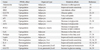
However, the activation of PPARγ results in the repression of the genes encoding leptin, tumor necrosis factor-α (TNF-α), and interleukin-6.32-35 PPARγ decreases serum free fatty acid level and increases the number of small adipocytes, with a concomitant decrease in the number of large adipocytes in white adipose tissue (WAT). In addition to the role of PPARγ in adipose tissue, PPARγ directly activates the genes of the glucose-sensing apparatus in the liver and pancreatic β-cells. TZDs increase the expression of the genes encoding glucokinase (LGK and βGK) and glucose transporter 2 (GLUT2) in the liver36,37 and pancreatic β-cells, respectively (see Table 1 for summary).38,39 The transcriptional activity of PPARγ is subject to control at various levels; i.e., via modification of the receptor itself or interactions with other proteins.
In this review, we will limit our discussion to the regulation of PPARγ activity by various interacting proteins including coregulators, and by post-translational modifications (PTMs) that result in transcriptional regulation of PPARγ target genes.

INTERACTING PROTEINS MODULATING TRANSCRIPTIONAL ACTIVITIES OF PPARγ

The transcriptional activity of PPARγ is principally modulated by agonists, which recruit either coactivators or corepressors. In general, ligand-bound PPARγ recruits coactivators, whereas ligand-free PPARγ is bound to corepressors. These coregulators function as histone-modifying enzymes or bridging groups between the basal transcriptional machinery and PPARγ.40 Moreover, additional proteins are recruited to these coregulators that may affect tissue-specific activities of PPARγ.

Coactivators of PPARγ

Coactivators with histone acetyltransferase activity

Ligand-bound PPARγ undergoes conformational changes, providing contact sites for LXXLL motifs that are present in coactivators such as p160/steroid receptor coactivator-1 (SRC-1) and p300/CREB-binding protein (CBP).41 These coactivators have intrinsic histone acetyltransferase activities, which enhance the transcriptional activities of PPARγ. Members of the p160/SRC-1 family including SRC-1 (also known as NcoA-1), SRC-2 (also known as TIF2, GRIP-1, or NcoA-2), and SRC-3 (also known as p/CIP, ACTR, RAC-3, AIB-1, or TRAM-1), belong to this category.42 SRC-1 knockout (KO) mice showed increased WAT mass and a decrease in the expression of genes involved in thermogenesis in brown adipose tissue (BAT). These KO mice also showed decreased expression of the genes encoding uncoupling protein (UCP-1), PPARγ coactivator-1 (PGC-1α), and acyl-CoA oxidase, as well as those encoding enzymes involved in fatty acid oxidation.42 LXXLL motifs in SRC-1 interact directly with the AF-2 domain of PPARγ, recruiting CBP, which is required for PPARγ function.43 SRC2-/- mice exhibit increased insulin sensitivity and are resistant to the development of obesity. These mice show increased lipolysis and decreased fatty acid uptake and storage which are related to the reduction of PPARγ activity.42 When SRC-3 is deficient, corepressors such as nuclear receptor co-repressor (NCoR) and nuclear receptor interacting protein 1 (NRIP1 or RIP140) are recruited to the PPRE of the UCP1 gene, resulting in a decrease in its transcription.44 SRC-3 and SRC-1 double KO mice are resistant to high-fat diet-induced obesity, due to the decreased expression of PPARγ target genes.44 PGC-1α activates PPARγ by increasing the binding of SRC-1 both in vivo and in vitro,45 whereas SRC-2 attenuates the formation of the PGC-1α-PPARγ complex by competing with SRC-1.42 This study suggests that the ratio of SRC-2/SRC-1 could be a critical metabolic determinant in the development of obesity and insulin resistance.42
CBP/p300 indirectly increases the transcriptional activity of PPARγ through its interaction with PGC-1α. The docking of PGC-1α to PPARγ induces a conformational change in PGC-1α that promotes the binding of SRC-1 and CBP/p300.45 SRC-1 is also required for a functional interaction between CBP/p300 and PPARγ.43 CBP/p300 not only binds to the AF-2 domain of PPARγ in a ligand-dependent manner but also binds directly to the AF-1 domain in a ligand-independent manner,46 increasing the transcriptional activities of PPARγ46 and thereby inducing adipogenesis in NIH3T3 fibroblasts.47 The recruitment of PPARγ along with CBP/p300 to the aP2 gene promoter results in adipocyte differentiation.48

TRAP mediator complex

The thyroid hormone receptor-associated protein (TRAP) complex was first discovered in yeast and shown to be essential for RNA polymerase II-dependent transcription. TRAPs were first purified by affinity chromatography from cells overexpressing the thyroid hormone receptor. They are components of the TRAP/vitamin D receptor-interacting protein (DRIP)/activator-recruited cofactor/Mediator (Med) complex, functioning as mediators between RNA polymerase II and CBP/p300 or p160/SRC.49 TRAPs also interact with nuclear receptors, such as the vitamin D receptor (VDR), retinoic acid receptor α (RARα), RXRα, PPARα, and PPARγ, in a ligand-dependent manner.50 Both TRAP220 and TRAP100 interact with PPARγ through their respective LXXLL motifs.50
TRAP220 is also referred to as the PPAR-binding protein/DRIP205/Med1 subunit of the TRAP complex, functioning as a bridging protein between various mediator complexes and nuclear receptors.51 TRAP220-/- mice are embryonically lethal at day 11.5, suggesting that TRAP is essential for development. The ligand-dependent transcriptional activity of PPARγ is decreased in TRAP220-/- mouse embryonic fibroblasts (MEFs).51 TRAP220-/- MEF cells were not able to induce adipogenic genes via PPARγ. The PPARγ2-TRAP220 interaction is essential for adipogenesis52 and increases PPARγ-mediated transactivation of the promoter reporter construct.53 Although PPARγ acts by forming heterodimers with RXRα, treatment with the cognate PPARγ- and RXRα-selective ligands results in the recruitment of different coactivators. RXRα-specific ligands recruit SRC-1/p160 to PPARγ-RXR, whereas PPARγ ligands recruit TRAP220, but not SRC-1/p160.54
Regulation of PPARγ is achieved by the combinatorial actions of the coactivator and its ligands. Ligand-mediated selective recruitment of the coactivator may be responsible for fine-tuning of target gene expression.

The switching/sucrose nonfermenting (SWI/SNF) chromatin remodeling complex

The mating type SWI/SNF complex is an ATP-dependent chromatin remodeling enzyme that activates transcription by promoting the access of transcription factors to their cognate binding sites.55 The core components of the complex include either the Brg1 or Brm ATPases and several Brg1/Brm-associated factors (BAFs). Brg1 and/or Brm can interact with a number of different transcriptional regulatory proteins.56 For example, CCAAT-enhancer binding protein alpha (C/EBPα), a critical factor for adipogenesis, is known to interact with hBrm.57
The Brg1/Brm-associated factors (BAFs) family is an accessory subunit of the SWI/SNF complex, acting as a connector between transcription factors and SWI/SNF complexes.49 BAF180 binds PPARγ-RXRα. The factor contains six bromodomains that bind selectively to acetylated histone tails, an important protein modification for targeting the coregulator complex to chromatin.58 In addition, the presence of Brg1 and Brm in the PPARγ promoter suggests that these coregulator complexes may contribute to adipogenesis. Transcriptional regulation by PPARγ during adipogenesis critically depends on the SWI/SNF complex, which plays a key role in the formation of preinitiation complexes.56
BAF60c2 (a BAF of 60 kDa, subunit 2) is also known to interact with the LBD of PPARγ. The N-terminal of BAF60c binds to the C-terminal of PPARγ, and the C-terminal of BAF60c interacts with the N-terminal of PPARγ in a ligand-independent manner. BAF acts as an anchor between SWI/SNF complexes and PPARγ. BAF60c increases the transcriptional activity of PPARγ in the presence of ligand but does not affect adipocyte differentiation.59

Other interacting proteins

ADP-ribosylation factor (ARF6), a key regulator of the aP2 gene, is a novel transcription factor that is purified from BAT.60 ARF6 binding sites are present in the aP2 and PEPCK gene promoters.25,61 The PPARγ/RXRα heterodimer interacts with ARF6 during adipogenesis.60
Menin, encoded by the multiple endocrine neoplasia type 1 (MEN1) tumor suppressor gene, is involved in activation of gene transcription as a component of the mixed-lineage leukemia (MLL) 1/MLL2 (also known as KMT2A/B) protein complexes, and exhibits methyltransferase (HMT) activity.62 Ectopic expression of menin increases the transcription of PPARγ target genes, and knock down of menin inhibits the differentiation of 3T3L1 preadipocytes into mature adipocytes. Menin interacts directly with the AF-2 domain of PPARγ and enhances PPARγ-mediated transcriptional activities in a ligand-dependent fashion. Menin increases histone H3K4 methylation in the PPARγ target gene, Fabp4, through a direct interaction with the AF-2 domain of PPARγ.62
Multiprotein bridging factor-1 (MBF-1) is a cofactor that was first identified in Bombyx mori (Bm). It has been shown to interact with LXRα or PPARγ, and stimulate their ligand-dependent transcriptional activities.63 MBF-1 does not have either histone acetyltransferase or methyltransferase activity but interacts with transcription factor IID (TFIID). MBF-1 acts as a bridging protein between PPARγ and TFIID, increasing the transcriptional activity of PPARγ. Since MBF-1 is also known to interact with LXRα and liver receptor homolog 1 (LRH-1), a detailed investigation of the role of MBF-1 is important to understand its function in the context of lipid metabolism. The central domain of MBF-1 is necessary and critical for interaction with LRH-1, LXRα, and PPARγ.64
PPARγ and thromboxane synthase (TXS) are expressed in macrophages; therefore, they may be involved in atherogenesis. PPARγ binds to nuclear factor E2-related factor 2 (NRF2), which results in decreasing TXS gene expression by preventing the binding of NRF2 to the TXS gene. The suppression of TXS gene expression by PPARγ was increased by treatment with15-deoxy-Δ12,14-prostaglandin J2 and troglitazone.65 TXS is increased in an inflammatory model of hydronephrosis, which is characterized by infiltration of macrophages into the kidney, and produces thromboxane.66,67 Thromboxane inhibitors are shown to suppress the progression of experimental diabetic nephropathy in rats68 and ameliorate microalbuminuria in patients with T2DM.69 Hence, PPARγ ligands could be used as drugs for treating renal complications of T2DM.
PPAR-interacting protein (PRIP, also known as RAP250/ASC-2/TRBP/NRC) is expressed in the reproductive organs (testis, prostate, and ovary) and identified as a novel, direct interacting coactivator of PPARγ, RXRα, PPARα, RARα, estrogen receptor (ER), and thyroid hormone receptor β.70,71 Knock out of PRIP resulted in embryonic lethality and vascular dysfunction of the placenta.72 PRIP-/- MEFs exhibit repression of the transcriptional activity of RXRα rather than PPARγ activity. Although PRIP was isolated in the yeast two-hybrid screen using PPARγ as a bait, PRIP has a preference for RXRα over its heterodimeric partner, PPARγ.73
PPARγ-DBD interacting protein 1 (PDIP1) was isolated using the yeast two-hybrid system with the DBD and hinge regions of human PPARγ as bait. Two isoforms (α and β) of the PDIP1 gene are generated by alternative splicing. PDIP1 α and β increase the PPARγ-mediated transactivation of the PPRE, and treatment with PDIP1 siRNA significantly reduced the transcriptional activity of PPARγ. Because PDIP1 shows an expression pattern similar to that of CBP and TRAP220 during adipocyte differentiation, it might be involved in PPARγ-mediated adipogenesis.74
PGC-1α also binds to DBD and hinge regions of PPARγ in a ligand-independent fashion, similar to PDIP.75 PGC-1α was isolated from a BAT cDNA library and has been shown to increase the transcriptional activity of PPARγ on the UCP-1 gene. UCP-1 increases mitochondrial DNA content and β-oxidation.75 PGC-1α-deficient mice exhibit a reduced number of mitochondria and lower respiratory capacity, and fail to maintain core body temperature following exposure to cold.76 Overexpression of PGC-1α in WAT resulted in phenotypic changes into BAT.77,78 This phenotypic change provides a defense mechanism against obesity.79 In adipocytes, PPARγ acts as a master regulator of adipogenesis upregulating the aP2 and GyK genes. However, aP2 expression is increased in mature adipocytes whereas that of the GyK gene is not. PPARγ-mediated induction of GyK requires the recruitment of the PPARγ ligand and PGC-1α to PPARγ to replace corepressors with coactivators. In contrast, aP2 gene expression by PPARγ does not require its ligands. Differential regulation of target genes by ligands may determine the selective recruitment of coregulators.80 The interaction between PGC-1α and PPARγ induces a conformational change in PGC-1α, facilitating the recruitment of SRC-1 and CBP/p300.45 Although PGC-1α is known to interact with various nuclear receptors, PGC-1α is an essential cofactor for the transactivation of PPARγ, acting as a hub linking nutritional and hormonal signals to energy metabolism.81

Corepressors of PPARγ

Nuclear receptor co-repressor (NCoR) and silencing mediator of retinoid and thyroid hormone receptor (SMRT)

The PPARγ antagonist T0070907 covalently binds to PPARγ at Cys313 in helix 3, and was shown to decrease PPARγ activity in a cell-based reporter assay. T0070907 blocks the recruitment of the coactivator and promotes the recruitment of NCoR to PPARγ.82 In the absence of ligand, NCoR and silencing mediator of retinoid and thyroid hormone receptor (SMRT) are recruited to PPARγ, resulting in a decrease in its transcriptional activity. In cells treated with pioglitazone, SMRT and NCoR dissociate from PPARγ. In addition, treatment with siRNA against SMRT and NCoR increased adipogenesis and the accumulation of lipid droplets in 3T3L1 adipocytes.83
NAD-dependent deacetylase sirtuin-1 (SIRT1) is known to be responsible for calorie restriction and mobilizing WAT. SIRT1 activation by resveratrol decreases fat accumulation in differentiated adipocytes. SIRT1 represses PPARγ transcriptional activity by recruiting NCoR and SMRT.84 Since a reduction in fat accumulation is sufficient to extend life span in mice,85 the role of SIRT1 in fat mobilization constitutes a possible molecular pathway connecting calorie restriction to life extension.84
Adipocyte-specific NCoR knockout (AKO) mice exhibit an increase in the expression of PPARγ-responsive genes and a decrease in cyclin-dependent kinase (Cdk5)-mediated PPARγ Ser273 phosphorylation, resulting in constitutive activation of these genes. Although AKO mice show an increase in adiposity, they also exhibit improved systemic insulin sensitivity and glucose tolerance, and decreased adipose tissue inflammation. These studies suggest that the dominant function of adipocyte NCoR is to transrepress PPARγ and promote Cdk5-mediated PPARγ phosphorylation, similar to the effects of TZDs.86

Other interacting proteins

RIP140 is a liver protein that interacts with the AF-2 domain of PPARγ and also with PPARγ. BRL49653, a PPARγ ligand, strengthens the interaction between PPARγ and RIP140.87,88 Because RIP140 is generally known to inhibit nuclear receptor activity through competition with SRC-1, transrepression of PPARγ by RIP140 occurs indirectly.88 Although RIP140 inhibits the transcriptional activity of PPARγ, it does not affect adipogenesis. However, RIP140 KO mice showed increased UCP1 gene expression and resistance to high-fat diet-induced obesity and hepatic steatosis.89
The forkhead transcription factor Foxo1 was identified as a PPARγ-interacting protein that disrupts the binding of PPARγ to the target gene. In addition, PPARγ plays a negative role in the transactivation of Foxo1, suggesting that there is a reciprocal interaction between these factors. Ectopic expression of the constitutively active form of Foxo1 in preadipocytes prevents adipogenesis and heterozygous Foxo1 KO mice are less susceptible to diet-induced insulin resistance.90
The retinoblastoma protein (Rb) plays a negative role during mitotic clonal expansion in the cell cycle by increasing the transactivation of C/EBP.91,92 PPARγ has been shown to interact directly with Rb in 3T3L1 adipocytes, recruiting histone deacetylase HDAC3 which attenuates adipogenic gene expression. Dissociation of the PPARγ-Rb-HDAC3 complex by phosphorylation of Rb or inhibition of HDAC3 activity resulted in the activation of PPARγ.93
Lipin1 is known to be expressed in adipose tissue.94 The null mice of lipin1 show lipodystrophy with severely reduced adipose tissue mass.95 Lipin1 is increased in the later stages of adipocyte differentiation and increases transcriptional activity of PPARγ2 through direct protein-protein interaction.96
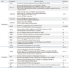
Small heterodimer partner (SHP) is an atypical orphan nuclear receptor that inhibits gluconeogenesis by interacting with Foxo1, hepatocyte nuclear factor 4, or C/EBPα.97,98 SHP is also known to increase PPARγ activity by interacting with PPARγ in a ligand-independent manner. SHP competes with NCoR for binding to the DBD/hinge region of PPARγ. It has been suggested that SHP may act as an endogenous activator of PPARγ.99 However, a contradictory report states that SHP represses the transcriptional activity of PPARγ and does not interact with PPARγ.100 SHP decreases LGK gene expression by inhibiting the transcriptional activity of LXRα and PPARγ via interaction with their common partner, RXRα. Thus, SHP may play a role in fine-tuning glucose homeostasis.100 The diverse functions of PPARγ cofactors are summarized in Table 2.

REGULATION OF PPARγ ACTIVITY BY POST-TRANSLATIONAL MODIFICATION

Phosphorylation

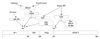
Phosphorylation of nuclear receptors is one of the principal modifications determining their transcriptional activities. Adipocyte differentiation is inhibited by growth factors101-103 and cytokines,104-106 which are known to phosphorylate PPARγ through their respective signaling pathway (Fig. 1). The site of phosphorylation is Ser112 in the N-terminal transactivation domain (AF-1), which is well conserved among species ranging from fish to man.107,108 Ser112 phosphorylation by mitogen-activated protein kinase (MAPK) results in a decrease in transcriptional activity and adipogenesis.109-112 MAPK is activated by extracellular signal-regulated kinase 1/2 (ERK1/2) that is stimulated by growth factors such as epidermal growth factor, platelet-derived growth factor, transforming growth factor-β, insulin, or the prostaglandin PGF2α.109,110,113-116 Phosphorylation of Ser112 by other signals including stress (UV, anisomysin) is mediated by c-Jun N-terminal kinase 1/2 and p38.107,113
Insulin plays a key role in adipogenesis.109 Although preadipocytes express a limited number of insulin receptors, the cells require insulin or insulin-like growth factor-1 for optimal differentiation.117,118 After maturation, large numbers of insulin receptors are expressed, transmitting insulin signals for the induction of lipogenic genes.119,120 Although insulin is a pivotal player in adipogenesis, Ras/MAPK activation by insulin represses PPARγ activity109 as shown in the growth factor-induced phosphorylation of PPARγ at Ser112.
Specifically, downstream tyrosine kinase-1 (Dok1), a multi-site adapter molecule in insulin receptor signaling,121-123 acts as a negative regulator of MAPK.124-126 In mice fed a high-fat diet, Dok1 expression is markedly increased in WAT. A lower mass of WAT is seen in Dok1-deficient mice than in wild-type mice, and the level of PPARγ phosphorylation was increased by ERK.127 These data suggest that an increase in Dok1 gene expression caused by a high-fat diet inhibits the insulin-mediated activation of Ras/MAPK signaling, resulting in increased PPARγ activity.127
In contrast to the MAPK-mediated phosphorylation of Ser112, the cyclin-dependent kinases Cdk7 and Cdk9 phosphorylate the same Ser112 in PPARγ and increase PPARγ activity.128,129 Trichothiodystrophy (TTD) is a rare autosomal recessive disease caused by mutations in the xeroderma pigmentosum (XP) group-D (XPD) gene. The clinical manifestations include immature sexual development, mental retardation, skeletal abnormalities, and dwarfism. A number of patients with TTD exhibit a lack of subcutaneous fat tissue mass. XPD helicase is a subunit of the transcription factor IIH (TFIIH) complex bridging the core-TFIIH [containing particular form of xeroderma pigmentosum B (XPB) helicase] subcomplex and the Cdk-activating kinase containing Cdk7.130 When the C-terminus of XPD is mutated, XPD helicase cannot perform nucleotide excision repair.131 In the process of transcription, Cdk7 in the TFIIH complex phosphorylates the C-terminal domain of the largest subunit of RNA polymerase II132 and nuclear receptors such as ER, VDR, and RARα.133-136 PPARγ phosphorylation by Cdk7 is decreased in XPD patients.128 The activity of a PPARγ promoter reporter was rescued by PPARγ-112S→E, a constitutively active form of PPARγ, in fibroblasts isolated from patients with TTD.128
In addition, Cdk9, a component of positive transcription elongation factor b, has been shown to participate in adipogenesis by directly interacting with PPARγ and phosphorylating Ser112.129 Overexpression of Cdk9 in 3T3L1 cells increased adipogenesis, whereas inhibition of Cdk9 by specific Cdk inhibitors or a dominant-negative Cdk9 mutant inhibited adipogenesis.129 These data suggest that the transcriptional activity of PPARγ is either activated or inhibited depending on the types of kinases involved.
In the adipose tissues of mice fed a high-fat diet, phosphorylation of Ser273 by Cdk5 results in a reduction of adiponectin gene expression, without affecting adipogenesis.137 Cdk5-mediated phosphorylation of PPARγ is blocked by full agonists such as rosiglitazone or partial agonists such as MRL24 or SR1664.137,138
Partial agonists, like MRL24 and SR1664, have been shown to have excellent anti-diabetic activity without increasing adipogenesis.137,138 These compounds are known to block the phosphorylation of Ser273 by Cdk5137,138 and can therefore potentially be used as therapeutic drugs for T2DM without causing weight gain and fluid retention, which are major side effects of full agonist-antidiabetic drugs.
It is worth note that strong PPARγ activators are not necessary to increase insulin sensitivity. Understanding the regulation of Ser273 phosphorylation in PPARγ could provide a hint for the development of drugs to treat T2DM that have fewer side effects.138

Sumoylation

SUMOylation is one of the post-translational modifications responsible for regulating the stability, nuclear-cytosolic distribution, and activity of transcription factors. Small ubiquitin-like modifier (SUMO) family proteins (SUMO-1, -2, and -3 in mammals) affect the interaction between target proteins and their substrates or the DNA that they bind. SUMO binds to proteins by forming isopeptide bonds between the C-terminal glycine residue of SUMO and the ε-amino group of a lysine in the target protein.139,140 Currently, a number of transcription factors including nuclear receptors, such as PPARs,141,142 LXR,143 glucocorticoid receptor,144 androgen receptor,145 and RXRα146 are known to be SUMOylated.
Selective modulation of the transcriptional activity of PPARγ by SUMOylation is now beginning to be understood.142,147 The transcriptional activities of PPARγ isoforms in the presence or absence of ligands are regulated by SUMOylation.142 PPARγ2 is SUMOylated by protein inhibitor of activated STAT 1 (PIAS1) or PIASx, belonging to the PIAS family, regardless of its ligand. PPARγ2 is SUMOylated at Lys107 in the AF-1 domain, and at Lys395 in the AF-2 domain (equivalent to Lys77 and Lys365 of PPARγ1, respectively). SUMOylation of PPARγ2 at Lys107 negatively regulates the transcriptional activity of PPARγ2, because the 107K→R mutation showed increased transcriptional activity.142 This observation is further supported by a promoter reporter assay performed using the variant PPARγ2 107K→R in NIH3T3 fibroblasts.148,149 Furthermore, fibroblast growth factor21 (FGF21)-KO mice exhibit impaired insulin sensitivity in adipocytes and reduced fat mass and adipocyte size. This phenomenon occurs because PPARγ2-induced adipogenesis is inhibited by SUMOylation in WAT. These results indicate that FGF21 is a key regulator of PPARγ2 in the context of SUMOylation.150 In addition, the transcriptional activity of PPARγ2 is increased by overexpressing SUMO1/sentrin/SMT3-specific peptidase 2 (SENP2), a SUMO-specific protease, in C2C12 myotubes.147 Interestingly, the inhibition of PPARγ2 transcriptional activity by SUMOylation is augmented when PPARγ2 is phosphorylated at Ser112.148,149 This indicates an interrelationship between the SUMOylation and phosphorylation of PPARγ2.
The SUMOylation of PPARγ1 at Lys365 (equivalent to Lys395 of PPARγ2) is important in the regulation of inflammatory gene expression. This SUMOylation mediates the transrepression of inflammatory genes like inducible nitric oxide synthase (iNOS) and TNF-α, which are regulated by nuclear factor kappa B in macrophages.151,152 In the basal state, iNOS gene is repressed by TBL1/TBLR1/HDAC3/NCoR complex. Treatment of lipopolysaccharide (LPS) resulted in the removal of HDAC3/NCoR from the complex in a TBL1/TBLR1 and Ubc5-dependent fashion, allowing activation of iNOS gene.148 When RAW264.7 macrophages or primary cultured macrophages were treated with LPS and rosiglitazone, PPARγ1 was found to be SUMOylated on Lys365 by Ubc9, which forms a complex with NCoR/HDAC3 on the promoters of the iNOS gene. Thus, the formation of the NCoR/HDAC3/SUMOylated PPARγ1 complex inhibits the ubiquitination of NCoR/HDAC3, resulting in the repression of the iNOS and TNF-α genes.151,152
Ligand-dependent SUMOylation of PPARγ1 therefore directly represses the promoters of inflammatory genes by stabilizing the NCoR and HDAC3 complexes. This mechanism demonstrates that the role of Lys365 SUMOylation of PPARγ1 is different from that of Lys107 SUMOylation of PPARγ2 in that Lys365 SUMOylation of PPARγ1 represses the expression of inflammatory genes in the presence of ligand.

Ubiquitination

The ubiquitin-proteasome system (UPS) is responsible for the degradation of a variety of intracellular proteins including transcription factors.153,154 Ubiquitin is well conserved between species, binding to target proteins in a sequential manner through the actions of three different cascading enzymes: an ubiquitin-activating enzyme (E1), an ubiquitin-conjugating enzyme (E2), and an ubiquitin protein ligase (E3).155 The polyubiquitinated proteins are recognized and degraded by the 26S proteasome.156 The role of the UPS with respect to transcriptional regulation is well documented.157 In the nucleus of adipocytes, the PPARγ2 protein level is decreased by the action of TZDs.158 Degradation occurs in a ubiquitin-dependent manner in the AF-2 domain of PPARγ.159 However, the AF-1 domains of PPARγ1 and PPARγ2 are degraded by the REGγ proteasome, a type of proteasome that degrades the target substrate in an ubiquitin and ATP-independent fashion.159-161
Degradation of PPARγ is also regulated by interferon-γ (IFN-γ) in adipocytes. Transcription of PPARγ is decreased by IFN-γ-activated STAT signaling.162 When Ser112 of PPARγ, which is known to be phosphorylated by ERK1/2, was replaced with Ala, degradation of the protein was decreased. In addition, U1026, an inhibitor of ERK1/2, decreased IFN-γ-induced PPARγ degradation.163 However, ERK1/2 is not known to be activated by IFN-γ or TZDs; thus, it is assumed that there might be an indirect relationship between the phosphorylation and ubiquitination of PPARγ.163
TNF-α is well known for its role in insulin resistance.164 Degradation of PPARγ is promoted by TNF-α in adipocytes. Treatment of adipocytes with TNF-α and cycloheximide yielded a 44-kDa sized fragment of PPARγ, which is also seen in the WAT or BAT of diabetic rats. However, the molecular link between this fragment and PPARγ degradation is not known.165 Proteasome-dependent PPARγ degradation is increased by resveratrol, a potent activator of SIRT1; however, the mechanism of SIRT1 requires further investigation.84,166

PERSPECTIVE

Regulation of PPARγ activity may be achieved through the interrelationship between agonists, PTM, and coregulators, rather than by the simple action of individual activators or inhibitors. Agonists can induce either coregulator exchange or PTM; the mechanisms of which require further study. Understanding the mechanistic complexity underlying the interactions of these regulators may help accelerate the development of therapeutic drugs against obesity, T2DM, and metabolic syndromes.

Figures and Tables

Fig. 1
Modulation of PPARγ activity by phosphorylation. Positions of phosphorylation sites in PPARγ and the implicated signaling pathways are indicated. Ser112 phosphorylation by growth factors, cytokines, and stress signals are related to decreased PPARγ activity, whereas phosphorylation by Cdk7 and Cdk9 is related to increased PPARγ activity. Obesity or high-fat diet-mediated phosphorylation of PPARγ at Ser273 is related to decreased insulin sensitivity. AF-1 and 2, activation function 1 and 2, respectively; Cdk5, 7 and 9, cyclin-dependent kinase 5, 7 and 9, respectively; DBD, DNA binding domain; Dok1, downstream of tyrosine kinase-1; ERK1/2, extracellular signal-regulated kinase 1/2; HFD, high fat diet; JNK, c-Jun N-terminal kinase; LBD, ligand binding domain; MAPK, mitogen-activated protein kinase; p38, p38 MAP kinase; Rosi, rosiglitasone; PPARγ, peroxisome proliferator-activated receptor gamma.
ymj-54-545-g001
Table 1
Selected PPARγ Target Genes Involved in Metabolism
ymj-54-545-i001

PPARγ, peroxisome proliferator-activated receptor gamma; CAP, Cbl-associated protein; IRS2, insulin receptor substrate 2; GLUT4, glucose transporter 4; LPL, lipoprotein lipase; GyK, glycerol kinase; TNF-α, tumor necrosis factor-α; IL-6, interleukin-6; GLUT2, glucose transporter 2; CD36, fatty acid translocase; ACS, acetyl-CoA synthetase; PCK2, phosphoenolpyruvate carboxykinase 2; GK, glucokinase.

Table 2
List of Regulatory Factors for PPARγ Activity
ymj-54-545-i002

PPARγ, peroxisome proliferator-activated receptor gamma; SRC-1, steroid receptor coactivator-1; PGC-1α, PPARγ coactivator-1; CBP, CREB-binding protein; TRAP, thyroid hormone receptor-associated protein; BAF, Brg1/Brm-associated factor; MBF-1, multiprotein bridging factor-1; NRF2, nuclear factor E2-related factor 2; PRIP, PPAR-interacting protein; PDIP, PPARγ-DBD interacting protein; SHP, small heterodimer partner; NCoR, nuclear corepressor; SMRT, silencing mediator of retinoid and thyroid hormone receptor; Rb, retinoblastoma protein; SHP, small heterodimer partner; DBD, DNA-binding domain; UCP, uncoupling protein; BAT, brown adipose tissue; AF-1, agonist-independent activation domain; AF-2, agonist-dependent activation domain; LBD, ligand-binding domain; PEPCK, phosphoenol pyruvate carboxykinase; RXR, retinoid X receptor; TFIID, transcription factor IID; TXS, thromboxane synthase; Cdk5, cyclin-dependent kinase; ARF6, ADP-ribosylation factor; RIP140, receptor-interacting protein 140.

ACKNOWLEDGEMENTS

We apologize to all the contributors in the field whose work could not be cited due to space limitations. This research was supported by Basic Science Research Program through the National Research Foundation of Korea (NRF) funded by the Ministry of Education, Science and Technology, Republic of Korea (2011-0030706 to Y.H. Ahn).

Notes

The authors have no financial conflicts of interest.

References

1. Evans RM, Barish GD, Wang YX. PPARs and the complex journey to obesity. Nat Med. 2004. 10:355–361.
crossref
2. Michalik L, Auwerx J, Berger JP, Chatterjee VK, Glass CK, Gonzalez FJ, et al. International Union of Pharmacology. LXI. Peroxisome proliferator-activated receptors. Pharmacol Rev. 2006. 58:726–741.
crossref
3. Kersten S, Desvergne B, Wahli W. Roles of PPARs in health and disease. Nature. 2000. 405:421–424.
crossref
4. Rosen ED, Hsu CH, Wang X, Sakai S, Freeman MW, Gonzalez FJ, et al. C/EBPalpha induces adipogenesis through PPARgamma: a unified pathway. Genes Dev. 2002. 16:22–26.
5. Jay MA, Ren J. Peroxisome proliferator-activated receptor (PPAR) in metabolic syndrome and type 2 diabetes mellitus. Curr Diabetes Rev. 2007. 3:33–39.
crossref
6. Vidal-Puig AJ, Considine RV, Jimenez-Liñan M, Werman A, Pories WJ, Caro JF, et al. Peroxisome proliferator-activated receptor gene expression in human tissues. Effects of obesity, weight loss, and regulation by insulin and glucocorticoids. J Clin Invest. 1997. 99:2416–2422.
crossref
7. Chawla A, Schwarz EJ, Dimaculangan DD, Lazar MA. Peroxisome proliferator-activated receptor (PPAR) gamma: adipose-predominant expression and induction early in adipocyte differentiation. Endocrinology. 1994. 135:798–800.
crossref
8. Tontonoz P, Hu E, Graves RA, Budavari AI, Spiegelman BM. mPPAR gamma 2: tissue-specific regulator of an adipocyte enhancer. Genes Dev. 1994. 8:1224–1234.
crossref
9. Kubota N, Terauchi Y, Miki H, Tamemoto H, Yamauchi T, Komeda K, et al. PPAR gamma mediates high-fat diet-induced adipocyte hypertrophy and insulin resistance. Mol Cell. 1999. 4:597–609.
crossref
10. Tontonoz P, Hu E, Spiegelman BM. Stimulation of adipogenesis in fibroblasts by PPAR gamma 2, a lipid-activated transcription factor. Cell. 1994. 79:1147–1156.
crossref
11. Rosen ED, Spiegelman BM. PPARgamma: a nuclear regulator of metabolism, differentiation, and cell growth. J Biol Chem. 2001. 276:37731–37734.
12. Ferré P. The biology of peroxisome proliferator-activated receptors: relationship with lipid metabolism and insulin sensitivity. Diabetes. 2004. 53:Suppl 1. S43–S50.
13. Nolan JJ, Ludvik B, Beerdsen P, Joyce M, Olefsky J. Improvement in glucose tolerance and insulin resistance in obese subjects treated with troglitazone. N Engl J Med. 1994. 331:1188–1193.
crossref
14. Kumar S, Boulton AJ, Beck-Nielsen H, Berthezene F, Muggeo M, Persson B, et al. Troglitazone Study Group. Troglitazone, an insulin action enhancer, improves metabolic control in NIDDM patients. Diabetologia. 1996. 39:701–709.
crossref
15. Hamza MS, Pott S, Vega VB, Thomsen JS, Kandhadayar GS, Ng PW, et al. De-novo identification of PPARgamma/RXR binding sites and direct targets during adipogenesis. PLoS One. 2009. 4:e4907.
16. Lefterova MI, Zhang Y, Steger DJ, Schupp M, Schug J, Cristancho A, et al. PPARgamma and C/EBP factors orchestrate adipocyte biology via adjacent binding on a genome-wide scale. Genes Dev. 2008. 22:2941–2952.
crossref
17. Nielsen R, Pedersen TA, Hagenbeek D, Moulos P, Siersbaek R, Megens E, et al. Genome-wide profiling of PPARgamma: RXR and RNA polymerase II occupancy reveals temporal activation of distinct metabolic pathways and changes in RXR dimer composition during adipogenesis. Genes Dev. 2008. 22:2953–2967.
crossref
18. Iwaki M, Matsuda M, Maeda N, Funahashi T, Matsuzawa Y, Makishima M, et al. Induction of adiponectin, a fat-derived antidiabetic and antiatherogenic factor, by nuclear receptors. Diabetes. 2003. 52:1655–1663.
crossref
19. Combs TP, Wagner JA, Berger J, Doebber T, Wang WJ, Zhang BB, et al. Induction of adipocyte complement-related protein of 30 kilodaltons by PPARgamma agonists: a potential mechanism of insulin sensitization. Endocrinology. 2002. 143:998–1007.
crossref
20. Baumann CA, Chokshi N, Saltiel AR, Ribon V. Cloning and characterization of a functional peroxisome proliferator activator receptor-gamma-responsive element in the promoter of the CAP gene. J Biol Chem. 2000. 275:9131–9135.
crossref
21. Smith U, Gogg S, Johansson A, Olausson T, Rotter V, Svalstedt B. Thiazolidinediones (PPARgamma agonists) but not PPARalpha agonists increase IRS-2 gene expression in 3T3-L1 and human adipocytes. FASEB J. 2001. 15:215–220.
crossref
22. Wu Z, Xie Y, Morrison RF, Bucher NL, Farmer SR. PPARgamma induces the insulin-dependent glucose transporter GLUT4 in the absence of C/EBPalpha during the conversion of 3T3 fibroblasts into adipocytes. J Clin Invest. 1998. 101:22–32.
crossref
23. Dana SL, Hoener PA, Bilakovics JM, Crombie DL, Ogilvie KM, Kauffman RF, et al. Peroxisome proliferator-activated receptor subtype-specific regulation of hepatic and peripheral gene expression in the Zucker diabetic fatty rat. Metabolism. 2001. 50:963–971.
crossref
24. Desvergne B, Wahli W. Peroxisome proliferator-activated receptors: nuclear control of metabolism. Endocr Rev. 1999. 20:649–688.
crossref
25. Tontonoz P, Hu E, Devine J, Beale EG, Spiegelman BM. PPAR gamma 2 regulates adipose expression of the phosphoenolpyruvate carboxykinase gene. Mol Cell Biol. 1995. 15:351–357.
crossref
26. Guan HP, Li Y, Jensen MV, Newgard CB, Steppan CM, Lazar MA. A futile metabolic cycle activated in adipocytes by antidiabetic agents. Nat Med. 2002. 8:1122–1128.
crossref
27. Kim HJ, Jung TW, Kang ES, Kim DJ, Ahn CW, Lee KW, et al. Depot-specific regulation of perilipin by rosiglitazone in a diabetic animal model. Metabolism. 2007. 56:676–685.
crossref
28. Nagai S, Shimizu C, Umetsu M, Taniguchi S, Endo M, Miyoshi H, et al. Identification of a functional peroxisome proliferator-activated receptor responsive element within the murine perilipin gene. Endocrinology. 2004. 145:2346–2356.
crossref
29. Motojima K, Passilly P, Peters JM, Gonzalez FJ, Latruffe N. Expression of putative fatty acid transporter genes are regulated by peroxisome proliferator-activated receptor alpha and gamma activators in a tissue- and inducer-specific manner. J Biol Chem. 1998. 273:16710–16714.
crossref
30. Schoonjans K, Peinado-Onsurbe J, Lefebvre AM, Heyman RA, Briggs M, Deeb S, et al. PPARalpha and PPARgamma activators direct a distinct tissue-specific transcriptional response via a PPRE in the lipoprotein lipase gene. EMBO J. 1996. 15:5336–5348.
crossref
31. Martin G, Schoonjans K, Lefebvre AM, Staels B, Auwerx J. Coordinate regulation of the expression of the fatty acid transport protein and acyl-CoA synthetase genes by PPARalpha and PPARgamma activators. J Biol Chem. 1997. 272:28210–28217.
crossref
32. Saraf N, Sharma PK, Mondal SC, Garg VK, Singh AK. Role of PPARg2 transcription factor in thiazolidinedione-induced insulin sensitization. J Pharm Pharmacol. 2012. 64:161–171.
crossref
33. Kallen CB, Lazar MA. Antidiabetic thiazolidinediones inhibit leptin (ob) gene expression in 3T3-L1 adipocytes. Proc Natl Acad Sci U S A. 1996. 93:5793–5796.
crossref
34. Jiang C, Ting AT, Seed B. PPAR-gamma agonists inhibit production of monocyte inflammatory cytokines. Nature. 1998. 391:82–86.
crossref
35. Sigrist S, Bedoucha M, Boelsterli UA. Down-regulation by troglitazone of hepatic tumor necrosis factor-alpha and interleukin-6 mRNA expression in a murine model of non-insulin-dependent diabetes. Biochem Pharmacol. 2000. 60:67–75.
crossref
36. Kim HI, Kim JW, Kim SH, Cha JY, Kim KS, Ahn YH. Identification and functional characterization of the peroxisomal proliferator response element in rat GLUT2 promoter. Diabetes. 2000. 49:1517–1524.
crossref
37. Kim SY, Kim HI, Park SK, Im SS, Li T, Cheon HG, et al. Liver glucokinase can be activated by peroxisome proliferator-activated receptor-gamma. Diabetes. 2004. 53:Suppl 1. S66–S70.
38. Kim HI, Cha JY, Kim SY, Kim JW, Roh KJ, Seong JK, et al. Peroxisomal proliferator-activated receptor-gamma upregulates glucokinase gene expression in beta-cells. Diabetes. 2002. 51:676–685.
crossref
39. Kim HI, Ahn YH. Role of peroxisome proliferator-activated receptor-gamma in the glucose-sensing apparatus of liver and beta-cells. Diabetes. 2004. 53:Suppl 1. S60–S65.
40. Wu SC, Zhang Y. Minireview: role of protein methylation and demethylation in nuclear hormone signaling. Mol Endocrinol. 2009. 23:1323–1334.
crossref
41. Nolte RT, Wisely GB, Westin S, Cobb JE, Lambert MH, Kurokawa R, et al. Ligand binding and co-activator assembly of the peroxisome proliferator-activated receptor-gamma. Nature. 1998. 395:137–143.
crossref
42. Picard F, Géhin M, Annicotte J, Rocchi S, Champy MF, O'Malley BW, et al. SRC-1 and TIF2 control energy balance between white and brown adipose tissues. Cell. 2002. 111:931–941.
crossref
43. McInerney EM, Rose DW, Flynn SE, Westin S, Mullen TM, Krones A, et al. Determinants of coactivator LXXLL motif specificity in nuclear receptor transcriptional activation. Genes Dev. 1998. 12:3357–3368.
crossref
44. Wang Z, Qi C, Krones A, Woodring P, Zhu X, Reddy JK, et al. Critical roles of the p160 transcriptional coactivators p/CIP and SRC-1 in energy balance. Cell Metab. 2006. 3:111–122.
crossref
45. Puigserver P, Adelmant G, Wu Z, Fan M, Xu J, O'Malley B, et al. Activation of PPARgamma coactivator-1 through transcription factor docking. Science. 1999. 286:1368–1371.
crossref
46. Gelman L, Zhou G, Fajas L, Raspé E, Fruchart JC, Auwerx J. p300 interacts with the N- and C-terminal part of PPARgamma2 in a ligand-independent and -dependent manner, respectively. J Biol Chem. 1999. 274:7681–7688.
crossref
47. Takahashi N, Kawada T, Yamamoto T, Goto T, Taimatsu A, Aoki N, et al. Overexpression and ribozyme-mediated targeting of transcriptional coactivators CREB-binding protein and p300 revealed their indispensable roles in adipocyte differentiation through the regulation of peroxisome proliferator-activated receptor gamma. J Biol Chem. 2002. 277:16906–16912.
crossref
48. Qi C, Surapureddi S, Zhu YJ, Yu S, Kashireddy P, Rao MS, et al. Transcriptional coactivator PRIP, the peroxisome proliferator-activated receptor gamma (PPARgamma)-interacting protein, is required for PPARgamma-mediated adipogenesis. J Biol Chem. 2003. 278:25281–25284.
crossref
49. Viswakarma N, Jia Y, Bai L, Vluggens A, Borensztajn J, Xu J, et al. Coactivators in PPAR-Regulated Gene Expression. PPAR Res. 2010. 2010:pii: 250126.
crossref
50. Yuan CX, Ito M, Fondell JD, Fu ZY, Roeder RG. The TRAP220 component of a thyroid hormone receptor-associated protein (TRAP) coactivator complex interacts directly with nuclear receptors in a ligand-dependent fashion. Proc Natl Acad Sci U S A. 1998. 95:7939–7944.
crossref
51. Zhu Y, Qi C, Jia Y, Nye JS, Rao MS, Reddy JK. Deletion of PBP/PPARBP, the gene for nuclear receptor coactivator peroxisome proliferator-activated receptor-binding protein, results in embryonic lethality. J Biol Chem. 2000. 275:14779–14782.
crossref
52. Ge K, Guermah M, Yuan CX, Ito M, Wallberg AE, Spiegelman BM, et al. Transcription coactivator TRAP220 is required for PPAR gamma 2-stimulated adipogenesis. Nature. 2002. 417:563–567.
crossref
53. Zhu Y, Qi C, Jain S, Rao MS, Reddy JK. Isolation and characterization of PBP, a protein that interacts with peroxisome proliferator-activated receptor. J Biol Chem. 1997. 272:25500–25506.
crossref
54. Yang W, Rachez C, Freedman LP. Discrete roles for peroxisome proliferator-activated receptor gamma and retinoid X receptor in recruiting nuclear receptor coactivators. Mol Cell Biol. 2000. 20:8008–8017.
crossref
55. Sudarsanam P, Winston F. The Swi/Snf family nucleosome-remodeling complexes and transcriptional control. Trends Genet. 2000. 16:345–351.
56. Salma N, Xiao H, Mueller E, Imbalzano AN. Temporal recruitment of transcription factors and SWI/SNF chromatin-remodeling enzymes during adipogenic induction of the peroxisome proliferator-activated receptor gamma nuclear hormone receptor. Mol Cell Biol. 2004. 24:4651–4663.
crossref
57. Pedersen TA, Kowenz-Leutz E, Leutz A, Nerlov C. Cooperation between C/EBPalpha TBP/TFIIB and SWI/SNF recruiting domains is required for adipocyte differentiation. Genes Dev. 2001. 15:3208–3216.
crossref
58. Lemon B, Inouye C, King DS, Tjian R. Selectivity of chromatin-remodelling cofactors for ligand-activated transcription. Nature. 2001. 414:924–928.
crossref
59. Debril MB, Gelman L, Fayard E, Annicotte JS, Rocchi S, Auwerx J. Transcription factors and nuclear receptors interact with the SWI/SNF complex through the BAF60c subunit. J Biol Chem. 2004. 279:16677–16686.
crossref
60. Tontonoz P, Graves RA, Budavari AI, Erdjument-Bromage H, Lui M, Hu E, et al. Adipocyte-specific transcription factor ARF6 is a heterodimeric complex of two nuclear hormone receptors, PPAR gamma and RXR alpha. Nucleic Acids Res. 1994. 22:5628–5634.
crossref
61. Graves RA, Tontonoz P, Spiegelman BM. Analysis of a tissue-specific enhancer: ARF6 regulates adipogenic gene expression. Mol Cell Biol. 1992. 12:1202–1208.
crossref
62. Dreijerink KM, Varier RA, van Beekum O, Jeninga EH, Höppener JW, Lips CJ, et al. The multiple endocrine neoplasia type 1 (MEN1) tumor suppressor regulates peroxisome proliferator-activated receptor gamma-dependent adipocyte differentiation. Mol Cell Biol. 2009. 29:5060–5069.
crossref
63. Li FQ, Ueda H, Hirose S. Mediators of activation of fushi tarazu gene transcription by BmFTZ-F1. Mol Cell Biol. 1994. 14:3013–3021.
crossref
64. Brendel C, Gelman L, Auwerx J. Multiprotein bridging factor-1 (MBF-1) is a cofactor for nuclear receptors that regulate lipid metabolism. Mol Endocrinol. 2002. 16:1367–1377.
crossref
65. Ikeda Y, Sugawara A, Taniyama Y, Uruno A, Igarashi K, Arima S, et al. Suppression of rat thromboxane synthase gene transcription by peroxisome proliferator-activated receptor gamma in macrophages via an interaction with NRF2. J Biol Chem. 2000. 275:33142–33150.
crossref
66. Tsutsumi E, Takeuchi K, Abe T, Takahashi N, Kato T, Taniyama Y, et al. Rat kidney thromboxane synthase: cDNA cloning and gene expression regulation in hydronephrotic kidney. Prostaglandins. 1997. 53:423–431.
crossref
67. Needleman P, Kulkarni PS, Raz A. Coronary tone modulation: formation and actions of prostaglandins, endoperoxides, and thromboxanes. Science. 1977. 195:409–412.
crossref
68. Hora K, Oguchi H, Furukawa T, Hora K, Tokunaga S. Effects of a selective thromboxane synthetase inhibitor OKY-046 on experimental diabetic nephropathy. Nephron. 1990. 56:297–305.
crossref
69. Giustina A, Perini P, Desenzani P, Bossoni S, Ianniello P, Milani M, et al. Long-term treatment with the dual antithromboxane agent picotamide decreases microalbuminuria in normotensive type 2 diabetic patients. Diabetes. 1998. 47:423–430.
crossref
70. Caira F, Antonson P, Pelto-Huikko M, Treuter E, Gustafsson JA. Cloning and characterization of RAP250, a novel nuclear receptor coactivator. J Biol Chem. 2000. 275:5308–5317.
crossref
71. Zhu Y, Kan L, Qi C, Kanwar YS, Yeldandi AV, Rao MS, et al. Isolation and characterization of peroxisome proliferator-activated receptor (PPAR) interacting protein (PRIP) as a coactivator for PPAR. J Biol Chem. 2000. 275:13510–13516.
crossref
72. Antonson P, Schuster GU, Wang L, Rozell B, Holter E, Flodby P, et al. Inactivation of the nuclear receptor coactivator RAP250 in mice results in placental vascular dysfunction. Mol Cell Biol. 2003. 23:1260–1268.
crossref
73. Zhu YJ, Crawford SE, Stellmach V, Dwivedi RS, Rao MS, Gonzalez FJ, et al. Coactivator PRIP, the peroxisome proliferator-activated receptor-interacting protein, is a modulator of placental, cardiac, hepatic, and embryonic development. J Biol Chem. 2003. 278:1986–1990.
crossref
74. Tomaru T, Satoh T, Yoshino S, Ishizuka T, Hashimoto K, Monden T, et al. Isolation and characterization of a transcriptional cofactor and its novel isoform that bind the deoxyribonucleic acid-binding domain of peroxisome proliferator-activated receptor-gamma. Endocrinology. 2006. 147:377–388.
crossref
75. Puigserver P, Wu Z, Park CW, Graves R, Wright M, Spiegelman BM. A cold-inducible coactivator of nuclear receptors linked to adaptive thermogenesis. Cell. 1998. 92:829–839.
crossref
76. Leone TC, Lehman JJ, Finck BN, Schaeffer PJ, Wende AR, Boudina S, et al. PGC-1alpha deficiency causes multi-system energy metabolic derangements: muscle dysfunction, abnormal weight control and hepatic steatosis. PLoS Biol. 2005. 3:e101.
77. Wu Z, Puigserver P, Andersson U, Zhang C, Adelmant G, Mootha V, et al. Mechanisms controlling mitochondrial biogenesis and respiration through the thermogenic coactivator PGC-1. Cell. 1999. 98:115–124.
crossref
78. Tiraby C, Langin D. Conversion from white to brown adipocytes: a strategy for the control of fat mass? Trends Endocrinol Metab. 2003. 14:439–441.
crossref
79. Kopecky J, Clarke G, Enerbäck S, Spiegelman B, Kozak LP. Expression of the mitochondrial uncoupling protein gene from the aP2 gene promoter prevents genetic obesity. J Clin Invest. 1995. 96:2914–2923.
crossref
80. Guan HP, Ishizuka T, Chui PC, Lehrke M, Lazar MA. Corepressors selectively control the transcriptional activity of PPARgamma in adipocytes. Genes Dev. 2005. 19:453–461.
crossref
81. Liu C, Lin JD. PGC-1 coactivators in the control of energy metabolism. Acta Biochim Biophys Sin (Shanghai). 2011. 43:248–257.
crossref
82. Lee G, Elwood F, McNally J, Weiszmann J, Lindstrom M, Amaral K, et al. T0070907, a selective ligand for peroxisome proliferator-activated receptor gamma, functions as an antagonist of biochemical and cellular activities. J Biol Chem. 2002. 277:19649–19657.
crossref
83. Yu C, Markan K, Temple KA, Deplewski D, Brady MJ, Cohen RN. The nuclear receptor corepressors NCoR and SMRT decrease peroxisome proliferator-activated receptor gamma transcriptional activity and repress 3T3-L1 adipogenesis. J Biol Chem. 2005. 280:13600–13605.
crossref
84. Picard F, Kurtev M, Chung N, Topark-Ngarm A, Senawong T, Machado De Oliveira R, et al. Sirt1 promotes fat mobilization in white adipocytes by repressing PPAR-gamma. Nature. 2004. 429:771–776.
crossref
85. Blüher M, Kahn BB, Kahn CR. Extended longevity in mice lacking the insulin receptor in adipose tissue. Science. 2003. 299:572–574.
crossref
86. Li P, Fan W, Xu J, Lu M, Yamamoto H, Auwerx J, et al. Adipocyte NCoR knockout decreases PPARγ phosphorylation and enhances PPARγ activity and insulin sensitivity. Cell. 2011. 147:815–826.
crossref
87. Powell E, Kuhn P, Xu W. Nuclear Receptor Cofactors in PPARgamma-Mediated Adipogenesis and Adipocyte Energy Metabolism. PPAR Res. 2007. 2007:53843.
88. Treuter E, Albrektsen T, Johansson L, Leers J, Gustafsson JA. A regulatory role for RIP140 in nuclear receptor activation. Mol Endocrinol. 1998. 12:864–881.
crossref
89. Leonardsson G, Steel JH, Christian M, Pocock V, Milligan S, Bell J, et al. Nuclear receptor corepressor RIP140 regulates fat accumulation. Proc Natl Acad Sci U S A. 2004. 101:8437–8442.
crossref
90. Nakae J, Kitamura T, Kitamura Y, Biggs WH 3rd, Arden KC, Accili D. The forkhead transcription factor Foxo1 regulates adipocyte differentiation. Dev Cell. 2003. 4:119–129.
crossref
91. Higgins C, Chatterjee S, Cherington V. The block of adipocyte differentiation by a C-terminally truncated, but not by full-length, simian virus 40 large tumor antigen is dependent on an intact retinoblastoma susceptibility protein family binding domain. J Virol. 1996. 70:745–752.
crossref
92. Chen PL, Riley DJ, Chen Y, Lee WH. Retinoblastoma protein positively regulates terminal adipocyte differentiation through direct interaction with C/EBPs. Genes Dev. 1996. 10:2794–2804.
crossref
93. Fajas L, Egler V, Reiter R, Hansen J, Kristiansen K, Debril MB, et al. The retinoblastoma-histone deacetylase 3 complex inhibits PPARgamma and adipocyte differentiation. Dev Cell. 2002. 3:903–910.
crossref
94. Péterfy M, Phan J, Xu P, Reue K. Lipodystrophy in the fld mouse results from mutation of a new gene encoding a nuclear protein, lipin. Nat Genet. 2001. 27:121–124.
crossref
95. Reue K, Péterfy M. Mouse models of lipodystrophy. Curr Atheroscler Rep. 2000. 2:390–396.
crossref
96. Koh YK, Lee MY, Kim JW, Kim M, Moon JS, Lee YJ, et al. Lipin1 is a key factor for the maturation and maintenance of adipocytes in the regulatory network with CCAAT/enhancer-binding protein alpha and peroxisome proliferator-activated receptor gamma 2. J Biol Chem. 2008. 283:34896–34906.
crossref
97. Yamagata K, Daitoku H, Shimamoto Y, Matsuzaki H, Hirota K, Ishida J, et al. Bile acids regulate gluconeogenic gene expression via small heterodimer partner-mediated repression of hepatocyte nuclear factor 4 and Foxo1. J Biol Chem. 2004. 279:23158–23165.
crossref
98. Park MJ, Kong HJ, Kim HY, Kim HH, Kim JH, Cheong JH. Transcriptional repression of the gluconeogenic gene PEPCK by the orphan nuclear receptor SHP through inhibitory interaction with C/EBPalpha. Biochem J. 2007. 402:567–574.
crossref
99. Nishizawa H, Yamagata K, Shimomura I, Takahashi M, Kuriyama H, Kishida K, et al. Small heterodimer partner, an orphan nuclear receptor, augments peroxisome proliferator-activated receptor gamma transactivation. J Biol Chem. 2002. 277:1586–1592.
crossref
100. Kim TH, Kim H, Park JM, Im SS, Bae JS, Kim MY, et al. Interrelationship between liver X receptor alpha, sterol regulatory element-binding protein-1c, peroxisome proliferator-activated receptor gamma, and small heterodimer partner in the transcriptional regulation of glucokinase gene expression in liver. J Biol Chem. 2009. 284:15071–15083.
crossref
101. Hauner H, Röhrig K, Petruschke T. Effects of epidermal growth factor (EGF), platelet-derived growth factor (PDGF) and fibroblast growth factor (FGF) on human adipocyte development and function. Eur J Clin Invest. 1995. 25:90–96.
crossref
102. Navre M, Ringold GM. Differential effects of fibroblast growth factor and tumor promoters on the initiation and maintenance of adipocyte differentiation. J Cell Biol. 1989. 109(4 Pt 1):1857–1863.
crossref
103. Serrero G, Mills D. Physiological role of epidermal growth factor on adipose tissue development in vivo. Proc Natl Acad Sci U S A. 1991. 88:3912–3916.
crossref
104. Torti FM, Torti SV, Larrick JW, Ringold GM. Modulation of adipocyte differentiation by tumor necrosis factor and transforming growth factor beta. J Cell Biol. 1989. 108:1105–1113.
crossref
105. Ron D, Brasier AR, McGehee RE Jr, Habener JF. Tumor necrosis factor-induced reversal of adipocytic phenotype of 3T3-L1 cells is preceded by a loss of nuclear CCAAT/enhancer binding protein (C/EBP). J Clin Invest. 1992. 89:223–233.
crossref
106. Berg M, Fraker DL, Alexander HR. Characterization of differentiation factor/leukaemia inhibitory factor effect on lipoprotein lipase activity and mRNA in 3T3-L1 adipocytes. Cytokine. 1994. 6:425–432.
crossref
107. Camp HS, Tafuri SR, Leff T. c-Jun N-terminal kinase phosphorylates peroxisome proliferator-activated receptor-gamma1 and negatively regulates its transcriptional activity. Endocrinology. 1999. 140:392–397.
crossref
108. van Beekum O, Fleskens V, Kalkhoven E. Posttranslational modifications of PPAR-gamma: fine-tuning the metabolic master regulator. Obesity (Silver Spring). 2009. 17:213–219.
crossref
109. Hu E, Kim JB, Sarraf P, Spiegelman BM. Inhibition of adipogenesis through MAP kinase-mediated phosphorylation of PPARgamma. Science. 1996. 274:2100–2103.
crossref
110. Reginato MJ, Krakow SL, Bailey ST, Lazar MA. Prostaglandins promote and block adipogenesis through opposing effects on peroxisome proliferator-activated receptor gamma. J Biol Chem. 1998. 273:1855–1858.
crossref
111. Shao D, Rangwala SM, Bailey ST, Krakow SL, Reginato MJ, Lazar MA. Interdomain communication regulating ligand binding by PPAR-gamma. Nature. 1998. 396:377–380.
crossref
112. Ristow M, Müller-Wieland D, Pfeiffer A, Krone W, Kahn CR. Obesity associated with a mutation in a genetic regulator of adipocyte differentiation. N Engl J Med. 1998. 339:953–959.
crossref
113. Adams M, Reginato MJ, Shao D, Lazar MA, Chatterjee VK. Transcriptional activation by peroxisome proliferator-activated receptor gamma is inhibited by phosphorylation at a consensus mitogen-activated protein kinase site. J Biol Chem. 1997. 272:5128–5132.
crossref
114. Tang X, Guilherme A, Chakladar A, Powelka AM, Konda S, Virbasius JV, et al. An RNA interference-based screen identifies MAP4K4/NIK as a negative regulator of PPARgamma, adipogenesis, and insulin-responsive hexose transport. Proc Natl Acad Sci U S A. 2006. 103:2087–2092.
crossref
115. Camp HS, Tafuri SR. Regulation of peroxisome proliferator-activated receptor gamma activity by mitogen-activated protein kinase. J Biol Chem. 1997. 272:10811–10816.
crossref
116. Zhang B, Berger J, Zhou G, Elbrecht A, Biswas S, White-Carrington S, et al. Insulin- and mitogen-activated protein kinase-mediated phosphorylation and activation of peroxisome proliferator-activated receptor gamma. J Biol Chem. 1996. 271:31771–31774.
crossref
117. Schmidt W, Pöll-Jordan G, Löffler G. Adipose conversion of 3T3-L1 cells in a serum-free culture system depends on epidermal growth factor, insulin-like growth factor I, corticosterone, and cyclic AMP. J Biol Chem. 1990. 265:15489–15495.
crossref
118. Hauner H. Complete adipose differentiation of 3T3 L1 cells in a chemically defined medium: comparison to serum-containing culture conditions. Endocrinology. 1990. 127:865–872.
crossref
119. Rubin CS, Lai E, Rosen OM. Acquisition of increased hormone sensitivity during in vitro adipocyte development. J Biol Chem. 1977. 252:3554–3557.
crossref
120. Lawrence JC Jr. Signal transduction and protein phosphorylation in the regulation of cellular metabolism by insulin. Annu Rev Physiol. 1992. 54:177–193.
crossref
121. Carpino N, Wisniewski D, Strife A, Marshak D, Kobayashi R, Stillman B, et al. p62(dok): a constitutively tyrosine-phosphorylated, GAP-associated protein in chronic myelogenous leukemia progenitor cells. Cell. 1997. 88:197–204.
crossref
122. Hosomi Y, Shii K, Ogawa W, Matsuba H, Yoshida M, Okada Y, et al. Characterization of a 60-kilodalton substrate of the insulin receptor kinase. J Biol Chem. 1994. 269:11498–11502.
crossref
123. Yamanashi Y, Baltimore D. Identification of the Abl- and rasGAP-associated 62 kDa protein as a docking protein, Dok. Cell. 1997. 88:205–211.
crossref
124. Di Cristofano A, Niki M, Zhao M, Karnell FG, Clarkson B, Pear WS, et al. p62(dok), a negative regulator of Ras and mitogen-activated protein kinase (MAPK) activity, opposes leukemogenesis by p210(bcr-abl). J Exp Med. 2001. 194:275–284.
crossref
125. Wick MJ, Dong LQ, Hu D, Langlais P, Liu F. Insulin receptor-mediated p62dok tyrosine phosphorylation at residues 362 and 398 plays distinct roles for binding GTPase-activating protein and Nck and is essential for inhibiting insulin-stimulated activation of Ras and Akt. J Biol Chem. 2001. 276:42843–42850.
crossref
126. Yamanashi Y, Tamura T, Kanamori T, Yamane H, Nariuchi H, Yamamoto T, et al. Role of the rasGAP-associated docking protein p62(dok) in negative regulation of B cell receptor-mediated signaling. Genes Dev. 2000. 14:11–16.
crossref
127. Hosooka T, Noguchi T, Kotani K, Nakamura T, Sakaue H, Inoue H, et al. Dok1 mediates high-fat diet-induced adipocyte hypertrophy and obesity through modulation of PPAR-gamma phosphorylation. Nat Med. 2008. 14:188–193.
crossref
128. Compe E, Drané P, Laurent C, Diderich K, Braun C, Hoeijmakers JH, et al. Dysregulation of the peroxisome proliferator-activated receptor target genes by XPD mutations. Mol Cell Biol. 2005. 25:6065–6076.
crossref
129. Iankova I, Petersen RK, Annicotte JS, Chavey C, Hansen JB, Kratchmarova I, et al. Peroxisome proliferator-activated receptor gamma recruits the positive transcription elongation factor b complex to activate transcription and promote adipogenesis. Mol Endocrinol. 2006. 20:1494–1505.
crossref
130. Roy R, Adamczewski JP, Seroz T, Vermeulen W, Tassan JP, Schaeffer L, et al. The MO15 cell cycle kinase is associated with the TFIIH transcription-DNA repair factor. Cell. 1994. 79:1093–1101.
crossref
131. Zurita M, Merino C. The transcriptional complexity of the TFIIH complex. Trends Genet. 2003. 19:578–584.
crossref
132. Lu H, Zawel L, Fisher L, Egly JM, Reinberg D. Human general transcription factor IIH phosphorylates the C-terminal domain of RNA polymerase II. Nature. 1992. 358:641–645.
crossref
133. Chen D, Riedl T, Washbrook E, Pace PE, Coombes RC, Egly JM, et al. Activation of estrogen receptor alpha by S118 phosphorylation involves a ligand-dependent interaction with TFIIH and participation of CDK7. Mol Cell. 2000. 6:127–137.
crossref
134. Drané P, Compe E, Catez P, Chymkowitch P, Egly JM. Selective regulation of vitamin D receptor-responsive genes by TFIIH. Mol Cell. 2004. 16:187–197.
crossref
135. Keriel A, Stary A, Sarasin A, Rochette-Egly C, Egly JM. XPD mutations prevent TFIIH-dependent transactivation by nuclear receptors and phosphorylation of RARalpha. Cell. 2002. 109:125–135.
crossref
136. Rochette-Egly C, Adam S, Rossignol M, Egly JM, Chambon P. Stimulation of RAR alpha activation function AF-1 through binding to the general transcription factor TFIIH and phosphorylation by CDK7. Cell. 1997. 90:97–107.
crossref
137. Choi JH, Banks AS, Estall JL, Kajimura S, Boström P, Laznik D, et al. Anti-diabetic drugs inhibit obesity-linked phosphorylation of PPARgamma by Cdk5. Nature. 2010. 466:451–456.
crossref
138. Choi JH, Banks AS, Kamenecka TM, Busby SA, Chalmers MJ, Kumar N, et al. Antidiabetic actions of a non-agonist PPARγ ligand blocking Cdk5-mediated phosphorylation. Nature. 2011. 477:477–481.
crossref
139. Johnson ES. Protein modification by SUMO. Annu Rev Biochem. 2004. 73:355–382.
crossref
140. Geiss-Friedlander R, Melchior F. Concepts in sumoylation: a decade on. Nat Rev Mol Cell Biol. 2007. 8:947–956.
crossref
141. Pourcet B, Pineda-Torra I, Derudas B, Staels B, Glineur C. SUMOylation of human peroxisome proliferator-activated receptor alpha inhibits its trans-activity through the recruitment of the nuclear corepressor NCoR. J Biol Chem. 2010. 285:5983–5992.
crossref
142. Ohshima T, Koga H, Shimotohno K. Transcriptional activity of peroxisome proliferator-activated receptor gamma is modulated by SUMO-1 modification. J Biol Chem. 2004. 279:29551–29557.
crossref
143. Ghisletti S, Huang W, Ogawa S, Pascual G, Lin ME, Willson TM, et al. Parallel SUMOylation-dependent pathways mediate gene- and signal-specific transrepression by LXRs and PPARgamma. Mol Cell. 2007. 25:57–70.
crossref
144. Tian S, Poukka H, Palvimo JJ, Jänne OA. Small ubiquitin-related modifier-1 (SUMO-1) modification of the glucocorticoid receptor. Biochem J. 2002. 367(Pt 3):907–911.
crossref
145. Poukka H, Karvonen U, Janne OA, Palvimo JJ. Covalent modification of the androgen receptor by small ubiquitin-like modifier 1 (SUMO-1). Proc Natl Acad Sci U S A. 2000. 97:14145–14150.
crossref
146. Choi SJ, Chung SS, Rho EJ, Lee HW, Lee MH, Choi HS, et al. Negative modulation of RXRalpha transcriptional activity by small ubiquitin-related modifier (SUMO) modification and its reversal by SUMO-specific protease SUSP1. J Biol Chem. 2006. 281:30669–30677.
crossref
147. Chung SS, Ahn BY, Kim M, Kho JH, Jung HS, Park KS. SUMO modification selectively regulates transcriptional activity of peroxisome-proliferator-activated receptor γ in C2C12 myotubes. Biochem J. 2011. 433:155–161.
crossref
148. Shimizu M, Yamashita D, Yamaguchi T, Hirose F, Osumi T. Aspects of the regulatory mechanisms of PPAR functions: analysis of a bidirectional response element and regulation by sumoylation. Mol Cell Biochem. 2006. 286:33–42.
crossref
149. Yamashita D, Yamaguchi T, Shimizu M, Nakata N, Hirose F, Osumi T. The transactivating function of peroxisome proliferator-activated receptor gamma is negatively regulated by SUMO conjugation in the amino-terminal domain. Genes Cells. 2004. 9:1017–1029.
crossref
150. Dutchak PA, Katafuchi T, Bookout AL, Choi JH, Yu RT, Mangelsdorf DJ, et al. Fibroblast growth factor-21 regulates PPARγ activity and the antidiabetic actions of thiazolidinediones. Cell. 2012. 148:556–567.
crossref
151. Pascual G, Fong AL, Ogawa S, Gamliel A, Li AC, Perissi V, et al. A SUMOylation-dependent pathway mediates transrepression of inflammatory response genes by PPAR-gamma. Nature. 2005. 437:759–763.
crossref
152. Jennewein C, Kuhn AM, Schmidt MV, Meilladec-Jullig V, von Knethen A, Gonzalez FJ, et al. Sumoylation of peroxisome proliferator-activated receptor gamma by apoptotic cells prevents lipopolysaccharide-induced NCoR removal from kappaB binding sites mediating transrepression of proinflammatory cytokines. J Immunol. 2008. 181:5646–5652.
crossref
153. Conaway RC, Brower CS, Conaway JW. Emerging roles of ubiquitin in transcription regulation. Science. 2002. 296:1254–1258.
crossref
154. Ciechanover A. The ubiquitin-proteasome pathway: on protein death and cell life. EMBO J. 1998. 17:7151–7160.
crossref
155. Pickart CM. Mechanisms underlying ubiquitination. Annu Rev Biochem. 2001. 70:503–533.
crossref
156. Ciechanover A, Orian A, Schwartz AL. Ubiquitin-mediated proteolysis: biological regulation via destruction. Bioessays. 2000. 22:442–451.
crossref
157. Muratani M, Tansey WP. How the ubiquitin-proteasome system controls transcription. Nat Rev Mol Cell Biol. 2003. 4:192–201.
crossref
158. Hauser S, Adelmant G, Sarraf P, Wright HM, Mueller E, Spiegelman BM. Degradation of the peroxisome proliferator-activated receptor gamma is linked to ligand-dependent activation. J Biol Chem. 2000. 275:18527–18533.
crossref
159. Kilroy GE, Zhang X, Floyd ZE. PPAR-gamma AF-2 domain functions as a component of a ubiquitin-dependent degradation signal. Obesity (Silver Spring). 2009. 17:665–673.
crossref
160. Li X, Lonard DM, Jung SY, Malovannaya A, Feng Q, Qin J, et al. The SRC-3/AIB1 coactivator is degraded in a ubiquitin- and ATP-independent manner by the REGgamma proteasome. Cell. 2006. 124:381–392.
crossref
161. Zhou P. REGgamma: a shortcut to destruction. Cell. 2006. 124:256–257.
162. Waite KJ, Floyd ZE, Arbour-Reily P, Stephens JM. Interferon-gamma-induced regulation of peroxisome proliferator-activated receptor gamma and STATs in adipocytes. J Biol Chem. 2001. 276:7062–7068.
crossref
163. Floyd ZE, Stephens JM. Interferon-gamma-mediated activation and ubiquitin-proteasome-dependent degradation of PPARgamma in adipocytes. J Biol Chem. 2002. 277:4062–4068.
crossref
164. Borst SE. The role of TNF-alpha in insulin resistance. Endocrine. 2004. 23:177–182.
165. He F, Doucet JA, Stephens JM. Caspase-mediated degradation of PPARgamma proteins in adipocytes. Obesity (Silver Spring). 2008. 16:1735–1741.
166. Floyd ZE, Wang ZQ, Kilroy G, Cefalu WT. Modulation of peroxisome proliferator-activated receptor gamma stability and transcriptional activity in adipocytes by resveratrol. Metabolism. 2008. 57:7 Suppl 1. S32–S38.
TOOLS
Similar articles