

 PDF
PDF ePub
ePub Citation
 Print
Print


INTRODUCTION
18-Fluoredeoxyglucose position emission tomography and computed tomography (F-18FDG PET/CT) has been useful in the evaluation of malignant disorders and has been used in cancer screening.1 However, F-18FDG uptake was not found to be specific only for cancer, but also for benign diseases such as infection and inflammation, particularly granulomatous diseases.2,3
Tuberculousoccurs frequently in patients with cancers,2 and the prevalence in our country is relatively high compared to other countries.3 Therefore, false positive cases are observed in practice during oncologic workup with F-18FDG PET/CT. Intense FDG uptake of benign diseases have sometimes been reported to be difficult to differentiate from malignant disease.1,4 We report a case of intense F-18 FDG uptake related to extensive tuberculous lymphadenitis mimicking distant lymph node metastasis.
CASE REPORT
A 62-year-old woman was diagnosed with malignant melanoma on the left sole of the foot and underwent wide excision of the sole melanoma with inguinal lymphadenectomy. She was well without evidence of tumor recurrence 4 years after surgery.
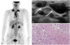
After 4 years, she complained of a palpable right neck mass. F-18FDG PET/CT was performed for the recurrent melanoma evaluation. A F-18FDG PET/CT showed markedly increased FDG uptakes (SUV max: 12.4) in the bilateral neck, mediastinum, hila, left parasternal, and abdominal lymph nodes, which were suspicious for metastases (Fig. 1A).
Plain chest X-ray and PET/CT of both lungs showed normal.
Ultrasonography (US) showed multiple enlarged cervical lymph nodes considered to be suspicious for metastasis (Fig. 1B), and US guided fine needle aspiration was performed for diagnosing the cause of cervical lymphadenopathy.
Cytology result was negative for malignancy, and immunocytochemistry result for HMB45 that reacts against an antigen present in melanomas was also negative. Therefore, excisional lymph node biopsy was performed to discover the reason behind the image-pathologic discordance.
Excisional lymph node biopsy for cervical lymph node showed chronic granulomatous lymphadenitis with caseous necrosis (Fig. 1C) and tuberculosis polymerase chain reaction was positive, compatible with tuberculosis lymphadenitis. Finally, tuberculosis lymphadenitis was diagnosed by excisional biopsy.
DISCUSSION
F-18 FDG PET/CT has been useful in the evaluation of malignant disorders and has been extensively used in cancer screening.1 However, F-18FDG uptake is not specific for cancer diagnosis and there have been reports of benign diseases being observed with F-18FDG uptake such as infection and inflammation, particularly granulomatous diseases.4,5
Because of the high glucose use in granulomatous diseases, false positive FDG uptake in patients with tuberculosis is expected to some extent. A few cases with increased FDG uptake of tuberculous lymphadenitis have been reportedin various anatomical locations in patients with previous cancer history, with those uptakes being misdiagnosed as malignant diseases.1,6,7 However, our case was very unusual in that it was very difficult to consider tuberculosis lymphadenitis as a first impression. First, markedly increased FDG uptakes were observed in the extensive organs such as neck, mediastinum, and abdominal lymph nodes, a pattern unlike other reported cases.1,6,7 Another confounding factor was the patient's past history; the diagnosis and treatment of malignant melanoma on the left sole of the foot. Malignant melanoma is a highly malignant tumor and about one third of cases end up suffering from distant metastasis.8 Moreover, frequently affected organs for distant metastases are lymph nodes.8 Fine needle aspiration biopsy of lymph node is a simple and accurate diagnostic method, however, it sometimes gives a negative result for only malignancy instead of a specific benign pathology.9 In such situations, further diagnostic biopsies such as core needle biopsy or excisional biopsy should be considered for accurate diagnosis.9
Tuberculous lymphadenitis can show increased FDG uptake on F-18 FDG PET/CT and images similar to multiple enlarged lymph nodes considered to be suspicious for metastasis on US. Therefore, tuberculosis should also be considered in the differential diagnosis of increased FDG uptake in patients with previous cancer history.
 XML Download