

 PDF
PDF ePub
ePub Citation
 Print
Print


INTRODUCTION
The prognosis of advanced colorectal cancer and of recurrent disease has improved over the last ten years due to improvements in systemic therapies; for example, overall survival in metastatic colorectal cancer has increased from 12 to 20 months over the past decade.1 Many chemotherapeutic agents have been introduced to treat advanced colorectal cancer, and patients with advanced disease are now being managed more aggressively. As a result, physicians now are able to concentrate more on the planning of treatment strategies. In palliative chemotherapy, patients are evaluated based on their response to treatment at certain points in time, and thereby, decisions concerning the continuation of treatment are made. Traditionally these decisions have been made with the aid of conventional imaging and physical examination findings and symptoms.
In practice, the main objective of disease management is to identify disease progression during therapy at the earliest opportunity and to adjust treatment accordingly. Some studies have been undertaken to explore the efficacy of carcinoembryonic antigen (CEA) monitoring for assessing disease status as a substitute for conventional imaging studies, but consensus has not been reached regarding the merits of this approach. Accordingly, this study was undertaken to evaluate the effectiveness of CEA monitoring in discerning disease status. In particular, we analyzed relations between tumor marker levels and computed tomography (CT) findings in terms of sensitivity, specificity, positive predictive value, negative predictive value, and diagnostic accuracy.
MATERIALS AND METHODS
Patients
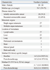
Between January 2005 and December 2007, 48 patients with initially unresectable metastatic colorectal cancer (n=26, 54.2%) or recurrent unresectable metastatic colorectal cancer (n=22, 45.8%) underwent palliative chemotherapy at our institute. Medical records were retrospectively reviewed. Twenty-eight patients were male and twenty were female. Median patient age was 58.5 years (range 33-75). Twenty-seven of the patients had single organ metastasis and twenty-one had more than two. Twenty-seven had lymph node metastasis, twenty-three liver metastasis, fifteen lung metastasis, three peritoneal carcinomatosis, one bone metastasis, one adrenal gland metastasis, and one brain and scalp metastasis. Before treatment, median CEA level was 16.5 ng/mL and median carbohydrate antigen (CA) 19-9 level was 31.7 IU/mL. The demographic and clinical characteristics of the patients are shown in Table 1.
Treatment
All patients received oxaliplatin, folinic acid, and 5-fluorouracil (FOLFOX-4) chemotherapy. On day 1, oxaliplatin 85 mg/m2 and folinic acid 200 mg/m2 were intravenously injected over 120 minutes as a mixed infusion, followed by a bolus of 5-FU (400 mg/m2, i.v.) and 5-FU (600 mg/m2, i.v. over 22 hours). On day 2, folinic acid 200 mg/m2 was infused over 120 minutes and followed by a bolus of 5-FU (400 mg/m2) and 5-FU (600 mg/m2 continuous infusion over 22 hours). This cycle was repeated every two weeks unless disease progression or unacceptable toxicity occurred.
Assessments of tumor response
Measurement of tumor markers and response evaluation
Serum CEA levels and CA 19-9 levels were measured using an immunoradiometric assay. Baseline tumor marker levels were measured prior to chemotherapy and were followed-up after 3 cycles of chemotherapy, following the guidelines set by the National Health Insurance Corporation of Korea. The reference ranges for CEA and CA 19-9 were 0-5.0 ng/mL and 0-37 IU/mL, respectively. Serum levels before and after chemotherapy were compared. Complete response (CR) was defined as the normalization of tumor marker level to within the reference range. Partial response (PR) was defined as at least a 30% decrease in tumor marker level from baseline, and progressive disease (PD) was defined as at least a 20% increase. Other responses were defined as stable disease (SD).
Response evaluation by CT
CT was performed prior to chemotherapy and after 3 cycles of chemotherapy. Tumor responses were evaluated according to the Response Evaluation Criteria for Solid Tumors (RECIST) criteria.2 CR was defined as the disappearance of all target lesions. PR was defined as at least a 30% decrease in the sum of the diameters of target lesions, in reference to the sum of baseline diameters. PD was defined as at least a 20% increase in the sum of the diameters of target lesions, which also demonstrated an absolute increase of at least 5 mm, taking as reference the smallest sum observed. Other responses were defined as SD.
RESULTS
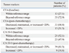
Serum CEA and CA 19-9 levels were measured at baseline in all 48 patients before starting chemotherapy. Initially, CEA was elevated in 35 (72.9%) and CA 19-9 in 23 (47.9%). All 23 in the latter group exhibited elevated CEA and CA 19-9 levels. After three cycles of chemotherapy, serum CEA and CA 19-9 levels were re-measured. CEA levels decreased, were maintained, or increased by <20% versus baseline in 33 patients, and increased by >20% in 15. CA 19-9 levels decreased, were maintained, or increased by <20% in 39 patients, and increased by >20% in 9. Changes in tumor marker levels are summarized in Table 2.
According to CT findings, 24 patients, 10 patients and 14 patients showed PR, SD, and PD after chemotherapy. No patient showed CR.
CEA assessment
According to CEA assessment, 14 of the 48 (29.2%) patients showed a complete or partial response (CR+PR) to treatment and 15 (31.3%) showed PD. According to the CT assessment, 24 (50.0%) exhibited a complete or partial response (CR+PR) to treatment and 14 (29.2%) showed PD. A breakdown of the results is presented in Table 3.
The sensitivity, specificity and diagnostic accuracy of CEA measurement for predicting disease progression (PD) were 50.0%, 76.5%, and 68.8%, respectively. The accuracy of CEA measurement for predicting complete or partial response (CR+PR), non-progression (CR+PR+SD), and disease progression (PD) is presented in Table 4.
When the patients were dichotomized into the initially normal CEA group and initially elevated CEA group (defined as a CEA level of >5.0 ng/mL), the sensitivity and diagnostic accuracy for predicting disease progression were higher in the initially elevated CEA group (sensitivity=66.7% vs. 20.0%; diagnostic accuracy=71.4% vs. 61.5%). The accuracy of CEA measurement for predicting disease progression according to the initial CEA level is provided in Table 5.
CA 19-9 assessment
According to CA 19-9 assessment, 8 (16.7%) of the 48 patients showed a complete or partial response (CR+PR) to treatment and 9 (18.8%) showed PD. Cross comparisons of CA19-9 assessment and CT assessment results are provided in Table 6.
The sensitivity and specificity of CA 19-9 measurement for predicting complete or partial response (CR+PR, determined by CT) were 29.2% and 95.8%, respectively; the diagnostic accuracy of CA 19-9 measurement for predicting response (CR+PR, by CT) was 62.5%. The sensitivity and specificity of CA 19-9 measurement for predicting disease progression (PD, by CT) were 28.6% and 85.3%, respectively; the diagnostic accuracy for predicting disease progression (PD, by CT) was 68.8%. A summary of the above is provided in Table 7.
Combined analysis of CEA and CA 19-9
For the combined analysis of CEA and CA 19-9, responses were defined as CR or PR for both tumor markers. Disease progression was defined as PD for at least one tumor marker. Accordingly, 5 (10.4%) of the 48 study subjects responded to treatment and 17 (35.4%) showed disease progression (PD). Cross comparisons of the combined CEA and CA 19-9 assessment with CT assessment are shown in Table 8 and 9.
The sensitivity and specificity of the combined CEA and CA 19-9 assessment for predicting response (CR+PR) as padetermined by CT were 20.8% and 100%. The positive predictive value and diagnostic accuracy of the combined CEA and CA 19-9 assessment for predicting response (CR+PR, by CT) were 100% and 60.4%. Its sensitivity and specificity for predicting disease progression (PD, by CT) were 50.0% and 70.6%, respectively, and its positive predictive value and diagnostic accuracy for predicting disease progression (PD, by CT) were 41.2% and 64.6%. The above results are summarized in Table 10.
DISCUSSION
Many treatment options have been proposed for metastatic colorectal cancer.3-7 Nowadays patients with surgically unresectable metastatic colorectal cancer have a greater chance of being treated with chemotherapeutic agents, and, as a result, these patients exhibit substantially greater survivals than comparable patients of a few decades ago. The proper evaluation of tumor responses is important to maximize the therapeutic effects and to minimize the adverse effects of these chemotherapeutic agents. To date, tumor response evaluations have been generally performed using imaging studies, such as CT scans or MRI, according to World Health Organization (WHO) response criteria8 or the RECIST criteria.2 Further to imaging modalities, tumor markers could be used if they are proved to accurately reflect tumor responses.
CEA level in colorectal cancer is known to be well associated with preoperative tumor extent, prediction of tumor outcomes, and recurrence.9 However, concerning the role of CEA in assessing tumor responses to chemotherapy, no consensus has yet been reached, although some authors have examined the efficacy of CEA monitoring for the evaluation of tumor response in palliative chemotherapy.9-14
Ward, et al.10 measured the levels of the tumor markers of CEA, CA-195, and CA-242 in 33 patients undergoing 5-FU-based chemotherapy for metastatic colorectal cancer to determine whether these markers could be used to accurately monitor disease course and reduce the need for imaging. For the CEA assessment, they defined a positive response as a decrease in CEA level of >15% from baseline, and PD as an increase in CEA level of >15% from baseline. Before treatment, CEA was found to be elevated in 85% of the patients. The WHO criteria were used for the imaging study. In this previous study, the authors compared CEA levels and CT scans as well as calculated the sensitivity, specificity, and positive predictive value of CEA measurement for predicting tumor response, which were 100%, 79%, and 77% for predicting positive response, respectively, and 74%, 100%, and 100% for predicting PD. According to their analysis, a fall in CEA level was highly sensitive for predicting positive responses, although its specificity was low, and a rise in CEA was found to be highly specific for predicting progressive disease. They suggested although falling levels of markers overestimate tumor response, rising tumor markers invariably heralded disease progression, and thus, they concluded that CEA is useful for monitoring patients treated for advanced colorectal cancer.
Wang, et al.9 measured CEA levels in 136 patients with metastatic colorectal cancer undergoing oral tegafur-uracil- or intravenous 5-FU-based systemic chemotherapy regimens. To assess CEA level, they defined complete responses as a normalization of CEA level within the reference range (<5.0 ng/mL), partial responses as a reduction of >50%, and progressive disease as an increase of >50%. Pretreatment CEA was found to be elevated in 110 patients. For the imaging study, they used WHO criteria. Sensitivity, specificity, and the positive predictive value of CEA measurement were 72%, 86%, and 53% for predicting positive response, and 81%, 88%, and 85% for predicting PD. They concluded that CEA might be helpful for monitoring chemotherapeutic response in clinical practice, and that a change in CEA level better predicts progressive disease than positive response.
In the present study, we evaluated the usefulness of tumor marker assessment for predicting tumor response after oxaliplatin-based chemotherapy by comparing tumor marker levels and CT findings. Our finding of a sensitivity of 76.5% for predicting non-progression (CR+PR+SD) was comparable to the findings of previous studies. However, we found that CEA measurement had a lower sensitivity and lower positive predictive value for predicting disease progression (PD) than for predicting non-progression (CR+PR+SD), which was the opposite of that found in a previous study.9
In our study, the sensitivity of CEA measurement for predicting progressive disease was relatively low (50.0%) in the overall patients. However, when we categorized the patients into two groups according to baseline CEA level, there was a noticeable distinction between the two groups. The sensitivity of CEA measurement for predicting disease progression was very low in the initially normal CEA group, while it was comparatively high in the initially elevated CEA group (20.0% vs. 66.7%). In brief, the results of the present study suggest that CEA measurement can be useful for monitoring tumor progression during palliative chemotherapy in only patients with initially elevated CEA level.
Our analysis of the use of CA 19-9 levels showed an even lower sensitivity and a lower positive predictive value for predicting progressive disease than CEA levels. In one report, it was concluded that available evidence is insufficient to warrant the use of CA 19-9 for monitoring treatment in colorectal cancer.15 Our data also suggest that CA 19-9 monitoring has less merit than CEA monitoring for determining treatment response. Our analysis also revealed that progressive disease was underestimated in the assessment of CEA and CA 19-9 in combination.
Several authors have investigated the efficacy of CEA monitoring for the evaluation of tumor responses in palliative chemotherapy.9-14 Many clinicians hope that CEA monitoring can play a role in the evaluation of tumor response in daily clinical practice, because it has obvious advantages over imaging studies, which include cost, convenience, and the possibility of evaluating total tumor burden. Some authors have suggested that tumor marker monitoring could be employed as a primary evaluation tool before imaging studies, because tumor markers are sufficiently sensitive.10 Moreover, others have suggested that CEA monitoring might be helpful for determining tumor response when tumor burdens are difficult to assess by imaging.9
The present study demonstrated the usefulness and the limitations of CEA monitoring for the evaluation of tumor response during palliative chemotherapy. Based on the findings of the present study, we suggest that CEA monitoring is only likely to be useful in patients with an initially elevated CEA level, and that its use may result in the underestimation of disease progression in patients with an initially normal CEA level. Regarding the reason why CEA monitoring underestimates disease progression in patients with an initially normal CEA level, the heterogeneity of CEA expression in colorectal cancer should be considered. Although many colorectal cancers, up to 90% of primary colorectal cancers, produce CEA, some do not show an elevated CEA level at diagnosis.16 Furthermore, CEA levels in circulation are known to be dependent on several factors, such as tumor grade, liver status, tumor site, and tumor ploidy status.16,17 Accordingly, when a factor associated with a low CEA level is present, pre-treatment CEA and post-treatment CEA levels could be normal. Consequently, in cases with an initially normal CEA level, the post-treatment CEA level could remain normal despite disease progression, and thus, disease progression would be underestimated based on CEA considerations alone.
Summarizing, the present study revealed the merits and demerits of CEA monitoring during oxaliplatin-based palliative chemotherapy for colorectal cancer. For patients with an initially elevated CEA level, CEA monitoring was found to be useful for evaluating tumor progression. However, for patients with an initially normal CEA level, CEA monitoring was found likely to underestimate disease progression and should not be considered sufficient for evaluating tumor response.


 XML Download