

 PDF
PDF ePub
ePub Citation
 Print
Print


INTRODUCTION
Hemophagocytic lymphohistiocytosis (HLH) comprises the inherited form, primary HLH, and secondary HLH, which usually is virus or malignancy associated. Primary HLH often occurs in infancy and includes familial hemophagocytic lymphohistiocytosis (FHL) and the immune deficiency syndromes associated with HLH (Chediak-Higashi syndrome, Griscelli syndrome, and X-linked lymphoproliferative syndrome).1 FHL is a potentially lethal disorder of immune dysregulation that requires prompt and accurate diagnosis to initiate life-saving immunosuppressive therapy and to prepare for hematopoietic stem cell transplantation.2 Five subtypes of FHL have been identified so far, with mutations in the following genes: unknown (FHL1), PRF1 (FHL2), UNC13D (FHL3), STX11 (FHL4), and STXBP2 (FHL5), respectively.3-6 Functional tests with resting NK cells and/or CTL cells, including perforin detection and degranulation assay quantifying CD107a surface expression with flow cytometry, can direct genetic analyses. Perforin expression is constantly reduced or absent in most FHL2 patients, while patients with FHL3, FHL4 or FHL5 can be screened by a degranulation assay result below 5%.7 In this paper, we describe the case of a Chinese male neonate who presented with HLH and had one identical, novel, heterozygous mutation in the UNC13D gene.
CASE REPORT
An eight-day old Chinese neonate presented with progressive thrombocytopenia and was admitted to our hospital on February 15, 2011. His body temperature fluctuated around 38.5℃. At the time of admission, his liver and spleen had enlarged 3 cm and 4 cm below his costal margins, respectively. No rashes were noticed on his skin. Auscultation on his lungs and heart was normal. No CNS symptoms and signs were found. The following laboratory data were recorded: WBC of 7.65×10⁹/L; Hb of 129g/L; platelet count of 18×109/L; feritin >1500 ng/mL; bilirubin total/direct 120.3 µmol/L/32.6 µmol/L; AST of 76 IU/L; ALT of 33 IU/L; fibrinogen of 1.24 g/L; PT/APTT of 12.8/41.8 sec; thrombin time of 17.7 sec; D-dimer of 563 µg/L; triglyceride of 2.24 mmol/L and an LDH of 617 IU/L. Abdominal B-mode ultrasound examination showed massive hepatosplenomegaly. A bone marrow biopsy revealed numerous hemophagocytic histiocytes consistent with a diagnosis of HLH.8 The parents of this patient also had given birth to a daughter two years before, who died several hours after her birth because of "suspected infection".
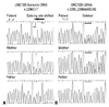
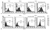
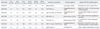
FHL-related gene analysis using genomic DNA showed that the patient had novel, compound heterozygous mutations in the UNC13D gene, including c.-250C>T, c.1+30G>A, c.279C>T, c.888G>C9, c.18+36A>G, c.20-48T>C, c.1977C>T, c.2296CA>T (Fig. 1A), c.24-46C>T, c.26-9_26-8insC, c.2599A>G, c.28+48C>T and c.3198A>G. Gene analysis using cDNA with a pair of primers for UNC13D-Sequence Coding for Amino Acids in Protein (CDS). showed a frameshift mutation of (c.2295_2298del GCAG, p.Glu765Aspfs*27) (Fig. 1B) in UNC13D, which was consistent with the genomic DNA sequencing results for c.2296C>T, as the splicing site shifted in the transcription process based on the "GT-AG" splicing law. These genomic DNA and cDNA sequencing results for the patient were confirmed by the sequencing results of his parents. We also sequenced other FHL-related genes for this patient and his parents, including PRF1, STX11 and STXBP2, but no mutation was found except for one synonymous change in PRF1 gene (c.900C>T, p.His300His). In the acute phase of HLH, the perforin expression percentage with resting NK cell by flow cytometry of this patient was 14.81% (normal range 70.57±21.32%) (Fig. 2A) and surface CD107a level after K562 stimulation was 0.51% (normal range 14.1±7.2%) (Fig. 2B), both of which were significantly reduced. However, when his condition of HLH was controlled, his level of perforin expression returned to normal (55.72%), whereas his damaged function of degranulation never recovered on repeated testing (never over 1%). Th1/Th2 cytokine levels, quantitatively determined by the cytometric bead array (CBA) kit-BD™ CBA Human Th1/Th2 Cytokine Kit II (BD Biosciences, San Jose, CA, USA), were successively monitored during the course of the disease in this patient. We also detected bacteria and viruses in his blood, with almost all negative results except for EBV-VCA IgG (Table 1). Bacteria and viruses were also detected in his parents, and all were showed negative results.
He received HLH-2004 chemotherapy, including etoposide (VP-16), dexamethasone, cyclosporine A and intrathecal methotrexate, and responded well. Four days after initial administration, his platelets began to rise steadily and the spleen gradually shrank back to its normal position. During the non-hospitalized period, he was followed-up mainly by telephone. Although an intensive search for a HLA-matched donor among candidates in the Chinese Hematopoietic Stem Cell Bank with over 1.3 million donors was conducted, no proper donor was found. The patient remained on the immunosuppressive therapy until his death in October 2011 because of gastrointestinal hemorrhage.
DISCUSSION
HLH in different ethnicities has been described in the literature, but few patients of Chinese descent have been reported.1 Yoon, et al.10 reported that FHL3 accounted for 89% of cases of FHL in Korea, and approximately 20% to 25% in Japan, indicating that UNC13D mutations may be responsible for a large part of HLH patients in Asian countries. Here, we clearly showed a neonate with HLH harboring compound heterozygous mutations (c.2295_2298delGCAG, p.Glu765Aspfs*27) in exon 23 of the UNC13D gene by bidirectional sequencing. So far as we know, such mutation in UNC13D has never been reported in the literature. In many published reports, compound heterozygous mutations were common in FHL patients. Although FHL belongs to the autosomal recessive form of the disease, whether an autosomal dominant trait in FHL patients exists or not remains to be defined. Our patient showed a clear heterozygous mutation, and family history of a sister who died previously may be partial evidence for the possibility of such a genetic defect.11 Results on degranulation assay constantly below 5% also suggested that the patient had a diagnosis of FHL.7 The mutations in the UNC13D gene of this patient were all inherited from his mother, yet the mother had no presentations of HLH with a mild reduction of degranulation function (4.76%), indicating that EBV infection plays an important role in FHL onset. However, due to the fact that our gene analysis was carried out using conventional sequencing of exons and splice-sites, possibilities of deep intronic mutations and other genetic aberrations cannot be definitely excluded. Another interesting question is that the neonate presented with a low level of perforin expression with NK cells in the acute phase of his disease. However, when his condition of HLH was controlled, his level of perforin expression returned to normal. These results indicate that the abnormality of perforin expression in NK cells is not a de novo defect, but rather a temporary phenomenon.
A specific Th1/Th2 cytokine pattern to diagnose HLH was first reported by our group with excellent clinical application.12,13 During the acute phase of HLH without complicating bacterial infection, both IFN-γ and IL-10 levels should be increased, while IL-6 remains normal or only slightly elevated. When HLH patients enters remission, the levels of IFN-γ and IL-10 both decline, usually with IFN-γ declining faster than IL-10. According to our previous studies, IL-6 levels are much higher in microbiologically documented infection patients than in patients with non-sepsis.13 When IFN-γ, IL-10 and IL-6 levels return to normal or close to normal, the HLH related conditions of the patient can be considered to be in control. In this regard, we were able to successively monitor Th1/Th2 cytokine levels in the present patient during the course of HLH, e.g. acute phase of HLH, exaggerated status of HLH, relieved status of HLH and controlled HLH, indicating that the cytokine patterns were consistent with HLH clinical phases. Cytokine profile monitoring could be an important biomarker for monitoring HLH status and allow us to make an earlier diagnosis through which to administer more effective treatment.
 XML Download