

 PDF
PDF ePub
ePub Citation
 Print
Print


INTRODUCTION
In the past, radiologic studies of joint disorders focused mainly on the static morphologic depiction of joint internal derangements. However, some joint disorders may not show definite abnormalities in a static radiologic study, but will still have dormant abnormalities that are aggravated with joint movement, which triggers the need for radiologic imaging of dynamic joint movement. The wrist joint in particular requires four-dimensional (4D) dynamic joint imaging because the wrist is an exceedingly complex and versatile structure, consisting of a radius, ulna, eight carpals, and five metacarpals all engaged with each other. Each of these carpal bones exhibits multiplanar motion involving significant out-of-plane rotation of bone rows, which is prominent during radio-ulnar deviation. The kinematics of these carpal bones have been not fully elucidated.1 Thus, studies using 4D wrist imaging were conducted to determine the proper modality and to investigate carpal kinematics.
Videofluoroscopy and plain radiography are used to diagnose midcarpal instability,2-4 but their diagnostic ability is limited due to their relatively low resolution, which can be problematic when diagnosing overlapping carpal bones. Ultrasound is useful for the detection of scapholunate ligament tears,5,6 but its utility in evaluating carpal instability is unproven.1 Magnetic resonance imaging provides accurate anatomical images of the main extrinsic ligaments in palmar midcarpal instabilities,7 but its image acquisition time is not appropriate for dynamic joint imaging. Multi-detector CT (MDCT) has potential for dynamic 4D joint imaging due to recent advances in CT technology resulting in a relatively short image acquisition time.8 This can provide early detection and insights into the functional pathophysiology of carpal instability. Many studies have been performed with CT to validate functional joint imaging of the wrist.9-11 Assuming that there is acceptable spatial resolution, shorter temporal resolution improves dynamic images and makes them more realistic. To date, temporal resolution has been reduced to 0.5 seconds with 256-MDCT.11
The dual source CT has two detectors arranged at a 90 degree angle to each other, rotating at the same time. Having two detectors can reduce the minimum detector rotation angle required for image acquisition, which in turn improves temporal resolution.12 The z-coverage length of a 128-channel dual source CT is 3.8 cm, which is shorter than that of higher channeled MDCT, but seems to be enough to cover eight carpal bones. It was assumed that improved dynamic wrist images with shorter temporal resolution could be obtained using dual source CT. The purpose of this study was to evaluate the feasibility of real-time kinematography with 4D dynamic functional wrist joint imaging using dual source CT.
MATERIALS AND METHODS
This study was approved by the institutional review board and informed consent was obtained. Two healthy young female volunteers (26 and 27 years old) were enrolled in the study. They practiced radioulnar deviation and pronation-supination of the wrist joint, which was later recorded using dynamic imaging with an optimized dual-source CT protocol in order to validate the feasibility of real-time kinematography.
Volunteer education and positioning
Before scanning, volunteers were taught how to make slow and continuous radioulnar and supination-pronation hand movements during a scan time of four or ten seconds. Repetitive exercises were performed in the CT gantry before scanning. The volunteers were positioned prone in the scanner with one hand stretched out forward, and a scan range of 3.8 cm (the maximum z-axis coverage of the CT scanner during one rotation) was selected to cover the carpal bones. Patients were informed of scan time by an audible timer, which resulted in even and continuous movements of the wrist during scanning.
CT scanning protocol and image reconstruction
A dual source CT scanner (SOMATOM Definition Flash, Siemens Medical, Forchheim, Germany) was used. The scanner consists of two detectors with 0.28 s and 0.33 s rotation time. The z-axis coverage length was 3.8 cm with 128 slices [64 (128)×0.6 cm=3.8 cm]. The optimal temporal resolution can reach up to 75 ms, which is equivalent to one fourth of the minimum gantry rotation time (0.28 s).12
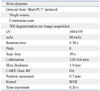
The scan protocol is presented in Table 1. For improved depiction of real carpal bone movement, the scan protocol was optimized and focused on temporal resolution rather than spatial resolution. To minimize radiation exposure, parameters related to spatial resolution, were reduced to minimum requirement for 3D carpal bone reconstruction, which were 100 kv and 80 mA for scanning with CARE Dose 4D on.
Conventional continuous scan mode without table increments (pitch value of 0) was chosen for optimal temporal resolution, which was modified from the premade body perfusion CT protocol. However, dual source scanning and a smaller rotation angle less than 360 degrees per image acquisition were not available in conventional scan mode due to hardware pre-settings. Dual source is available only with fixed table increments for cardiac imaging protocol. Thus, it was inevitable to use only one detector among dual detectors, and 360 degree of rotation per image acquisition. To achieve better temporal resolution, the detector with 0.28 s rotation time was chosen as single source, instead of the detector with 0.33 s rotation time. The "series splitting" option was selected when scanning for image reconstruction.
Kinematic image reconstruction was performed using 3D reconstruction software (Inspace, Siemens). The series splitting function was used to reconstruct one snapshot of motion 3D kinematic animation. Reconstruction also focused on temporal resolution to depict real carpal movement as much as possible with minimal gaps between the motions. Among the parameters used in reconstruction, time increments were adjusted to maximize the temporal resolution. A time increment is defined as the interval from the scan starting point of one series to that of the next series (Fig. 1). Time increments on the scanner used in this study can be set at 0.05 s intervals, such as 0.10 s, 0.15 s, 0.20 s and so on. To minimize the gap between motions and make the dynamic images smoother, time interval 0.3 s was chosen which is the closest value to the temporal resolution of 0.28 s.
RESULTS
Cine images of radioulnar deviation and pronation-supination movement were captured (Figs. 2, 3 and 4). Two series of images were produced according to the complete cycle of joint movement over 4 s and 10 s. Thirty-three images were obtained in 10 s during one motion cycle and ten images were obtained in 4 s during another motion cycle. Initial images were obtained with a joint movement cycle of 10 s. By combining the benefits of shorter temporal resolution (0.28 s) and theoptimumtime interval (0.30 s), the scan time was gradually reduced to the minimum time required to obtain acceptable motion images.
The motion images of one radioulnar and pronation-supination motion cycle over 10 s (Supplementary video 1 and 2) show smooth stream of movement with the interaction between individual carpal bones clearly depicted. When images are obtained over 4 s, the margin and shape of carpal bones and their interaction during motion are well-visualized in the images of radioulnar deviation (Supplementary video 3). However, the carpal bones in the images of radioulnar deviation are not fully covered in 3.8 cm of z-coverage length. This is due to the unintended forearm movement that accompanies wrist movement, which is partially caused by the greater difficulty of isolated wrist movement in 4 s compared to 10 s. The quality of the image showing pronation-supination over 4 s (Supplementary video 4) was lower than the images showing radioulnar deviation over 4 s due to motion artifact. And the margin and contour of the carpal bones was blurred.
Previous studies using 256 MDCT and this study using dual source CT are compared in Table 2. Compared to the previous study using 256 MDCT, we used dual source CT and obtained dynamic wrist images with improved temporal resolution, reduced to 0.28 s. Radiation dose was also reduced in our study to 0.48 mSv in 10 second images and 0.19 mSv in 4 second images, compared to 0.5 mSv radiation dose of the previous study.
DISCUSSION
4D kinematic images of carpal movement were obtained using dual source CT with a temporal resolution of 0.28 s in one detector, and an optimal time increment of 0.3 s. Although this study was unable to reduce temporal resolution to 75 ms with simultaneous utilization of two detectors, temporal resolution still decreased to about half of that reported in previous studies.9,11
The wrist is an exceedingly complex and versatile structure, consisting of a radius, ulna, eight carpals, and five metacarpals engaged with each other. The study of carpal kinematics, which investigates this dynamic inter-carpal positional relationship, dates back to the late 19th century. However there is still no theory or model that fully explains wrist anatomy and function.1 According to carpal ring theory, which is one recognized theory of carpal kinematics, the eight carpal bones can be divided into proximal and distal carpal bones that form a ring. Each bone in the ring exhibits multiplanar motion involving significant out-of-plane rotation of bone rows.13 Especially during radio-ulnar deviation, the proximal carpal bones not only move in the radio-ulnar plane, but also in the flexion-extension plane.1 In addition to multiplanar motion, hysteresis is an another characteristic of carpal motion. Hysteresis occurs when the specific motion of an individual carpal bone varies slightly depending on the direction in which the wrist moves.14 Thus, evaluating the real carpal movement in three-dimensional images of a moving carpal bone is challenging.
Other than depicting normal carpal kinematics, 4D imaging of the wrist also has clinical potential for visualization of dynamic pathophysiology. In diseases related to carpal motion, such as dynamic carpal instability, diagnoses are made based on clinical history and signs because there are no accepted diagnostic imaging criteria.15,16 The diagnosis of ulnar impaction is also made based on clinical symptoms and ulnar positive variance on a plain radiograph.17,18 On the other hand, dynamic wrist imaging can provide direct evidence of pathology that only occurs temporarily during motion, such as abnormal arrangement of carpal bones or impaction of the ulnar styloid process during motion.
Thus, many studies have been performed with CT to validate functional joint imaging of the wrist. Sun, et al.9 measured the continuous motion of the wrist from full flexion to full extension using two-dimensional CT images. However, 2D reconstruction cannot be applied to radio-ulnar deviation of the wrist, which shows multiplanar movement. Tay, et al.10 performed functional joint imaging of a cadaveric wrist using retrospectively gated spiral CT, in which the cadaveric wrist was mounted on a motion stimulator setup for 30 cycles of radioulnar deviation of the wrist per minute. This study has limitations in vivo because of the poor reproducibility of 30 cycles of wrist movement per minute, as well as the hysteresis of carpal movement. Functional joint imaging using 256-MDCT has also been performed in healthy volunteers by Kalia, et al.11 In that study, the temporal resolution of the moving wrist image was 0.5 s, equal to the gantry rotation time of CT. Images were acquired without any apparatus, during 10 seconds of one complete cycle of joint motion, which seems to be much slower than the typical carpal motion in daily life.
This is the first study that used in vivo functional imaging of wrist joint movement to obtain multiple images in a short scan time. Although dual source scanning was not possible, 0.28 s gantry rotation time provided smooth and flawless functional imaging compared to recent images obtained using 256-MDCT. Thirty-three series were obtained in 10 s during one motion cycle and ten series were obtained in 4 s during another motion cycle. One series was scanned with a temporal resolution of 0.28 s, which is equal to the rotation time of the detector. Kalia, et al.11 used the most advanced kinematographic scanning technique and obtained ten images in 10 s of one motion using 256 MDCT, with a temporal resolution of 0.5 s and a radiation dose of 0.5 mSv. The radiation dose was also reduced in our study, especially in 4 s motion images, to less than a half (0.19 mSv) compared to the previous study using 256 MDCT. A remarkably shortened scan time, such as 4 s of radioulnar motion, would significantly increase patient compliance, and consequently, guarantee the reproducibility of joint motion and allow to reduce radiation dosage (Table 2).
Volunteers were better able to follow directions with 4 s scanning than 10 s scanning, regardless of radioulnar motion or pronation-supination motion. Successful functional joint imaging relies on the accuracy and reliability of captured joint kinematics. In terms of reliability, it is advantageous to reduce a motion cycle from 10 s to 4 s.
Four seconds of supination-pronation yielded reduced image quality due to motion artifact when compared to 10 s of radioulnar deviation, 10 s of supination-pronation, and 4 s of radioulnar deviation. This is because of the relatively longer distance of individual carpal bone movement in supination-pronation than in radioulnar deviation. Considering the reasonable image quality of 10 s pronation-supination capture images, scan time might be further reduced from 10 s to 4 s.
There are several limitations of this study in terms of validating functional carpal joint imaging using dual source CT. First, the z-axis is limited to 3.8 cm. Although this was sufficient to cover carpal bones in most cases, it is necessary to use an experienced operator who sets an appropriate scan range when prescribing a topogram to cover all carpal bones during the scan. Second, the assessment of image quality in this study was quite subjective. This study focused on the initial experience of dynamic wrist image acquisition using dual source CT, and is limited in further analysis of acquired dynamic images. Several methods have been suggested to quantify the complicated movement of carpal bones,19-22 and further investigation is required for quantification and analysis of carpal kinematics. Third, in conventional scan mode, hardware limitations prevent simultaneous dual source scanning and acquisition at less than a 360 degree angle of rotation per image (these hardware settings are available only in cardiac mode). With utilization of a dual source scanner and reduced rotation angle, temporal resolution will be reduced to 75 ms. Temporal resolution is the principal factor to reproduce realistic images of joint movement. MDCT with faster gantry works also for better temporal resolution, but it may be easier to reduce temporal resolution by half with a reduced rotation angle and dual source scan, resulting in a 90 degree minimum rotation angle per image. Thus, dual source CT has future potential for use in functional joint imaging.
In conclusion, using one detector of dual source CT, acceptable 4D cine images of carpal bone movement were obtained in a shorter scan time with reduced radiation exposure than in previous studies. This is a meaningful step toward depiction of normal carpal kinematics and dormant abnormality in dynamic carpal instability.


 XML Download