

 PDF
PDF ePub
ePub Citation
 Print
Print


INTRODUCTION
It is a common assumption that auras provide clues to localize focal seizure onset.1,2 Patients with partial epilepsy who have certain structural abnormalities, which are determined using magnetic resonance images (MRIs), have a high probability that their seizures arise from the lesions or their adjacent regions, especially when the MRI findings correlate with electroencephalography (EEG) results.3-5 Several studies have already correlated aura symptoms with definite seizure onset zones, especially in patients who have had prolonged EEG monitoring evaluations or surgical treatments.1,2,6-10 Since most of the patients in these studies had drug-resistant epilepsy, the generalizations that were made based on the data that were acquired in these highly selective patients become questionable when applying them to the all partial epilepsy patients, although the localization and lateralization of the epileptic focus may be accurate. The values as well as the features of auras for localizing and/or lateralizing epileptic focus in lesional epilepsy patients were investigated in this study, according to the epilepsy localization and lateralization on an outpatient basis.
MATERIALS AND METHODS
Study population
Patients were identified who had visited the outpatient epilepsy clinic of Severance Hospital between 2000 and 2007 and had lesions confined to a single lobe based on MRI results. All subjects had scalp EEGs and brain MRIs, and they also provided a well-documented history of their symptoms. Subjects were excluded if they met any of the following criteria: 1) were mentally retarded and could not describe their aura symptoms; 2) scalp EEG findings were discordant with lobar localization and lateralization of the lesion, based on the MRI results; 3) had definitely different semiological features according to seizure events suggesting multiple lobar origins of the seizure onset; 4) seizures occurred only when they were sleeping; or 5) had arachnoid cysts in the temporal lobe or only subcortical lesions, based on the brain MRI results. After exclusions were made, a total of 276 partial epilepsy patients with single lobar lesions, based on their MRI results, were enrolled in the study. There were 180 males and 96 females.
Localization and lateralization of lesions
The seizure onset zone was assumed to be at the lesion although the exact seizure onset zone was not confirmed by intracranial EEG evaluations or surgical outcomes. We classified the localization of lesions into mesial temporal, lateral temporal, frontal, parietal, and occipital lobes. The central sulcus and the collateral sulcus were defined as the border between the frontal and parietal lobe and between the mesial and lateral temporal lobe, respectively. According to the localization of lesions, the subjects were considered as having mesial temporal lobe epilepsy (MTLE), lateral temporal lobe epilepsy (LTLE), frontal lobe epilepsy (FLE), parietal lobe epilepsy (PLE), or occipital lobe epilepsy (OLE). The subjects with lesions that are limited to the temporal lobe were regarded as having either MTLE or LTLE, according to the involvement of the mesial temporal structures. The subjects with asymmetrical hippocampal sclerosis (HS) with lateralization of a smaller hippocampus were also included in this study.
Classification of auras
Based on the well-documented history of the symptoms that the subjects recorded, we classified aura symptoms into 13 categories: epigastric auras; autonomic auras; emotional auras; vestibular auras; psychic (experiential) auras; visual auras; somatosensory auras; dysphasic auras; olfactory auras; auditory auras; whole body sensations; cephalic sensations; and a final category that includes symptoms that cannot be categorized into any of the other categories. Dysphasia cannot be a pure subjective sensation (definition of aura) but was included as aura symptom because it is not found in the majority of cases by others. Epigastric auras included nauseous feelings with or without an uprising sensation. Symptoms, including non-specific dizziness without a spinning sensation, headache, pressure on the head, and lightheadedness, were regarded as cephalic sensations, and not as vestibular auras. An urge to urinate, shortness of breath, sialorrhea, vomiting or retching, palpitation, gooseflesh, or generalized cold or warm feeling in the body was considered as an autonomic aura. Symptoms including feelings, thoughts, and distortion of memory, such as déjà vu or jamais vu type illusions, were classified as psychic auras. Psychic auras were subdivided into two categories, which were the memory-related and memory-unrelated categories. Memory-related psychic auras included déjà vu, jamais vu, and flashbacks. Memory-unrelated psychic auras included forced thinking, being in a dreamy state, having the feeling of being absorbed into something, and the feeling of being aware of the thoughts of another person. Symptoms involving emotional elements, such as fear, anxiety, depression, thoughts of dying, unpleasant feelings, feelings of being tired, euphoria, elation, and pleasure, were classified as emotional auras. Emotional auras were also subdivided into two different types of feelings, which were either good feelings or bad feelings. Euphoria, elation, or pleasure were considered as good feelings, and anxiety, depression, unpleasant feelings, the feeling of being tired, or fear were considered as bad feelings. Tingling, pain, or electrical sensations that arose on either side of the body were regarded as a somatosensory aura. If these sensations diffusely arose in the body without showing clear laterality, the sensations were then regarded as whole body sensations. Visual auras included all of the visual phenomena that subjects experienced. Visual auras included visual illusions, elementary visual hallucinations, and complex visual hallucinations. Transient deficit in reaching the target under visual guidance, which is termed optic ataxia, was regarded as a visual illusion. Auras that were classified into the miscellaneous category included symptoms of unexpressible feelings, fuzzy sensations, and premonitions of impending seizures. We also assessed the presence or absence of aura symptoms, as well as the number of auras reported by each subject.
Statistical analyses
We used either the Student's t-test or the analysis of variance (ANOVA) to analyze the continuous variables. The χ2 test or Fisher's exact test was performed in order to analyze the categorical variables, which allowed us to compare the aura characteristics between different epilepsy localization and lateralization, as well as the occurrence of each aura category between the subjects with specific lobar epilepsies and those without. The Spearman's correlation analysis was used to find pairs of closely related aura categories. Strength of correlation was estimated using the Spearman's rank correlation coefficient (ρ). All statistical tests were performed with the Statistical Package for Social Sciences (SPSS) version 18 (SPSS Inc., Chicago, IL, USA). The significance level was defined as p<0.05. This study was approved by the Institutional Review Board of the Severance Hospital.
RESULTS
Of the 276 cases with partial epilepsy that were reviewed, there were 134 cases (48.6%) of MTLE, 55 cases (19.9%) of LTLE, 65 cases (23.6%) of FLE, 17 cases (6.2%) of PLE, and 5 cases (1.8%) of OLE. There were 159 cases (57.6%) of left-sided epilepsy and 117 cases (42.4%) of right-sided epilepsy. One hundred seventy-six (63.8%) subjects experienced at least one aura, and 67 (38.1%) of them reported having two or more aura categories. A total of 271 auras were reported. Age at seizure onset ranged from 1 to 77 years of age (mean 25.8 years).
Suspected pathologies of the lesions that were identified using the MRI were as follows: HS was the most common etiology and was found in 111 (40.2%) of the subjects; atrophic lesions, which were related to trauma, cerebrovascular accidents, central nervous system infections, or were of unknown origin, occurred in 84 (30.4%) subjects; vascular malformations were identified in 26 (9.4%) subjects; foreign tissue in 20 (7.2%) subjects; and malformations of the cortical development in 23 (8.3%) of the subjects.
Frequencies of aura categories
There were 102 (76.1%) subjects who experienced at least one aura in MTLE, 36 (65.5%) in LTLE, 22 (33.8%) in FLE, 11 (64.7%) in PLE, and 5 (100%) in OLE. The average number of aura categories per subject was 1.19±0.09 for MTLE, 1.02±0.13 for LTLE, 0.48±0.10 for FLE, 0.88±0.21 for PLE, and 1.80±0.37 for OLE, respectively. ANOVA with Bonferroni's post-hoc analysis revealed that the number of auras in subjects with MTLE (p<0.001), LTLE (p=0.017) and OLE (p=0.024) was significantly higher than that in subjects with FLE.
Differences in aura characteristics among lobar epilepsies
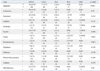
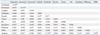
The relationship of 13 aura categories with the 5 types of lobar epilepsy was investigated by the χ2 test or Fisher's exact test (Table 1). Epigastric (p<0.001), autonomic (p=0.030), psychic (p=0.008), and visual (p=0.002) auras showed different distributions among the 5 categories of lobar epilepsy. Olfactory auras occurred only in TLE, of which 4 were MTLE subjects and 2 LTLE subjects. Cephalic sensations, emotional auras, and dysphasic auras did not have a definite localizing value in this study.
The χ2 test or Fisher's exact test analyzing the presence/absence of each aura category between patients with specific lobar epilepsy and those without (Table 1) revealed that somatosensory auras and whole body sensations were more frequent in subjects with PLE than those without. Three (17.6%) of the 17 subjects with PLE reported having somatosensory auras, while only 6 (2.3%) of the 259 subjects without PLE had these types of auras (p=0.013), and 2 (11.8%) of the 17 subjects with PLE had whole body sensations, while only 4 (1.5%) of the 259 subjects without PLE reported having whole body sensations (p=0.046). Epigastric (p<0.001), autonomic (p=0.001), and psychic (p=0.002) auras were more frequent in subjects with MTLE than those without. A total of 34 (25.4%), 19 (14.2%), and 24 (17.9%) of the 134 subjects with MTLE had epigastric, autonomic, and psychic auras, respectively. Ten (7.0%), 4 (2.8%), and 8 (5.6%) of the 142 subjects without MTLE had epigastric, autonomic, and psychic auras, respectively. Visual auras were more common in subjects with PLE (p=0.018) and OLE (p=0.036) than in subjects without PLE or OLE, as 4 (23.5%) of 17 with PLE and 2 (40.0%) of 5 with OLE reported visual auras, while only 14 (5.4%) of 259 subjects without PLE and 16 (5.9%) of 271 subjects without OLE reported having visual auras.
Comparison of aura between subjects with MTLE and those with LTLE
We also compared auras between MTLE and LTLE subjects with the χ2 test or Fisher's exact test. Autonomic auras were significantly more common in subjects with MTLE than those with LTLE (p=0.041), in which 19 (14.2%) of the 134 subjects with MTLE had autonomic auras, while only 2 (3.6%) of the 55 subjects with LTLE had these auras. Although statistical significance was not reached, epigastric (p=0.079) and psychic (p=0.182) auras seemed to be more common in MTLE subjects than in LTLE subjects. A total of 34 (25.4%) and 24 (17.9%) of the 134 subjects with MTLE reported having epigastric auras and psychic auras, respectively, while 7 (12.7%) and 5 (9.1%) of 55 subjects with LTLE reported having epigastric auras and psychic auras, respectively. On the contrary, while statistical significance was not reached, visual (p=0.158) and vestibular (p=0.149) auras seemed to be more common in LTLE subjects, since 5 (9.1%) and 3 (5.5%) of 55 subjects with LTLE had visual and vestibular auras, respectively, while only 5 (3.7%) and 2 (1.5%) of the 134 subjects with MTLE had these types of auras, respectively.
Differences of aura characteristics between the subjects with left-sided and right-sided epilepsy
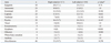
Thirteen aura categories were compared between the subjects with left-sided epilepsy and those with right-sided epilepsy by the χ2 test or Fisher's exact test (Table 2). Dysphasic auras (p<0.001) were more frequently found in subjects with left-sided epilepsy, in which 15 (9.4%) of the 159 subjects with left-sided epilepsy had dysphasic auras, while none of the 117 subjects with right-sided epilepsy had this type of aura. On the contrary, although statistical significance was not reached, vestibular (p=0.102) and olfactory (p=0.086) auras were more common in subjects with right-sided epilepsy, as 7 (6.0%) and 5 (4.3%) of 117 subjects with right-sided epilepsy had vestibular and olfactory auras, respectively, while only 3 (1.9%) and 1 (0.6%) of 159 subjects with left-sided epilepsy had these two types of auras, respectively. The frequencies of the other categories of auras were not different between the subjects with right-sided epilepsy and those with left-sided epilepsy.
Subtype analysis of psychic, visual, and emotional auras
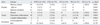
The subtype analysis of the psychic aura showed that the memory-related psychic auras had a significantly different distribution among the 5 categories of lobar epilepsy (p=0.021), in which 16 (11.9%) of the 134 subjects with MTLE and 1 (20.0%) of 5 subjects with OLE had memory-related psychic auras, while 2 (3.6%) of 55 subjects with LTLE, 1 (1.5%) of 65 subjects with FLE, and none of 17 subjects with PLE had this type of aura (χ2 test or Fisher's exact test) (Table 3). Among the 6 subjects who had déjà-vu illusions, 5 had lesions in the mesial temporal lobe, and 1 subject had the lesion in the frontal lobe. The frequencies of memory-unrelated type psychic auras were not different among the 5 categories of lobar epilepsy.
With regard to visual auras, the elementary visual hallucinations and visual illusions both showed significantly different distributions, although the significance of this is not clear due to too few subjects that had each subtype of visual aura. Two (40.0%) of 5 subjects with OLE and 2 (11.8%) of 17 subjects with PLE had elementary visual hallucinations, while 2 (1.5%) of 134 subjects with MTLE, 2 (3.6%) of 55 subjects with LTLE, and 2 (3.1%) of 65 subjects with FLE had this type of hallucinations (p=0.004). Whereas 1 (20.0%) of 5 subjects with OLE and 2 (11.8%) of 17 subjects with PLE had visual illusions, 4 (3.0%) of 134 subjects with MTLE, 2 (3.6%) of 55 subjects with LTLE, and none of 65 subjects with FLE had them (p=0.031). Although statistical significance was not reached, the frequency of complex visual hallucinations tended to be different among the 5 categories of lobar epilepsy, in which complex visual hallucinations (n=2) were observed in LTLE (n=1) and PLE (n=1), but not in MTLE, FLE, or OLE (Fisher's exact test, p=0.064).
Emotional auras were subdivided into either good feelings or bad feelings. Good feelings were observed only in those with left-sided LTLE (n=2), while bad feelings were observed in all of the lobar epilepsies studied (Table 3 and 4). No remarkable hemispheric preference was found in any of the other subtypes of psychic, visual, and emotional auras (Table 4).
Relationships between categories of auras
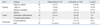
Spearman's correlation analysis showed that 5 pairs of aura categories had concurrent tendencies among the 13 aura categories (Table 5). The pairs of aura categories were epigastric and autonomic auras (p=0.01, ρ=0.155), autonomic and emotional auras (p=0.007, ρ=0.161), visual and vestibular auras (p<0.001, ρ=0.263), auditory and vestibular auras (p=0.021, ρ=0.139), and whole body sensation and auditory auras (p=0.002, ρ=0.19). A significant association between autonomic and emotional auras was observed in left-sided epilepsy subjects (p=0.01, ρ=0.204), but not in right-sided epilepsy subjects (p=0.292, ρ=0.098). Of the two subtypes of emotional auras, bad feelings showed concurrent tendency with autonomic auras (p=0.004, ρ=0.172) in left-sided epilepsies (p=0.005, ρ=0.223), but not in right-sided epilepsies (p=0.292, ρ=0.098). Among the 3 subtypes of visual auras, only elementary visual hallucinations (p<0.001, ρ=0.274) and visual illusions (p=0.002, ρ=0.183) were concurrent with vestibular auras. However, the strength of correlation of all of these items was considered weak because the correlation coefficients were all lower than 0.3.
DISCUSSION
In this study, we investigated the localizing and lateralizing values of auras in subjects with a unilateral single lobar lesion, based on MRI results on an outpatient basis, but not in surgically treated patients. The proportion of subjects with TLE in our study (68.5%) seemed to be relatively higher than what would be expected in a general epilepsy population. This might be explained by the dominance of HS in lesional epilepsy, the small number of subjects with lesions that were confined to either the occipital or parietal lobes that were of relatively small size, and exclusion of subjects with parietal or occipital lesions (epilepsies) but temporal interictal epileptiform discharges (IEDs) on EEG.11,12
The frequency of auras in our study subjects was 64%. This percentage is lower than 81% that was reported in Palmini and Gloor's2 study of 179 subjects with mixed lobar epilepsies, who underwent prolonged EEG monitoring or surgical treatment. However, our percentage is identical to 64% that was reported in a study that had investigated the relationship between auras and the lateralization of the EEG abnormalities for 290 subjects with TLE on an outpatient basis, which is a population that could be expected to have a higher frequency of auras.13 The mean number of auras and the proportion of subjects with at least one aura were significantly lower in subjects with FLE, and this finding was consistent with a previously published study.2
Localizing value of auras
In the present study, the association between epigastric auras and MTLE is consistent with the previously published findings.2,13,14 Epigastric auras are occasionally observed in subjects with extratemporal epilepsy, particularly FLE subjects,15 and can be reproduced by the stimulation of the extratemporal lobe, as well as the temporal lobe.16 This is in agreement with our findings that epigastric auras were also observed in three FLE subjects, but not in any of the PLE or OLE subjects.
Autonomic symptoms, such as sweating, sialorrhea, palpitation, and respiratory difficulty, were included as symptoms of autonomic auras, while we categorized nauseous feelings as a symptom of epigastric auras. However, epigastric and autonomic auras were classified as only one category of viscerosensory aura in previous studies.1,2 In this study, autonomic auras also showed a pattern of distribution similar to epigastric auras. However, the autonomic auras were more frequently reported by subjects with MTLE than those with LTLE.
Psychic auras correspond to the experiential auras discussed in Palmini and Gloor's study,2 except that, the complex visual and auditory hallucinations were categorized as visual and auditory auras, respectively in our study. Psychic auras auras were frequently reported by subjects with TLE. Similar to the results of Palmini and Gloor's study, in which three patients with occipital and two patients with frontal lobe seizure focus showed experiential auras,2 two subjects with FLE and one subject with OLE in our study also had psychic auras. Considering the fact that psychic symptoms were reproduced by electrical stimulation of the temporal lobe,2,17-21 we speculate that psychic auras in the subjects with OLE and FLE may result from the spread of seizure discharge to the temporal lobe structures.
When the psychic auras were subdivided into memory-related type and memory-unrelated type, only the memory-related type had a tendency to be more common in MTLE subjects. This may be due to the memory-related structures, such as the hippocampus or the amygdala, which are located in the mesial temporal lobe.
The association of visual auras with PLE and OLE is consistent with what has been previously reported.2 In the present study, complex visual hallucinations were regarded as visual auras that were then subdivided into three subcategories; visual illusions, elementary visual hallucinations, and complex visual hallucinations. Among the three subtypes of visual auras that were studied, visual illusions and elementary visual hallucinations were associated more with PLE and OLE. Our finding that complex visual hallucinations were observed in LTLE and PLE subjects, but not in OLE subjects is in agreement with the findings of a previous study on visual auras.9 Although elementary visual hallucinations were not reported in FLE in previous studies,2,22 two subjects with FLE in this study had their lesions near the parietal lobe and had reported nonspecific features of elementary visual hallucinations, such as darkening and orange sunshine in their entire visual field.
In the present study, emotional auras were observed in all of the categories of lobar epilepsy. The two patients in our study who reported euphoria as their auras had left-sided LTLE. This localizing and lateralizing value requires further investigation with more patients in order to confirm this finding.
All six subjects with olfactory auras in this study had TLE. Among these subjects, two had atrophic lesions in the temporal pole area. The other four subjects had lesions in the mesial temporal region, which commonly involved the amygdala and the types of lesions were a benign tumor, HS, cortical dysplasia, and an atrophic lesion. Olfactory auras were produced by the stimulation of the mesial temporal structures, which include the uncus and the amygdala or the olfactory bulb,19 and are associated with MTLE and mesial temporal pathology.6,10 In this study, the olfactory auras in the subjects with LTLE may be due to the spread of the seizure activity to the mesial temporal lobe structure or due to the involvement of the mesial temporal lobe structure related to head trauma, which cannot be resolved with a MRI.
Of the four subjects with auditory auras, three had lesions in the temporal lobe, and the other one subject had a lesion in the parietal lobe. The subject with PLE had a focal infarction in the left parietal lobe and an elementary auditory hallucination that presented itself as a buzzing sound. This finding is different from previous reports in that all subjects with auditory aura had TLE.1,2 Since this patient also experienced complex visual hallucinations and whole body sensations, the auditory aura may be due to the spread of seizure discharge from the parietal to the temporal lobe structure.
In the analysis of aura frequency between the subjects with specific lobar epilepsy and those without, whole body sensations and somatosensory auras were found to be more common in PLE subjects. This finding is consistent with previously published results that the somatosensory auras are associated with PLE patients.2
Vestibular, cephalic, and dysphasic auras had no definite localizing value in this study. Cephalic auras, which have been reported to have a significant association with FLE in a previous study,2 were diffusely distributed in all of the categories of lobar epilepsy. Penfield and Jasper23 have reported that electrical stimulation in either the parietal or temporal regions were observed to produce a sensation of vertigo. In this study, however, the vestibular auras were reported in all lobar epilepsies, which is consistent with the findings of another previously published study.2 Vertigo is thought to be caused by the mismatch between visuospatial and vestibular information,24 which could explain the significant correlation between vestibular and visual auras in our study.
Lateralizing value of auras
Previous studies on the lateralizing value of auras failed to confirm their findings.1,2 Only in an EEG-clinical correlation study, Gupta, et al.13 were able to observe that autonomic and psychic auras were more frequently associated with right-sided EEG abnormalities in TLE patients. In the present study, only the dysphasic auras were found to correlate with the side on which the lesion was present, verified by the brain MRI. The correlation of dysphasic auras with left-sided epilepsy seems to be plausible since the language centers are usually located in the left hemisphere of the brain. However, previous studies did not categorize this type of aura into its own aura categorization.1,2,13 In our study, psychic auras and déjà vu illusions did not show any significant preference for lateralization. In Palmini and Gloor's2 study, there was a strong trend of experiential auras and déjà vu illusions to originate from the right temporal lobe. Some stimulation studies showed that electrical stimulation of the right temporal lobe yielded more experiential responses and déjà vu illusions than that of the left lobe.17,18
In this study, vestibular auras tended to occur more frequently in subjects with right-sided epilepsy (seven out of ten subjects), although previous studies did not find lateralizing value of vestibular auras.1,2,13 Considering the dominant right hemispheric influence on visuospatial information processing, our data reflect the possibility that the right hemisphere may play a significant role in the expression of the vestibular aura. Furthermore, our present study showed that the olfactory auras tended to be more common in right-sided epilepsy (p=0.087), in which five of the six subjects with olfactory auras had right-sided epilepsies. However, there has been no significant preponderance for lateralization in previous studies.2,6,10,13
In this study, there was a significant correlation between autonomic auras and the emotional auras, particularly bad feeling in left-sided epilepsy patients. Lee, et al.25 found that patients with left-sided TLE exhibited autonomic hyperarousal when viewing negative emotional slides relative to controls and those with right-sided TLE. They suggested that the dysfunction of the left mesial temporal lobe structures may result in autonomic hyperarousal and a release of the unrestrained negative emotional tendencies of the right hemisphere. Morrow, et al.26 also reported less prominent autonomic arousal to emotionally loaded visual materials in persons with non-dominant hemisphere damage than in the control subjects or those with dominant hemisphere damage. Therefore, the association of autonomic auras and emotional auras in left-sided epilepsy patients might be explained in a similar context.
There are major limitations in this study. Patients with only lesional epilepsy were selected. Therefore, without intracranial EEG evaluations or surgical outcomes, the possibility of false localization of the epileptogenic zone by lesion exists, especially in patients with atrophic lesions that are related to infectious, traumatic, or unknown etiology. The exact nature of auras was not confirmed by video-EEG monitoring in most patients. Patients with an extratemporal lesion (epilepsy) who could show temporal IEDs were excluded in this study, which may reflect too small number of patients with OLE, thus preventing appropriate analysis of OLE. These limitations may make the present results difficult to apply to overall partial epilepsy. However, the analysis of auras in patients with a lesion highly suggestive of epileptogenic focus may provide some clinical value.
In conclusion, our results suggest that the previously known localizing value of auras in surgical epilepsy patients can be applied to patients with partial epilepsy on an outpatient basis. In addition, with a broader range of subjects, we were able to find the lateralizing values of dysphasic auras and the laterality-specific concurrent tendency of autonomic-emotional auras.
 XML Download