

 PDF
PDF ePub
ePub Citation
 Print
Print


INTRODUCTION
The adrenal glands have gained several advantages especially because of laparoscopic surgery due to its small size and deep location in the abdomen, and the laparoscopic adrenalectomy has emerged as a standard surgical procedure in the vast indications of the diverse adrenal diseases.1-3 Recently, advanced laparoscopic skills accumulated from experience and new developments in laparoscopic instruments have led to the introduction of single port laparoscopic adrenalectomy.
Herein, we present two cases of initial right and left transumbilical single port laparoscopic adrenalectomies.
CASE REPORT
Case 1 (right adrenalectomy)
A 38-year-old female patient was admitted to the emergency room with epistaxis. Evaluation revealed that she had hypokalemia (2.2 mmol/L; reference range: 3.5-5.5 mmol/L) and hypertension (220/130 mm Hg). High serum aldosterone level (1174 pg/mL; reference range: 10-105 pg/mL) was revealed, and computed tomography (CT) discovered a 2.5-cm sized well-defined mass on the right adrenal gland. She was diagnosed with primary hyperaldosteronism, based on the right adrenal adenoma.
The patient was placed in a left lateral kidney position, which was established by flexing the operative table with elevation of the kidney bar on just over the iliac crest.4
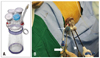
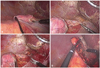
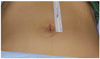
A 2 cm vertical transumbilical skin incision was made and deepened into the peritoneum. The Glove port (Nelis, Seoul, Korea) (Fig. 1A) was inserted through the incision, and the inner ring placed below the peritoneum using the Kelly and the outer ring was rolled down to fix the port on the abdominal wall. At this point, the trocar channel for laparoscope should be placed in the 6-o'clock position (Fig. 1B). The pneumoperitoneum was established, and a 10 mm-45 cm long-45 degree forward oblique bariatric laparoscope was inserted. A snake retractor was inserted to retract and elevate the right lobe of liver medially by the second assistant, which allowed the right adrenal gland visible (Fig. 1B). An articulating laparoscopic coagulator was inserted to dissect the right triangular ligament and inferior border of the liver. The retroperitoneum covering the adrenal gland and inferior vena cava was incised using the articulating hook coagulator with an aid of a 45 cm long straight laparoscopic grasper (Fig. 2A). The adrenal vein could then be identified at the medial side of the adrenal gland and carefully isolated using the articulating dissector (Fig. 2B) and ligated using 5 mm endo-clips (Fig. 2C). Once the adrenal vein was controlled, the tissues around the adrenal gland were completely dissected using the articulating hook coagulator with clipping of vasculatures (Fig. 2D). The specimen was retrived using the endopouch and the umbilical wound was closed without a drain (Fig. 3). There was no estimated blood loss and the total operating time was 60 minutes.
The patient resumed an oral diet the next day and was discharged on the 5th postoperative day in good general condition.
Case 2 (left adrenalectomy)
A 31-year-old female patient was admitted with edema of the extremities, central weight gain and muscle weakness for a period of one month. Her 24 hour urine free cortisol level was 1372 µg/day (reference range: 55.5-286.0 µg/day). A CT scan revealed a 2.9-cm sized left adrenal mass, and she was diagnosed with primary adrenal Cushing's syndrome.
The patient was placed in a right lateral kidney position and prepared in the same manner as the patient in case 1.
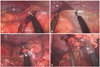
The white line of Toldt was dissected using an articulating laparoscopic hook coagulator with an aid of the laparoscopic grasper (Fig. 4A). The lienorenal ligament was incised, and the dissection was deepened through the plane between the colonic mesentery and Gerota's fascia (the superomedial side of the kidney) retracting the pancreas and spleen medially with the laparoscopic grasper (Fig. 4B). After the left renal vein was identified, the left adrenal vein was easily found (Fig. 4C) and ligated with 5 mm endoclips. After resection of the left adrenal vein, the tissues around the adrenal gland were completely dissected using the harmonic scalpel (Fig. 4D). The specimen was extracted using the endopouch and drain tube was not indwelled. There was no estimated blood loss, and the total operating time was 70 minutes. The postoperative course was uneventful and steroid was tapered. She was discharged on the 4th postoperative day.
DISCUSSION
Although several published studies have introduced single port laparoscopic adrenalectomy, the procedure is still in its infancy and have been performed via diverse methods.5-9
The transperitoneal approach using the vertical transumbilical incision used in the present study has several advantages; 1) providing sufficient working space and allowing early ligation of the adrenal vein6 compared with the retroperitoneal approach, 2) apparent cosmetic effect because the scar recedes into the umbilicus, 3) easy access to open and close without muscle splitting, and 4) easy wound extension.
The transperitoneal approach could be applied in a patient with bilateral adrenal lesions or other pathologic conditions of the abdominal organs such as the gallbladder, inguinal hernia, appendix, ovary, uterus, urinary system, and others.
However, the single port laparoscopic adrenalectomy requires some technical challenges. Working in a single and small fulcrum need instrument triangulation, which is essential for retraction and dissection. For the establishment of proper triangulation, one articulating main working instrument and counterintuitive movement of the long straight instrument in surgeon's other hand makes it possible. Crowding of instruments is an another major obstacle. Therefore, we used a 45 degree forward oblique laparoscope which enabled the surgeon to avoid a parallel between instruments. A 45 cm length bariatric laparoscope with 90 degree light cable adapter and long instruments helped to prevent external crowding, making a distance between the hands of operator and assistants. The Glove port (Nelis, Seoul, Korea) also plays a part. The port is composed of two rings (inner ring and outer ring to fix the port on the abdominal wall) and four trocar channels with gas insufflation and exsufflation lines. The two rings of the Glove port offers a relatively flexible fulcrum compared with other ports, and it's four trocar channels allows proper retraction of the adjacent organs, allowing an addition of a retractor or grasper whenever it is needed without additional port insertion: this is important for the safety and securing proper operative field. Furthermore, the trocar channel of the Glove port enables to use 10 mm laparoscope camera with high resolution and wide optic angle image, which makes a fine manipulation of the tissues possible and provides a comfort operative field for the surgeon.
As most of laparoscopic surgeons feel, a small disturbance in a setting of the instruments makes the operation impossible or difficult. In that point of view, our technique suggests many tips for successful and safe single port laparoscopic adrenalectomy, based on our accumulated experience of laparoscopic adrenalectomy and single port laparoscopic cholecystectomy using this Glove port.
We believe that the technique presented herein is technically feasible and safe. The potential benefits of this technique should be proven in the near future with careful selection of indications.
 XML Download