

 PDF
PDF ePub
ePub Citation
 Print
Print


INTRODUCTION
Lumbar spine bone mineral density (BMD) measured by dual energy X-ray absorptiometry (DXA) is known to be the best predictor of fracture risk:1-3 DXA beam is attenuated by bone and its surrounding soft tissue. However, it is not known how much of the attenuation is influenced by bone and soft tissue. DXA is based on the assumption of homogeneity of soft tissue composition in bone and its adjacent soft tissue. Thus, some inaccuracies can result from DXA measurements, because of it's assumptions on the homogeneity of soft tissue. The influence of ascites on lumbar spine bone density measurements was reported by some researchers,4,5 and the fat in the adipose capsules of the kidney has also been found to lead to overestimation of the bone mineral content (BMC) in spinal BMD DXA measurements.6 The shape of vertebrae as well as thickness and amount of its covering soft tissue varies not only between subjects but also within the same person. Therefore, identifying more influencing factors for DXA measurements are important for lumbar spine BMD interpretation in order to decrease DXA measurement error.
To our best knowledge, no study has been carried out to examine the change in DXA measured lumbar spine BMC and BMD due to changes in the different soft tissue composition of the abdominal area. The purpose of this study was to evaluate how similar changes in fat and extracellular fluid (ECF) volume in the abdominal area influence lumbar spine BMC and BMD, measured with DXA, and differences due to the type of fat (solid versus liquid) were also evaluated.
MATERIALS AND METHODS
Study subjects and design
Following informed consent, healthy volunteers (10 men and 10 women) were enrolled in the study. Control BMD of L1 to L4 was measured for each participant with a LUNAR iDXA scan (GE Healthcare, Waukesha, WI, USA). The scanning precision (coefficient of variance, CV%) was calculated from two repeated measurements with repositioning, which was performed on 20 healthy adults. The precision (CV%) of these measurements was 0.9% for lumbar spine BMD. The following 3 materials were then placed consecutively on the abdominal area, and measurements were repeated: lard 900 g to mimic solid fat (20×15×1.5 cm), oil 1.2 liters (25×15 cm) to mimic liquid fat (purely 100% fat), and water 1.4 liters (25×15 cm) to mimic ECF volume. The same procedure was applied using the Hologic anthropomorphic spine phantom with BMD measured twice under each condition. The phantom which was made of calcium hydroxyapatite enclosed in an epoxy resin was composed of four vertebrae to simulate soft tissue. Four vertebrae have similar densities and areas. All measurements were performed between 3 and 6 pm. The Ethical Committee of Ajou University Hospital approved this study.
Statistics
Data were analyzed using the statistical software package SPSS for Windows 13.0 version. Paired t-tests were used to examine the significance between changes in BMD measurements before and after placing lard, oil, and water on the abdominal area. The independent t-test was used to check for a significant difference in BMD changes in lumbar spine with and without exogenous lard and oil. Results were considered significant at a 2-tailed level of 0.05.
RESULTS
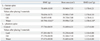
The average participants' age was 30.3±3.5 (mean±SD) years and the average body mass index (BMI) was 21.4±1.2 kg/m2. Tables 1 and 2 show BMC, bone area and BMD differences in lumbar spine and phantom with and without exogenous lard, oil, and water. Bone density of L1 to L4 in human subjects increased with the placement of lard and oil (0.006 g/cm2, p=0.301; 0.008 g/cm2, p=0.250, respectively); however, this was not statistically significant. In the case of water placement, BMC and BMD showed a statistically significant decrease (-0.675 g/cm2, p=0.035; -0.012 g/cm2, p=0.006, respectively). The average percentage of lumbar spine BMD change with and without exogenous lard, oil, and water showed increase of 0.51% and 0.67% and decrease of 1.02%, respectively. With the phantom, bone density decreased after placing both lard (-0.002 g/cm2, p=0.699) and water (-0.006 g/cm2, p=0.153) but there was no difference in bone density after placing oil (0 g/cm2, p=0.870). In men, BMD decreased statistically significantly only when water was placed (-0.019 g/cm2, p=0.040, data not shown). In women, when lard or oil was placed, BMD increased statistically significantly (0.016 g/cm2, p=0.039; 0.03 g/cm2, p=0.040 respectively, data not shown). Differences in lumbar spine BMD between lard and oil, representing solid fat and liquid fat respectively, were not statistically significant (p=0.607, data not shown).
DISCUSSION
The present results indicate that fat, oil, and water influence lumbar BMD values measured by DXA. Lumbar BMD showed a statistically significant decrease upon water placement on the abdominal area and tended to increase slightly with the placement of lard and oil. The average percentage of lumbar spine BMD change with and without exogenous lard, and oil, increased 0.51% and 0.67%, respectively, and decreased 1.02% with water. The difference in lumbar spine BMD change between lard and oil, representing solid fat and liquid fat, respectively, was not statistically significant. These materials showed similar influence on BMD, when measured with a DXA scanner made from Lunar Corp. (Madison, WI, USA) using a phantom particularly in the case of water placement. Consequently, these results suggest that if changes in fat and ECF volume of the human body are similar, ECF exerts a greater influence on lumbar BMD measurements by DXA than fat.
A study showed that total-body bone mineral change, simulated by homogenously placing 8.8 kgs of porcine lard on the bodies of women, increased approximately 1.3%,7 and an other study showed also that there were some changes in soft tissue heterogeneity with weight loss (11.3±6.9 kg), which falsely decreased the anterior-posterior (AP) spinal BMD 1-2% by DXA.8 On the other hand, there was a significant increase in the spine BMD of 4.2%4 after paracentesis (6.4±2.0 L) and there was a significant increase in the spine bone mineral content of 1.5% after drainage of peritoneal dialysate (1,908±508 mL),5 in support of the present results.
To our best knowledge, this is the first report on the impact of human body fluid changes, because of changes in abdominal soft tissue heterogeneity on the accuracy of BMD measurements, measured by DXA. A subtraction or addition of 1 kg of ECF to Reference Man would result in roughly 0.6% over- or under-estimation of total body fat mass in a theoretical model.9 However, there was no significant difference in the data obtained by DXA before and after dialysis.10,11 Of note is the fact that these studies measured body composition with DXA, but not BMD. Lumbar spine BMD could be measured in patients with fluid retention or removal. Significant ascites can cause falsely lower BMD measurements in the spine.4 There was a significant difference in the BMC of the spine before and after the drainage of dialyzate.5 ECF distorts tissue boundaries between bone and soft tissue, resulting in an increased measurement of soft tissue composition and hence a smaller difference between the soft tissue and bone compartments, and consequently underestimating spinal BMD.4 In regards to gender, lumbar BMD was significantly decreased by placing water on the abdominal area in men, but not women. However, these findings should further be confirmed in future by multiple DXA scans of the same subjects as well as a larger numbers of subjects.
In the case of fat and oil used to mimic abdominal fat, the present results show that spinal BMD measurements, following the placement of lard and oil on the abdominal area, were higher, though not significant, than without lard and oil. These results are supported by previous reports that heterogeneity in body fat distribution may theoretically cause inconsistency in the measurements of BMD by DXA due to extrapolation of thickness and fat % of soft tissue in non-bone pixels to bone pixels.12 The present results are also in accordance with a previous cross-sectional study of postmenopausal women with a wide range of BMI,13 which indicated that a difference in fat % between bone and none-bone pixels may lead to overestimation of AP spinal BMD. Higher BMD was obtained when an aluminum phantom surrounded by a 30 : 70 oil/water mixture was used to depict increased truncal thickness,14 and a significant increase in BMD measurement error was observed when ashed bone surrounded by an oil/water mixture representative of 50% body fat was scanned.15 The possible mechanism behind these results has been explained by the study, which showed, the greater the thickness of soft tissue, the greater the resulting X-ray attenuation and interference with bone edge detection, when layers of lard at a thickness of less than 9 cm were placed on a spine phantom.16 However, a previous study demonstrated different BMD's, depending on the brands of DXA scanners used to study the changes of bone mineral measurements during weight change.17 For example, with the Lunar instruments, the total-body BMD was reduced with weight loss, whereas it appeared to increase with the Hologic scanners, indicating that differences in the brands of DXA scanners used may influence BMD value, though there is no real bone change. As for the gender, lumbar BMD was significantly increased by placing lard and oil on the abdominal area of women. However, we found that BMD assessed by DXA using a phantom decreased with the placement of lard. It is quite possible that the thickness of lard used is responsible for these conflicting results.
The limitation of this study is its relatively small sample size compared with other studies, and this might be the reason of why lumbar BMD failed to show a significant decrease after placing water on the abdominal area of women. Nevertheless, the present study is the first pilot study to examine the impact of human body composition change on the accuracy of BMD measurements by DXA: body composition change may result from simultaneous heterogenous changes in abdominal soft tissue, i.e. changes in fat % and volume of soft tissue and ECF of the human body as they correspond to bone and non-bone pixels detected by DXA.
In conclusion, there are differences in spinal BMD measured by DXA before and after placement of oil and lard, especially water, on the abdominal area to mimic abdominal fat and ECF, respectively. If changes in fat and ECF volume are similar, ECF influences the changes in lumbar BMD measurements more than fat; therefore, lumbar BMD measurements need to be interpreted carefully when there is a change, especially in fluid composition, in the soft tissue of the abdominal area.

 XML Download