

 PDF
PDF ePub
ePub Citation
 Print
Print


INTRODUCTION
Cementless total hip arthroplasty was developed as an alternative method to cemented arthroplasty, in which aseptic loosening and subsequent mechanical failure were common problems.1-3 Bone ingrowth by direct contact between the host bone and the porous-coated surface of the prosthesis was expected to achieve stable biologic fixation and prevent component loosening.
Although "first-generation" cementless femoral components showed acceptable prosthetic stability due to bone ingrowth,4-6 problems were reported, including distal femoral osteolysis, stress-shielding, and activity-related thigh pain.7-13 Changes in prosthetic material and design produced second-generation cementless femoral components, using materials with a lower modulus of elasticity (such as titanium alloy) to minimize postoperative thigh pain and femoral stress-shielding. The porous-coated surface was limited to the proximal region of the stem to reproduce a load-transfer pattern similar to that of a normal femur. In addition, proximal porous-coating was applied in a circumferential pattern with no smooth channel that could act as a passage for polyethylene wear particles and cause distal diaphyseal osteolysis.14
The Harris-Galante (HG) Multilock (Zimmer, Warsaw, IN, USA) femoral stem is a second-generation femoral component that is newly characterized by use of titanium alloy with circumferential proximal porous coating. Although the stem produces relatively good short- to mid-term results,15-17 the collared and distal fluted design of the stem (retained from its predecessor) left room for improvement; hence its successor is a femoral prosthesis that is collarless and has a distal tapered design.
The purpose of the present study was to evaluate long-term clinical outcomes, radiographic results, and implant survival rates for patients followed up for a minimum of 10 years after a primary cementless total hip arthroplasty performed using a collared and distal fluted second-generation femoral prosthesis. The effectiveness of the collared and straight distal fluted geometry in the femoral prosthesis was also assessed.
MATERIALS AND METHODS
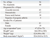
Between August 1991 and February 1996, a single surgeon performed 129 cementless total hip arthroplasties in 105 patients using the HG Multilock femoral stems and cementless acetabular components (Harris-Galante II; Zimmer). Eleven of these patients (15 hips) died within the 10-year postoperative period from conditions not related to the arthroplasty, and none underwent revision surgery before death. Fourteen patients (20 hips) were lost to follow-up within the 10-year period. All 14 patients were last contacted by telephone and had no complaints related to the hip replacement at their last follow up. The remaining 80 patients (94 hips) were available for clinical and radiologic analysis. The mean duration of follow-up was 14.3 years (range, 10 to 18.7 years). Only one patient (one hip) received revision surgery for a femoral component before the 10-year follow-up (six years post-operation); all the other hips were followed for a minimum of 10 years after the operation. Patient demographic data are shown in Table 1.
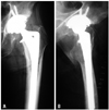
The HG Multilock femoral stem is a straight-collared femoral component made of Ti-6Al-4V alloy (Fig. 1). The implant's features include a proximal circumferential fiber-metal porous coating consisting of a titanium wire mesh that is diffusion-bonded to the substrate. The porous surface area is trapezoidal and extends into the metaphyseal-diaphyseal junction of the stem. The distal portion of the implant is polished and has fixation channels (flutes) that engage the diaphyseal cortex to enhance initial rotational stability.
A posterolateral approach without trochanteric osteotomy was used in all cases. A 28-mm cobalt-chromium alloy femoral head and a ram extruded GUR 4150 (Westlake Plastics, Lenni, PA, USA) ultra-high-molecular-weight polyethylene, machined and sterilized with γ-irradiation in air, was inserted in all patients. Prophylactic antibiotics (third-generation cephalosporin) were given to all patients intraoperatively unless contraindicated, and continued until 2 days after surgery. Anti-thromboembolic stockings were applied to all patients as prophylaxis for deep-vein thrombosis, but anti-coagulation agents were not routinely used except for those who had any risk factors for cardiovascular disease. Partial weight-bearing with two crutches was allowed for 6 weeks post-operation and full weight-bearing was allowed thereafter.
Preoperative clinical and radiographic evaluations were performed as well as postoperative exams at 6 weeks, 3 months, 1 year, and annually after that. The Harris Hip score18 was used for clinical evaluation of function and pain. The results were considered excellent for a Harris Hip score of 90-100, good for a score of 80-89, fair for a score of 70-79, and poor for a score below 70. Activity-related thigh pain was recorded separately. All intraoperative and postoperative complications were recorded.
Standard radiographs included an anteroposterior view of the pelvis and anteroposterior and lateral views of the proximal femur. Two independent observers (C.W.H. and I.H.Y.) who were not involved in the implantation evaluated all radiographs. The immediate postoperative radiograph was evaluated for the contact of the collar and the medial femoral cortical bone (calcar) using the method of Meding, et al.19 The locations of radiographic findings in the femur were recorded using the zones described by Gruen, et al.20 and Johnston, et al.21 Considering the structure of the HG Multilock stem, zones 1 and 2 were divided by the junction of the porous and smooth areas of the implant laterally, zone 4 included the distal smooth tip, zones 3 and 5 divided the remaining smooth portion of the stem laterally and medially, and zones 6 and 7 were divided by the junction of the porous and smooth surfaces of the stem medially.15 Femoral component fixation was graded as bone ingrowth, stable fibrous ingrowth, or unstable implant, using the criteria of Engh, et al.7 Endosteal bone bridging (spot weld) formation, presence of radiolucent lines at the bone-prosthesis interface, and osteolysis were evaluated. Proximal femoral remodeling (stress-shielding) was graded as first degree; calcar rounding only, second degree; calcar rounding and loss of medial cortical density at zone 7, third degree; more extensive resorption of cortical bone at zone 6 and 7, as fourth degree; and cortical resorption extended into the diaphysis.7 Intramedullary bone formation at the distal tip (bony pedestal) was considered 'stable' if the new bone formation was in direct contact with the distal stem tip and if no new radiolucencies or reactive lines formed around the stem tip.22 In contrast, a radiolucency and a line surrounding the distal stem was considered to indicate that the pedestal beneath the stem was unstable.22 Distal cortical hypertrophy was recorded. Change in the position of the stem to varus or valgus was determined by drawing a line through the longitudinal axis of the stem and another line through the longitudinal axis of the proximal femoral canal. Instability of the femoral stem was defined as subsidence of >2 mm or change of the stem position angle of >2°, or a continuous radiolucent line wider than 2 mm.7,23 Linear polyethylene wear was measured with the Picture Archiving Communication System (PACS, General Electrics, Milwaukee, WI, USA) according to the method by Livermore, et al.24
The statistical association between the collar-calcar contact and activity-related thigh pain, the status of stem fixation or the severity of stress-shielding was evaluated using the chi-square test and Fisher's exact test. The level of significance was p<0.05. Kaplan-Meier analysis of the survival of the femoral component was performed for all 129 hips.
The Institutional Review Board of Yonsei University College of Medicine approved this retrospective study.
RESULTS
Clinical results
The average Harris hip score improved from a preoperative 58 points (range, 4 to 93 points) to an 88-point average (range, 29 to 100 points) at the last follow up. Fifty-four hips (57%) had excellent outcomes; 21 hips (22%), good outcomes; 10 hips (11%), fair outcomes; and nine hips (10%), poor outcomes. Of the nine poor outcomes, the low Harris scores of 4 were not related to the total hip replacement; two were related to spinal stenosis, one was complicated with an ipsilateral knee problem, and one was associated with stroke. The other five poor outcomes were related to the acetabular cup or polyethylene liner. Activity-related thigh pain was reported in nine patients (9 hips; 10%). The pain was spontaneously relieved in six hips within the 2-year postoperative period and minor complaints in three hips did not compromise daily activities.
Postoperative dislocations occurred in six patients (six hips); five of these were treated with closed reduction, and re-dislocation was not reported. Only one patient had recurrent dislocation and required revision surgery to exchange the polyethylene liner and femoral head, and tighten the capsule two years after the index operation. The femoral component was stabilized during surgery in this patient.
Nondisplaced metaphyseal fractures (calcar cracks) during insertion of the femoral components occurred in nine patients (nine hips). All of these fractures were treated with two cerclage wirings, and all femoral components subsequently had stable ingrowth.
Radiographic assessment of the femoral components
Stable bone ingrowth occurred in all femoral stems except one (93 of 94 hips, 99%), and stable fibrous ingrowth occurred in the remaining one hip (1%). No loose stems were found upon radiographic assessment. Endosteal spot weld formation was seen in 93 hips (99%). Lateral porous surface (zone 1) was the most common site of endosteal spot weld formation (90 hips, 96%). Distal medial porous surface (distal zone 7) was the second most common site (75 hips, 80%).
Radiolucent lines in one or more zones were observed in 49 hips (52%) upon the last radiographic evaluation. The lines observed at the non-porous smooth surface were ≤1 mm in width, and were not progressive except as observed in one hip. An extensive radiolucent line was seen throughout the entire bone-prosthesis interface on both the anteroposterior and lateral radiographs in one hip, but the width was less than 1 mm, and subsidence or angle change of the stem was not detected. The prosthesis was therefore considered to have stable fibrous ingrowth. No femoral component had subsided or changed its varus-valgus alignment. Bone pedestals were present in 17 hips (18%), but none were accompanied by other findings of stem instability. Distal cortical hypertrophy was seen in three hips (3%).
Proximal femoral osteolysis was seen in 81 hips (86%). The mean time for first radiographic appearance of osteolysis was three years (range, 1 to 6 years) postoperatively. In 79 of the 81 hips, osteolytic lesions were restricted to the proximally located zones (zone 1 and 7) in the form of linear endosteal erosion. In the remaining two hips, however, relatively large osteolytic lesions (4×2 cm and 3×1 cm) were defined at the level of the greater trochanter (zone 1) in the 14- and 15-year postoperative follow-up radiographs, respectively. Although both stems appeared to be stable upon serial radiographic follow-up, the patients received revision surgery of the acetabular components for progression of acetabular osteolysis with severe polyethylene wear. The stability of the femoral stems was checked intraoperatively, and both stems were well-fixed to the femoral bones. Therefore, stem revisions were not performed, and only allogenous bone grafts for the femoral defect area were performed.
Proximal femoral stress-shielding was noted in 86 hips (91%), and eight hips (9%) showed no evidence of femoral stress-shielding upon the latest radiographic follow-up. Forty-six hips (49%) had first-degree stress-shielding and 39 hips (41%) had second-degree stress-shielding. Extensive cortical resorption was noted in one hip at the six-year postoperative point. In spite of stable bony ingrowth with endosteal spot weld in zone 7, the stem was revised because the patient was relatively young, male and highly active. At the latest clinical follow-up (15 years after index surgery, nine years after revision surgery), the patient had a 95-point Harris hip score and no complaints about his hip function.
Contact of the collar and femoral calcar was observed in 44 hips (47%), and collar-calcar contact showed no statistical correlation with thigh pain, the status of stem fixation or stress-shielding (p>0.05).
The linear wear rate was 0.20 mm/year (0.05 to 0.66 mm/year).
Revisions
Forty-four hips (47%) received revision surgery for any reason during the follow-up period (average 10.5 years, range 2.5-18.3 years after surgery). One hip received revision only of the femoral stem because of extensive stress-shielding. In 18 (19%) hips, only the polyethylene liner and femoral head were changed and 13 of these 18 liner-exchange surgeries were performed because of the locking mechanism failure in the Harris-Galante II acetabular prosthesis. The remaining 25 (27%) hips received acetabular component revisions to treat severe pelvic osteolysis with or without acetabular loosening. The stability of every femoral component was tested during the acetabular revision procedure and only one stem was confirmed to have been loosened, although there was no radiographic evidence of femoral loosening during the 15-year postoperative period (stable fibrous ingrowth status). Overall, only two femoral component revisions (one for severe stress-shielding and one for stem loosening) were performed.
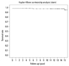
When femoral component revision for any reason was used as the end point, the cumulative survival rate of femoral prosthesis was 99% (95% confidence interval, 98% to 100%) at 10 years and 97% (95% confidence interval, 95% to 99%) at 15 years (Fig. 2).
DISCUSSION
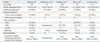
In the present study, stable bone ingrowth with endosteal spot weld formation was noted in 93 of 94 hips (99%) and the 10-year survival rate of the femoral component was 99%. These results compare favorably with those of other studies using the HG Multilock prosthesis (Table 2).15-17,25
Although first-generation cementless femoral prostheses produced stable bone ingrowth, studies also reported rates of diaphyseal osteolysis greater than 10%.11,26-28 Schmalzried, et al.13 described the concept of 'effective joint space', in which polyethylene wear particles can migrate to the distal bone-prosthesis interface via the passage with the lowest intra-articular pressure. Bobyn, et al.29 reported that in animal models, wear particles could migrate to the diaphyseal area more easily through the non-porous surface than through the porous surface. An important feature of second-generation cementless femoral prostheses is circumferential porous coating, which was expected to prevent distal migration of wear particles. For this reason, all osteolytic lesions in the present study were selected to be proximal to the porous-coated surface of the stem. Among these, however, two large osteolytic lesions were observed in the region of the greater trochanter in follow-up radiographs obtained more than 10 years post-operation. Although the osteolytic lesions were not accompanied by any diaphyseal extension or consequent stem loosening, prophylactic bone graft was performed to prevent periprosthetic fracture. In both cases, no radiographic abnormalities appeared before the 10-year postoperative follow-up. Meding, et al.30 also reported observing a large area of osteolysis in the region of the greater trochanter, without stem loosening, at the 10-year follow-up of another circumferential proximal porous-coated cementless femoral prosthesis. Long-term radiographic follow-up is therefore essential, even though notable osteolysis is not commonly associated with second-generation cementless femoral prostheses.
The known causes of activity-related thigh pain include micro-motion of the unstable stem tip and a difference in elastic modulus between the bone and the implant. A high rate (20-33%) of thigh pain was reported in previous studies of first-generation cementless femoral prostheses.31,32 In the present study, the rate of activity-related thigh pain was 10%, and pain persisted in only three hips (3%). These results are favorable compared to those of other studies (Table 2). The specific material and design features of the prosthesis may explain the low rate of thigh pain. The stem is composed of titanium alloy, which has increased biocompatibility and a lower modulus of elasticity than cobalt-chromium alloy. In addition, the close contact between the distal flutes of the stem and the femoral cortex through the line-to-line reaming process would promote early postoperative rotational stability, and the increased depth of the flutes with increasing stem diameter may have the effect of decreasing the diameter of the stem tip and attenuating bending and torsional stiffness similarly in the distal tapered femoral component.
Stress-shielding is a spontaneous reaction of the femur to the altered load-transfer pattern through the stably fixed prosthesis-bone interface. Engh, et al.7 reported a 12% rate of moderate to severe stress-shielding (third or fourth degree) after cementless total hip arthroplasty using an extensively porous-coated, cobalt-chromium alloy femoral stem. In the present study, we observed extensive stress-shielding in only one hip, perhaps because of the proximally limited porous coating and greater flexibility of the prosthetic material. Although the rate of severe stress-shielding was very low, the overall incidence of proximal femoral osteopenia, including minor involvement (first or second degree) was still high (91%). This may be due to the design feature of a straight (non-tapered) distal portion of the prosthesis. When early postoperative stability is obtained by tight distal fixation in a straight femoral stem, the viscoelasticity of the femoral metaphysis can relax the contact pressures at the proximal femoral area, resulting in diminished proximal load-transfer and consequent proximal femoral stress-shielding.33 In contrast, tapered geometry can lead to gradual subsidence of the stem into a tighter relationship with the bone, so that proximal load-transfer can be maintained.33 In support of this theory, Sano, et al.34 compared postoperative changes in femoral bone mineral density (BMD) between the distal tapered and straight fluted types of cementless stems, and found an early decrease of BMD that was recovered after 12 months in the tapered stem group, but a continued decrease in BMD without recovery in the fluted stem group. Moreover, a five-year follow-up study of the Versys Fiber Metal Taper (FMG; Zimmer), which is an advanced model of the HG Multilock prosthesis and which has a distal tapered geometry, gave a better result in terms of stress-shielding (21%).35
Meding, et al.19 showed that the collar has no effect in cementless femoral prostheses, and that in a stem with distal tapered geometry, the collar can block subsidence of the stem and interfere with tight fixation. Therefore, the collar of cementless prostheses should be improved, especially in those with tapered geometry. In the present study, collar-calcar contact showed no relationship to thigh pain, stem fixation, or stress-shielding.
During follow-up of this study, 44 revisions (47%) were performed, most of which (43 out of 44 cases) was due to problems in the locking mechanism of the Harris-Galante II acetabular cup, or pelvic osteolysis caused by polyethylene wear. Both of these problems can be resolved by modification of the cup design or development of an alternative bearing surface.
The retrospective design of our study and the relatively small number of cases (94 hips) available for analysis limit the general application of our results. Also, a direct comparison of the biomechanical performance of straight and tapered stems would have allowed us to address the problem of the straight stem design more specifically. However, because the present femoral prosthesis is no longer available, this long-term follow-up report may prove more valuable as a reference for studies of the newly developed, collarless and distal tapered femoral prosthesis.
In conclusion, this long-term follow-up of total hip arthroplasty using a second-generation cementless femoral prosthesis with collared and straight distal fluted geometry showed satisfactory results for a relatively young patient sample. However, the high rate of proximal stress-shielding and the minimal effect of the collar both indicate the need for some changes to the stem design, especially the collar and straight distal fixation channels.
 XML Download