

 PDF
PDF ePub
ePub Citation
 Print
Print


INTRODUCTION
Screening methods for scoliosis include general physical examinations as well as radiological study including chest radiographs. Forward bending test, angle of trunk rotation and Moire's tomography are the most used first step methods for primary evaluation,1 and the radiography image check is the most common further screening method. Screening with chest radiographs can provide information to physicians on the curve of the thoracic spine, and therefore, chest radiographs has been reported to be useful in scoliosis screening.1,2 However, scoliosis screening using chest radiograph has inherently been limited due to well-known problems such as ignorance of lumbar curve or chest radiograph dependence on arbitral posture. Today, the exact usefulness and limitation of screening programs by chest radiograph for early detection of scoliosis have so far not been examined. Therefore, we investigated the usefulness and restricted values of chest radiographs for screening program of scoliosis.
MATERIALS AND METHODS
Subject selection
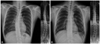
Korea engages in conscription and all men under a medical examination in preparation for this. This survey was conducted at the Regional Military Manpower Administration from April 2008 to May 2010. During this period, 419 men were selected for both chest radiographs and thoraco-lumbar standing radiographs (TLSR) to check the presence of scoliosis (Fig. 1). All examinees were 19 years old male. Their mean height, mean weight, and mean body mass index were 174.8±5.8 cm, 71.9±13.7 kg, and 23.2±3.9, respectively.
Image studies and analysis
The curvature was recorded using both chest radiograph and TLSR. The type of scoliosis was described with respect to the location and pattern of the curve or curves, such as thoracic curve, thoraco-lumbar curve, lumbar curve, and double major curve. The Cobb angle was the crossed angle on the perpendicular line from each end vertebrae that are the vertebrae at the upper and lower limits of the curve which tilted most severely toward the concavity of the curve.1,2 We considered normal spinal curvature to be a Cobb angle of less than 5 degrees, to compare between two different images although many studies defined as lesser than 10 degrees. Cobb angles in chest radiographs and TLSR were recorded by a radiologist, an orthopedic surgeon and a neurosurgeon, independently from each other. If the checked Cobb angle was differently depending on different physicians, the Cobb angle was rechecked, and the median angle was selected.
Statistical analysis
To estimate the usefulness of chest radiographs for scoliosis screening, the sensitivity and specificity of chest radiographs were calculated. The Cobb angle accuracy was defined more than five degrees as a meaningful difference between chest radiographs and TLSR. A statistical analysis was performed using SAS (version 9.1.3, SAS Institute, Inc., Cary, NC, USA). Student t-test was used to compare the tendency of changed curve type of right thoracic curve and left thoracic curve in chest radiographs. Scattered plot and coefficient of correlation were also used to check the distribution and relation of Cobb angle between chest radiographs and TLSR. Intraclass correlation was used for interobserver variability.
RESULTS
Interobserver variability
Cobb angles in chest radiographs and TLSR were recorded by a radiologist, an orthopedic surgeon and a neurosurgeon independently from each other. Intraclass correlation between a radiologist and an orthopedic surgeon was found to be 0.910, 0.927 between a radiologist and a neurosurgeon, and 0.925 between an orthopedic surgeon and a neurosurgeon. Cases in which the median Cobb angle was selected, because of more than 5 degrees of different Cobb angle by different physicians, was 23, but no cases exceed 10 degrees of Cobb angle difference.
Spinal curve pattern matching of chest radiographs and TLSR
A total of 419 examinees were examined using chest radiographs and TLSR. The overall matching rate with the focuson the pattern of spinal curvature of chest radiographs with TLSR was about 58.2% (244 among 419 cases) (Table 1). Abnormal thoracic curvature on chest radiograph was observed in 234 cases; 186 exhibited right thoracic curvature and 48 with left thoracic curvature. The thoracic curve patterns observed in chest radiographs were matched only in 122 cases (52.1%) to those in TLSR. Conversely, 110 cases (47.0%) exhibited a change in their final TLSR results with respect to spinal curvature such as normal spinal curve (15 cases, 6.4%), thoraco-lumbar curve (35 cases, 15.0%), lumbar curve (37 cases, 15.8%), and double major curve (23 cases, 9.8%). This tendency of chang of curve type was stronger in those with right thoracic curves than those with left thoracic curve on chest radiographs (p-value=0.001). The curve pattern match rate was 62% in the thoraco-lumbar curve (50 cases) on chest radiograph, and the others were shown as thoracic curve (2 cases, 4%) and lumbar curve (13 cases, 26%) on TLSR. Additionally, lumbar curves (19 cases) on chest radiograph were presented as thoraco-lumbar curve (1 case, 5.2%) on TLSR, and curve patterns were matched in 89.5%.
Cobb angle accuracy of chest radiographs and TLSR
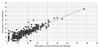
As for the Cobb angle accuracy, without consideration of curve type match, 263 cases (62.8%) were shown to have less than five degrees Cobb angle difference between chest radiograph and TLSR (Table 2). Conversely, the meaningful difference between chest radiographs and TLSR was observed in 156 cases (37.2%). A scatterplot with raw data and a corresponding fitted regression line shows the distribution and relation of the Cobb angle between chest radiograph and TLSR, and the plot is provided in Fig. 2. The mean difference in Cobb angle between chest radiographs and TLSR was 4.02 (0-24) with a coefficient of correlation of 0.903 (p<0.001). However, there was relatively high proportion of cases (9.5%) with greater than 10 degree differences in Cobb angles (Table 2).
Coincidence of spinal curve type and Cobb angle accuracy
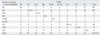
The coincidence of both types of spinal curves and accuracy of Cobb angle (a Cobb angle difference of less than five degree between chest radiograph and TLSR) was 27.9% (117 among 419 cases) (Table 1). Each accuracy according to the type of spinal curve was as follows: normal spinal curve, 64.7% (66 among 102 cases); right thoracic, 36.0% (67 among 186 cases); right thoraco-lumbar curve, 38.5% (5 among 13 cases); right lumbar curve, 80.0% (3 among 5 cases); double major curve convexity right to left, 38.5% (5 among 13 cases); double major curve convexity left to right, 0% (0 among 1 case); left thoracic curve, 35.4% (17 among 48 cases); left thoraco-lumbar curve, 40.5% (15 among 37 cases); and left lumbar curve, 35.7% (5 among 14 cases).
Sensitivity and specificity of chest radiographs
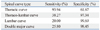
The sensitivity and specificity with chest radiograph and TLSR with resepct to spinal curvature are provided in Table 3. Regarding thoracic curve, the sensitivity was high (93.94%), but the specificity was low (61.67%). On the contrary, there was a low sensitivity and a high specificity in thoraco-lumbar, lumbar, and double major curvature. Right thoracic curvature on chest radiograph showed normal spinal curves (4.8%), right thoraco-lumbar curves (7.0%), double major curve convexity right to left (10.8%), left thoracic curve (1.1%), left thoraco-lumbar curve (10.8%), and left lumbar curve (16.1%) on the TLSR. This tendency of change of right thoracic curve from chest radiographs to TLSR may be categorized with respect to the pattern curve of S shape, with 83.3% of thoracic curves on chest radiograph belonging to this pattern (Table 4). Left thoracic curvature in chest radiograph was shown in TLSR as normal spinal curvature (12.5%), double major curve convexity left to right (6.3%), left lumbar curve (8.3%), right thoraco-lumbar curves (4.2%), and right lumbar curve (6.3%). Furthermore, 75.0% of them belonged to the inverted S shape curve (Table 5).
Congenital vertebral abnormal cases
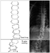
In this study, two cases of congenital vertebral abnormalities were observed, and their curve pattern on chest radiographs changed to other pattern on TLSR (Table 1). Right lumbar curve on TLSR was misinterpreted as left lumbar curve on chest radiograph, which was a congenital vertebral abnormality of the hemivertebra of 4th lumbar spine (Fig. 3). Similarly, a case of congenital vertebral abnormality, which had bilateral failure of segmentation from the 2nd to 4th lumbar spine, was presented as a right thoracic curve on chest radiograph but was finally represented as left lumbar curve on TLSR.
DISCUSSION
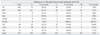
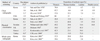
A literature review demonstrated that the prevalence of adolescent scoliosis with more than a 10 degree Cobb angle varied greatly from 0.3% to 12.6% because of different methods used in the screening of scoliosis as well as variation in the geography. Physical examinations are the most frequently used first step methods for primary evaluation, and the radiography image check is used for further screening. However, this step has often been neglected.2-6 Table 6 provides a summary of the prevalence of scoliosis and the proportion of scoliosis type according to the method used for scoliosis screening. The prevalence of scoliosis was relatively higher in studies screening using chest radiographs as opposed to those using physical examination. A higher proportion of thoracic and thoraco-lumbar curvature was prominent when screening with chest radiographs as compared to other types and was comparable to curve distribution observed when screening by physical examination. This tendency is not different from the current data. Our data showed a high proportion of thoracic and thoraco-lumbar curve scoliosis (a Cobb angle greater than 5 degrees) in screening by chest radiograph with thoracic in 55.8% of cases, thoraco-lumbar in 11.9%, lumbar in 4.5%, and double major curve in 3.3%. This proportion is similar to that of other studies using chest radiograph to screen for scoliosis.3-6 However, this distribution changed to thoracic 31.5%, thoraco-lumbar 19.3%, lumbar 21.5%, and double major curve 7.4% on TLSR, the finding that is more similar to the proportions in studies using physical examination or whole spine radiographs.7-14
The different proportions were arbitrarily created by the restricted field of sight on chest radiograph. On chest radiograph, the whole thoracic spine and upper lumbar spine can be included in the radiographic field, but the area below the mid lumbar area is easily excluded (Figs. 1 and 3). Chest radiographs have been very useful in detecting not only lung parenchyma disease but also scoliosis in the thoracic spine. Furthermore, the spinal alignment on chest radiograph could be flexible depending on the position, given the fact that most curves we observed on chest radiograph lacked consistency. In this study, a total of 19 cases were examined for normal spinal curvature in TLSR, although they were observed to have scoliosis on chest radiographs. A further limitation of chest radiographs is that the lumbar spinal curve is hidden, therefore, the observer cannot detect lumbar scoliosis. Furthermore, thoracic or thoraco-lumbar curves have been misunderstood as the S or inverted S shaped patterns (Table 4 and 5). The S curve patterns (222 cases; right thoracic curve, right thoraco-lumbar curve, double major curve convexity right to left, left lumbar curve of TLSR in Table 1) were more common than inverted S patterns (114 cases; left thoracic curve, left thoraco-lumbar curve, double major curve convexity left to right, right lumbar curve of TLSR in Table 1), and it made stronger tendency to change the curve type of right thoracic curves in chest radiographs than left thoracic curves (p<0.001). And, another reason for different result by chest radiographs and TLSR could be due to different position, inspiration/expiration difference, and posterior-anterior/posterior-anterior image difference. Although both chest radiographs and TLSR were taken by standing position, careful correction of radiographic position was carried out by TLSR. Chest radiographs can differently be checkable by inspiration or expiration, and it could contribute to the Cobb angle difference between chest radiographs and TLSR. Also, cinematography view could contribute to the difference as chest radiographs by posterior-anterior view and TLSR by anterior-posterior view.
In this study, the use of chest radiograph in scoliosis screening exhibited a high sensitivity and low specificity for thoracic curves, and very low sensitivity and high specificity for thoraco-lumbar, lumbar and double major curves (Table 3). This result indicates that chest radiographs are excellent in detecting thoracic type scoliosis, and poor in the detection of thoraco-lumbar, lumbar, and double major curves.
Sugita, et al.2 suggested that tuberculosis examination radiographs may be useful for scoliosis screening in high schools. They examined 2068 first year high school students who had chest radiographs taken, and found 24 cases with scoliosis involved a Cobb angle of more than 10 degrees. The correlation coefficient between the Cobb angle measured in the tuberculosis examination radiographs and in the total spinal radiographs taken by the hospital was 0.815 (p<0.001). In a recent study, the correlation coefficient between the Cobb angle measured in chest radiograph and TLSR was 0.903 (p<0.001). However, 37.2% of cases exhibited a greater than five degree difference in Cobb angle between chest radiograph and TLSR, and 9.5% exhibited a Cobb angle difference of more than five degrees (Table 2). Moreover, the coincidence of both types of spinal curve and accuracy of Cobb angle, with a difference of less than five degrees between chest radiograph and TLSR, was only 27.9%.
In conclusion, the coincidence of spinal curve type was 58.2% and the consentaneity of Cobb angle was 62.8% between chest radiographs and TLSR. The accuracy for using chest radiographs as scoliosis screening was only 27.9%. Furthermore, thoracic curve scoliosis was overestimated, and lumbar curve scoliosis was easily missed on chest radiographs. Scoliosis screening using chest radiography has limited values, nevertheless, it is useful method for detecting thoracic curve scoliosis.
 XML Download